
Key Product Details
Species Reactivity
Validated:
Human, Mouse, Rat
Cited:
Human, Mouse, Rat
Applications
Validated:
Immunohistochemistry, Western Blot, Immunocytochemistry
Cited:
Immunohistochemistry, Western Blot, Immunoprecipitation, Bioassay
Label
Unconjugated
Antibody Source
Polyclonal Goat IgG
Loading...
Product Specifications
Immunogen
E. coli-derived recombinant human AMPK alpha 1
Lys349-Gln559
Accession # Q13131
Lys349-Gln559
Accession # Q13131
Specificity
Detects human, mouse, and rat AMPK alpha 1 in Western blots. The antibody does not cross-react with recombinant human AMPK alpha 2.
Clonality
Polyclonal
Host
Goat
Isotype
IgG
Scientific Data Images for AMPK alpha 1 Antibody
AMPK alpha 1 in Human Liver.
AMPKa1 was detected in immersion fixed paraffin-embedded sections of human liver using 5 µg/mL Goat Anti-Human/Mouse/Rat AMPKa1 Antigen Affinity-purified Polyclonal Antibody (Catalog # AF3197) overnight at 4 °C. Tissue was stained with the Anti-Goat HRP-DAB Cell & Tissue Staining Kit (brown; Catalog # CTS008) and counterstained with hematoxylin (blue). Lower panel shows secondary antibody only control experiment. View our protocol for Chromogenic IHC Staining of Paraffin-embedded Tissue Sections.
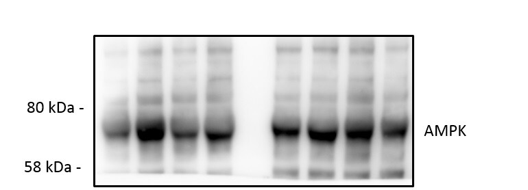
Detection of Human. Mouse, and Rat AMPK alpha 1 by Western Blot.
Western blot shows lysates of HeLa human cervical epithelial carcinoma cell line, C2C12 mouse myoblast cell line, and Rat-2 rat embryonic fibroblast cell line. PVDF membrane was probed with 2 µg/mL of Goat Anti-Human/Mouse/Rat AMPKa1 Antigen Affinity-purified Polyclonal Antibody (Catalog # AF3197) followed by HRP-conjugated Anti-Goat IgG Secondary Antibody (Catalog # HAF017). A specific band was detected for AMPKa1 at approximately 70 kDa (as indicated). This experiment was conducted under reducing conditions and using Immunoblot Buffer Group 1.
AMPK alpha 1 in MCF‑7 Human Cell Line.
AMPKa1 was detected in immersion fixed MCF-7 human breast cancer cell line using Goat Anti-Human/Mouse/Rat AMPKa1 Antigen Affinity-purified Polyclonal Antibody (Catalog # AF3197) at 5 µg/mL for 3 hours at room temperature. Cells were stained using the NorthernLights™ 557-conjugated Anti-Goat IgG Secondary Antibody (red; Catalog # NL001) and counterstained with DAPI (blue). Specific staining was localized to nuclei and cytoplasm. View our protocol for Fluorescent ICC Staining of Cells on Coverslips.
Detection of AMPK alpha 1 by Western Blot
TSPAN1 knockdown decreases cell proliferation in OCCC cell lines. (A) TSPAN1 expression was detected by real‐time PCR (left panel) and immunoblotting (right panel) in TOV‐21G and OVTOKO cells, with alpha ‐actinin as an internal loading control. Data are expressed as the mean ± standard error (SE); n = 3. Unpaired t‐test was performed. *P < 0.05, **P < 0.01. (B) Cell proliferation was determined via crystal violet staining at various times after transient transfection of siControl or siTSPAN1 in TOV‐21G and OVTOKO cells. Error bars represent mean ± SE; n = 3. Unpaired t‐test was performed. **P < 0.01, ***P < 0.001. (C) Colony forming assay was performed in TOV‐21G and OVTOKO cells. Upper panel shows representative images; lower panel shows the relative absorbance at 595 nm. Error bars represent mean ± SE; n = 3. Unpaired t‐test was performed. **P < 0.01, ***P < 0.001. (D) Protein expressions were detected using immunoblotting after transient transfection of siControl or siTSPAN1 in TOV‐21G and OVTOKO cells, with alpha ‐actinin as an internal loading control. Image collected and cropped by CiteAb from the following open publication (https://pubmed.ncbi.nlm.nih.gov/33331115), licensed under a CC-BY license. Not internally tested by R&D Systems.Applications for AMPK alpha 1 Antibody
Application
Recommended Usage
Immunocytochemistry
5-15 µg/mL
Sample: Immersion fixed MCF-7 human breast cancer cell line
Sample: Immersion fixed MCF-7 human breast cancer cell line
Immunohistochemistry
5-15 µg/mL
Sample: Immersion fixed paraffin-embedded sections of human liver
Sample: Immersion fixed paraffin-embedded sections of human liver
Western Blot
2 µg/mL
Sample: HeLa human cervical epithelial carcinoma cell line, C2C12 mouse myoblast cell line, and Rat‑2 rat embryonic fibroblast cell line
Sample: HeLa human cervical epithelial carcinoma cell line, C2C12 mouse myoblast cell line, and Rat‑2 rat embryonic fibroblast cell line
Reviewed Applications
Read 2 reviews rated 5 using AF3197 in the following applications:
Formulation, Preparation, and Storage
Purification
Antigen Affinity-purified
Reconstitution
Reconstitute at 0.2 mg/mL in sterile PBS. For liquid material, refer to CoA for concentration.
Loading...
Formulation
Lyophilized from a 0.2 μm filtered solution in PBS with Trehalose. *Small pack size (SP) is supplied either lyophilized or as a 0.2 µm filtered solution in PBS.
Shipping
Lyophilized product is shipped at ambient temperature. Liquid small pack size (-SP) is shipped with polar packs. Upon receipt, store immediately at the temperature recommended below.
Stability & Storage
Use a manual defrost freezer and avoid repeated freeze-thaw cycles.
- 12 months from date of receipt, -20 to -70 °C as supplied.
- 1 month, 2 to 8 °C under sterile conditions after reconstitution.
- 6 months, -20 to -70 °C under sterile conditions after reconstitution.
Calculators
Background: AMPK alpha 1
Long Name
AMP-activated Protein Kinase alpha 1
Alternate Names
PRKAA1
Entrez Gene IDs
5562 (Human)
Gene Symbol
PRKAA1
UniProt
Additional AMPK alpha 1 Products
Product Documents for AMPK alpha 1 Antibody
Certificate of Analysis
To download a Certificate of Analysis, please enter a lot or batch number in the search box below.
Note: Certificate of Analysis not available for kit components.
Product Specific Notices for AMPK alpha 1 Antibody
For research use only
Related Research Areas
Citations for AMPK alpha 1 Antibody
 Powered by Bioz
Customer Reviews for AMPK alpha 1 Antibody (2)
5 out of 5
2 Customer Ratings
Have you used AMPK alpha 1 Antibody?
Submit a review and receive an Amazon gift card!
$25/€18/£15/$25CAN/¥2500 Yen for a review with an image
$10/€7/£6/$10CAN/¥1110 Yen for a review without an image
Submit a review
Customer Images

Showing
1
-
2 of
2 reviews
Showing All
Filter By:
-
Application: Western BlotSample Tested: EndothelialSpecies: MouseVerified Customer | Posted 10/11/2019

-
Application: Western BlotSample Tested: MDA-MB-231 human breast cancer cell lineSpecies: HumanVerified Customer | Posted 10/17/2018
There are no reviews that match your criteria.
Protocols
Find general support by application which include: protocols, troubleshooting, illustrated assays, videos and webinars.
- Antigen Retrieval Protocol (PIER)
- Antigen Retrieval for Frozen Sections Protocol
- Appropriate Fixation of IHC/ICC Samples
- Cellular Response to Hypoxia Protocols
- Chromogenic IHC Staining of Formalin-Fixed Paraffin-Embedded (FFPE) Tissue Protocol
- Chromogenic Immunohistochemistry Staining of Frozen Tissue
- ClariTSA™ Fluorophore Kits
- Detection & Visualization of Antibody Binding
- Fluorescent IHC Staining of Frozen Tissue Protocol
- Graphic Protocol for Heat-induced Epitope Retrieval
- Graphic Protocol for the Preparation and Fluorescent IHC Staining of Frozen Tissue Sections
- Graphic Protocol for the Preparation and Fluorescent IHC Staining of Paraffin-embedded Tissue Sections
- Graphic Protocol for the Preparation of Gelatin-coated Slides for Histological Tissue Sections
- ICC Cell Smear Protocol for Suspension Cells
- ICC Immunocytochemistry Protocol Videos
- ICC for Adherent Cells
- IHC Sample Preparation (Frozen sections vs Paraffin)
- Immunocytochemistry (ICC) Protocol
- Immunocytochemistry Troubleshooting
- Immunofluorescence of Organoids Embedded in Cultrex Basement Membrane Extract
- Immunofluorescent IHC Staining of Formalin-Fixed Paraffin-Embedded (FFPE) Tissue Protocol
- Immunohistochemistry (IHC) and Immunocytochemistry (ICC) Protocols
- Immunohistochemistry Frozen Troubleshooting
- Immunohistochemistry Paraffin Troubleshooting
- Preparing Samples for IHC/ICC Experiments
- Preventing Non-Specific Staining (Non-Specific Binding)
- Primary Antibody Selection & Optimization
- Protocol for Heat-Induced Epitope Retrieval (HIER)
- Protocol for Making a 4% Formaldehyde Solution in PBS
- Protocol for VisUCyte™ HRP Polymer Detection Reagent
- Protocol for the Fluorescent ICC Staining of Cell Smears - Graphic
- Protocol for the Fluorescent ICC Staining of Cultured Cells on Coverslips - Graphic
- Protocol for the Preparation & Fixation of Cells on Coverslips
- Protocol for the Preparation and Chromogenic IHC Staining of Frozen Tissue Sections
- Protocol for the Preparation and Chromogenic IHC Staining of Frozen Tissue Sections - Graphic
- Protocol for the Preparation and Chromogenic IHC Staining of Paraffin-embedded Tissue Sections
- Protocol for the Preparation and Chromogenic IHC Staining of Paraffin-embedded Tissue Sections - Graphic
- Protocol for the Preparation and Fluorescent ICC Staining of Cells on Coverslips
- Protocol for the Preparation and Fluorescent ICC Staining of Non-adherent Cells
- Protocol for the Preparation and Fluorescent ICC Staining of Stem Cells on Coverslips
- Protocol for the Preparation and Fluorescent IHC Staining of Frozen Tissue Sections
- Protocol for the Preparation and Fluorescent IHC Staining of Paraffin-embedded Tissue Sections
- Protocol for the Preparation of Gelatin-coated Slides for Histological Tissue Sections
- Protocol for the Preparation of a Cell Smear for Non-adherent Cell ICC - Graphic
- R&D Systems Quality Control Western Blot Protocol
- TUNEL and Active Caspase-3 Detection by IHC/ICC Protocol
- The Importance of IHC/ICC Controls
- Troubleshooting Guide: Immunohistochemistry
- Troubleshooting Guide: Western Blot Figures
- Western Blot Conditions
- Western Blot Protocol
- Western Blot Protocol for Cell Lysates
- Western Blot Troubleshooting
- Western Blot Troubleshooting Guide
- View all Protocols, Troubleshooting, Illustrated assays and Webinars


