Vimentin is a 57 kDa class III intermediate filament (IF) protein that belongs to the intermediate filament family. It is the predominant IF in cells of mesenchymal origin such as vascular endothelium and blood cells (1-3). The human Vimentin cDNA encodes a 466 amino acid (aa) protein that contains head and tail regions with multiple regulatory Ser/Thr phosphorylation sites, and a central rod domain with three coiled-coil regions separated by linkers (1, 2). Human Vimentin shares 97-98% aa identity with mouse, rat, ovine, bovine, and canine Vimentin. Sixteen Vimentin coiled-coil dimers self-assemble to form intermediate (10-12 nm wide) filaments (4). These filaments then anneal longitudinally to form non-polarized fibers that support cell structure and withstand stress (4). IF fibers are highly dynamic, and half-life depends on the balance between kinase and phosphatase activity. For example, phosphorylation followed by dephosphorylation drives IF disintegration, followed by reorganization during mitosis (1, 5, 6). Interactions of head and tail domains link IFs with other structures such as actin and microtubule cytoskeletons (7). Vimentin is involved in positioning autophagosomes, lysosomes and the Golgi complex within the cell (8). It facilitates cell migration and motility by recycling internalized trailing edge integrins back to the cell surface at the leading edge (9-11). Vimentin helps maintain the lipid composition of cellular membranes, and caspase cleavage of Vimentin is a key event in apoptosis (8, 12). Phosphorylation promotes secretion of Vimentin by TNF-alpha -stimulated macrophages (13). Extracellular Vimentin has been shown to associate with several microbes, and appears to promote an antimicrobial oxidative burst (13, 14). Cell-associated Vimentin can also interact with NKp46 to recruit NK cells to tuberculosis-infected monocytes (15).

Key Product Details
Validated by
Knockout/Knockdown
Species Reactivity
Validated:
Human, Mouse, Rat
Cited:
Human, Mouse, Rabbit, Transgenic Mouse, Xenograft
Applications
Validated:
Knockout Validated, Multiplex Immunofluorescence, Immunohistochemistry, Western Blot, Intracellular Staining by Flow Cytometry, Immunocytochemistry, Simple Western, COMET
Cited:
Immunohistochemistry, Immunohistochemistry-Paraffin, Western Blot, Flow Cytometry, Immunofluorescence, Immunocytochemistry
Label
Unconjugated
Antibody Source
Monoclonal Rat IgG2A Clone # 280618
Loading...
Product Specifications
Immunogen
E. coli-derived recombinant human Vimentin
Ser2-Glu466
Accession # P08670
Ser2-Glu466
Accession # P08670
Specificity
Vimentin antibodies are ideal for immunocytochemistry colocalization studies in intermediate filaments. Detects human, mouse and rat Vimentin in Western blots.
Clonality
Monoclonal
Host
Rat
Isotype
IgG2A
Scientific Data Images for Vimentin Antibody (280618)
Detection of Vimentin in Human Colon via Multiplex Immunofluorescence staining on COMET™
Vimentin was detected in immersion fixed paraffin-embedded sections of human colon using Rat Anti-Human/Mouse/Rat Vimentin Monoclonal Antibody (Catalog # MAB2105) at 1 µg/mL at 37 ° Celsius for 4 minutes. Before incubation with the primary antibody, tissue underwent an all-in-one dewaxing and antigen retrieval preprocessing using PreTreatment Module (PT Module) and Dewax and HIER Buffer H (pH 9). Tissue was stained using the Alexa Fluor™ Plus 647 Goat anti-Rat IgG Secondary Antibody at 1:200 at 37 ° Celsius for 2 minutes. (Yellow; Lunaphore Catalog # DR647RT) and counterstained with DAPI (blue; Lunaphore Catalog # DR100). Specific staining was localized to the cytoplasm and cytoskeleton. Protocol available in COMET™ Panel Builder.
Detection of Vimentin in Human Liver via seqIF™ staining on COMET™
Vimentin was detected in immersion fixed paraffin-embedded sections of human liver using Rat Anti-Human/Mouse/Rat Vimentin Monoclonal Antibody (Catalog # MAB2105) at 1 µg/mL at 37 ° Celsius for 4 minutes. Before incubation with the primary antibody, tissue underwent an all-in-one dewaxing and antigen retrieval preprocessing using PreTreatment Module (PT Module) and Dewax and HIER Buffer H (pH 9; Epredia Catalog # TA-999-DHBH). Tissue was stained using the Alexa Fluor™ 647 Goat anti-Rat IgG Secondary Antibody at 1:200 at 37 ° Celsius for 2 minutes. (Yellow; Lunaphore Catalog # DR647RT) and counterstained with DAPI (blue; Lunaphore Catalog # DR100). Specific staining was localized to the cytoplasm and cytoskeleton. Protocol available in COMET™ Panel Builder.
Detection of Vimentin in Human Kidney via seqIF™ staining on COMET™
Vimentin was detected in immersion fixed paraffin-embedded sections of human kidney using Rat Anti-Human/Mouse/Rat Vimentin Monoclonal Antibody (Catalog # MAB2105) at 1 µg/mL at 37 ° Celsius for 4 minutes. Before incubation with the primary antibody, tissue underwent an all-in-one dewaxing and antigen retrieval preprocessing using PreTreatment Module (PT Module) and Dewax and HIER Buffer H (pH 9; Epredia Catalog # TA-999-DHBH). Tissue was stained using the Alexa Fluor™ 647 Goat anti-Rat IgG Secondary Antibody at 1:200 at 37 ° Celsius for 2 minutes. (Yellow; Lunaphore Catalog # DR647RT) and counterstained with DAPI (blue; Lunaphore Catalog # DR100). Specific staining was localized to the cytoplasm and cytoskeleton. Protocol available in COMET™ Panel Builder.
Detection of Vimentin in Mouse Kidney via seqIF™ staining on COMET™
Vimentin Antibody was detected in immersion fixed paraffin-embedded sections of Mouse Kidney using Rat Anti-Mouse Vimentin, Monoclonal Antibody (Catalog # MAB2105) at 10ug/mL at 37 ° Celsius for 4 minutes. Before incubation with the primary antibody, tissue underwent an all-in-one dewaxing and antigen retrieval preprocessing using PreTreatment Module (PT Module) and Dewax and HIER Buffer H (pH 9; Epredia Catalog # TA-999-DHBH). Tissue was stained using the Alexa Fluor™ 647 Goat anti-Rat IgG Secondary Antibody at 1:200 at 37 ° Celsius for 2 minutes. (Yellow; Lunaphore Catalog # DR647RT) and counterstained with DAPI (blue; Lunaphore Catalog # DR100). Specific staining was localized to the membrane. Protocol available in COMET™ Panel Builder.
Detection of Human Vimentin by Western Blot.
Western blot shows lysates of Jurkat human acute T cell leukemia cell line and K562 human chronic myelogenous leukemia cell line. PVDF membrane was probed with 2 µg/mL of Rat Anti-Human/Mouse/Rat Vimentin Monoclonal Antibody (Catalog # MAB2105) followed by HRP-conjugated Anti-Rat IgG Secondary Antibody (Catalog # HAF005). A specific band was detected for Vimentin at approximately 55 kDa (as indicated). This experiment was conducted under reducing conditions and using Immunoblot Buffer Group 1.
Detection of Mouse and Rat Vimentin by Western Blot.
Western blot shows lysates of MEF mouse embryonic feeder cells, NIH-3T3 mouse embryonic fibroblast cell line, Rat-2 rat embryonic fibroblast cell line, and NR8383 rat alveolar macrophage cell line. PVDF membrane was probed with 1 µg/mL of Rat Anti-Human/Mouse/Rat Vimentin Monoclonal Antibody (Catalog # MAB2105) followed by HRP-conjugated Anti-Rat IgG Secondary Antibody (Catalog # HAF005). A specific band was detected for Vimentin at approximately 55 kDa (as indicated). This experiment was conducted under reducing conditions and using Immunoblot Buffer Group 1.
Vimentin in NTera‑2 Human Cell Line.
Vimentin was detected in immersion fixed NTera-2 human testicular embryonic carcinoma cell line using Rat Anti-Human/Mouse/Rat Vimentin Monoclonal Antibody (Catalog # MAB2105) at 10 µg/mL for 3 hours at room temperature. Cells were stained using the NorthernLights™ 557-conjugated Anti-Rat IgG Secondary Antibody (yellow; Catalog # NL013) and counterstained with DAPI (blue). View our protocol for Fluorescent ICC Staining of Cells on Coverslips.
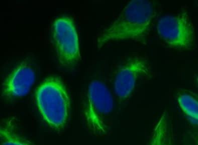
Vimentin in A549 Human Cell Line.
Vimentin was detected in immersion fixed A549 human lung carcinoma cell line using Rat Anti-Human/Mouse/Rat Vimentin Monoclonal Antibody (Catalog # MAB2105) at 10 µg/mL for 3 hours at room temperature. Cells were stained using the NorthernLights™ 493-conjugated Anti-Rat IgG Secondary Antibody (green; Catalog # NL015) and counterstained with DAPI (blue). View our protocol for Fluorescent ICC Staining of Cells on Coverslips.
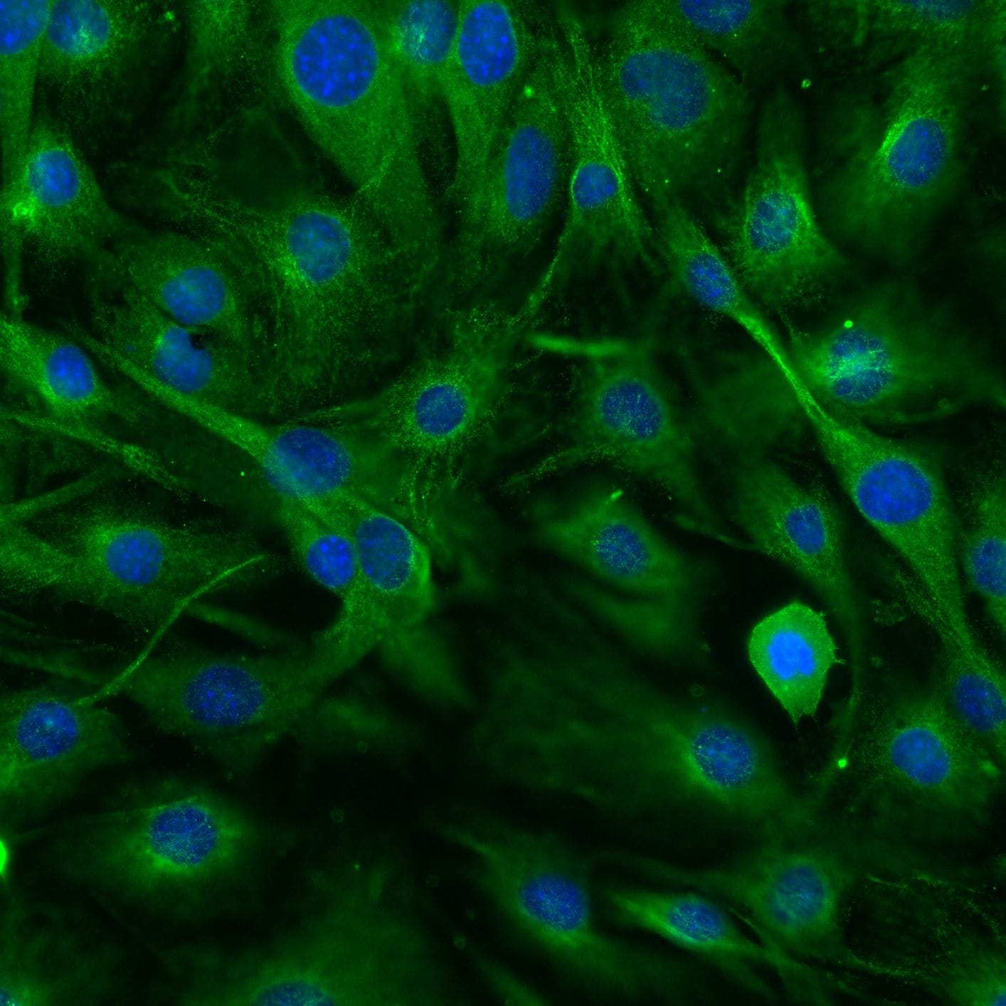
Vimentin in Mouse Cortical Stem Cells.
Vimentin was detected in immersion fixed mouse cortical stem cells using Rat Anti-Human/Mouse/Rat Vimentin Monoclonal Antibody (Catalog # MAB2105) at 10 µg/mL for 3 hours at room temperature. Cells were stained using the NorthernLights™ 557-conjugated Anti-Rat IgG Secondary Antibody (red; Catalog # NL013) and counterstained with DAPI (blue). Specific staining was localized to cytoskeleton. View our protocol for Fluorescent ICC Staining of Cells on Coverslips.
Vimentin in Rat Cortical Stem Cells.
Vimentin was detected in immersion fixed rat cortical stem cells using Rat Anti-Human/Mouse/Rat Vimentin Monoclonal Antibody (Catalog # MAB2105) at 10 µg/mL for 3 hours at room temperature. Cells were stained using the NorthernLights™ 557-conjugated Anti-Rat IgG Secondary Antibody (red; Catalog # NL013) and counterstained with DAPI (blue). Specific staining was localized to cytoskeleton. View our protocol for Fluorescent ICC Staining of Cells on Coverslips.
Vimentin in Human Tonsil.
Vimentin was detected in immersion fixed paraffin-embedded sections of human tonsil using Rat Anti-Human/Mouse/Rat Vimentin Monoclonal Antibody (Catalog # MAB2105) at 0.5 µg/mL for 1 hour at room temperature followed by incubation with the Anti-Rat IgG VisUCyte™ HRP Polymer Antibody (Catalog # VC005). Tissue was stained using DAB (brown) and counterstained with hematoxylin (blue). Specific staining was localized to cytoplasm. View our protocol for IHC Staining with VisUCyte HRP Polymer Detection Reagents.
Detection of Vimentin in A172 Human Cell Line by Flow Cytometry.
A172 human glioblastoma cell line was stained with Rat Anti-Human/Mouse/Rat Vimentin Monoclonal Antibody (Catalog # MAB2105, filled histogram) or isotype control antibody (Catalog # MAB006, open histogram) followed by anti-Rat IgG PE-conjugated secondary antibody (Catalog # F0105B). To facilitate intracellular staining, cells were fixed with Flow Cytometry Fixation Buffer (Catalog # FC004) and permeabilized with Flow Cytometry Permeabilization/Wash Buffer I (Catalog # FC005). View our protocol for Staining Intracellular Molecules.
Detection of Human Vimentin by Simple WesternTM.
Simple Western lane view shows lysates of Jurkat human acute T cell leukemia cell line, loaded at 0.2 mg/mL. A specific band was detected for Vimentin at approximately 58 kDa (as indicated) using 10 µg/mL of Rat Anti-Human/Mouse/Rat Vimentin Monoclonal Antibody (Catalog # MAB2105) followed by 1:50 dilution of HRP-conjugated Anti-Rat IgG Secondary Antibody (Catalog # HAF005). This experiment was conducted under reducing conditions and using the 12-230 kDa separation system.
Western Blot Shows Human Vimentin Specificity by Using Knockout Cell Line.
Western blot shows lysates of K562 human chronic myelogenous leukemia parental cell line and Vimentin knockout K562 cell line (KO). PVDF membrane was probed with 2 µg/mL of Rat Anti-Human/Mouse/Rat Vimentin Monoclonal Antibody (Catalog # MAB2105) followed by HRP-conjugated Anti-Goat IgG Secondary Antibody (HAF017). A specific band was detected for Vimentin at approximately 55 kDa (as indicated) in the parental K562 cell line, but is not detectable in knockout K562 cell line. GAPDH (MAB5718) is shown as a loading control. This experiment was conducted under reducing conditions and using Western Blot Buffer Group 1.
Detection of Human Vimentin by Immunocytochemistry/Immunofluorescence
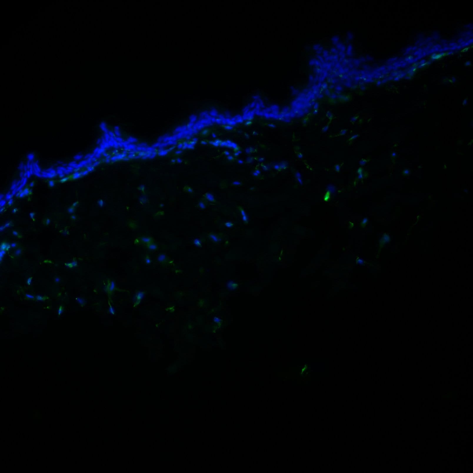
A very minimal population of human vimentin+ HSCs/myofibroblasts express a primary cilium, with none detected on CD31+ endothelial cells.Human ALD liver tissue was examined for the expression of primary cilia ( alpha -acetylated tubulin, green; gamma -tubulin, red) by vimentin+ (grey) HSCs/myofibroblasts (A) or CD31+ (grey) ECs (C). (A) The majority of vimentin+ cells were Pc-ve in the tissues examined. Representative image shown, displaying absence of Pc on vimentin+ cells. To confirm this result, ciliary protein Arl13b (green) was co-stained with vimentin (grey). Rare Arl13b ciliary structures (arrow) co-localised with vimentin+ cells. Final panel in A illustrates rare Pc+ ( alpha -acetylated tubulin, green; gamma -tubulin, red) vimentin+ (grey) HSCs/myofibroblasts, at the cirrhotic interface. (B) Number of vimentin+ Pc+ cells or vimentin+ Pcneg cells per FOV (n = 3 ALD samples, 8 FOV/sample). (C) No Pc were detected on CD31+ cells in the tissues examined (ALD n = 3, 8 FOV/sample). Representative image shown. All images obtained using confocal microscopy, 63x objective. DAPI, blue. White arrows illustrate Pc. * Non-specific liver autofluorescence. Image collected and cropped by CiteAb from the following publication (https://dx.plos.org/10.1371/journal.pone.0171480), licensed under a CC-BY license. Not internally tested by R&D Systems.
Detection of Human Vimentin by Immunocytochemistry/Immunofluorescence
Widespread GLI expression in human donor and cirrhotic liver.(A) Frozen (4 μm) human donor (n = 5), and cirrhotic liver sections [ALD (n = 6), NASH (n = 3), PBC (n = 1)] were screened for GLI2 (red) expression by immunofluorescence. Representative images taken at 5x or 40x (insets) objective shown. DAPI, blue. (B) qRT-PCR for GLI1 and GLI3 transcript in human donor or ALD samples. Mean±S.E.M. Significant (*) difference between means (One-sided student t-test, **p<0.005). Western blot for full-length GLI1 protein (>150 kDa) in donor (Don) or ALD patient samples. Densitometry analysis with GLI1 normalised to GAPDH (Image J). Mean±S.E.M; **p = 0.0093 (Two-sided student t-test). (C) Nuclear GLI2 (green) expression in EpCAM+ (red) LPCs in donor, ALD, PBC and NASH liver. (D) Nuclear GLI2 (green) expression demonstrated within CD31+ (red) ECs, CK18+ (red) hepatocytes, CD45+ (red) leukocytes and vimentin+ (red) HSCs/myofibroblasts, in ALD. 63x objective. (E) Maximum intensity projection illustrating close physical association between EpCAM+ LPCs (green) and vimentin+ HSCs/myofibroblasts (red), both of which express GLI2 (grey), in ALD tissue. Arrows indicate myofibroblasts directly contacting LPCs. Confocal microscopy, 63x objective. Quantitation (%) of EpCAM+ GLI2+ cells and vimentin+ GLI2+ cells within the same FOV (n = 3 ALD samples, 8 FOV/sample). Image collected and cropped by CiteAb from the following publication (https://dx.plos.org/10.1371/journal.pone.0171480), licensed under a CC-BY license. Not internally tested by R&D Systems.
Detection of Vimentin in Mouse Kidney.
Vimentin was detected in immersion fixed paraffin-embedded sections of mouse kidney using Rat Anti-Human/Mouse/Rat Vimentin Monoclonal Antibody (Catalog # MAB2105) at 5 µg/ml overnight at 4 °C. Before incubation with the primary antibody, tissue was subjected to heat-induced epitope retrieval using VisUCyte Antigen Retrieval Reagent-Basic (Catalog # VCTS021). Tissue was stained using the HRP-conjugated Anti-Rat IgG Secondary Antibody (Catalog # HAF005) and counterstained with hematoxylin (blue). Specific staining was localized to the cytoplasm. View our protocol for Chromogenic IHC Staining of Paraffin-embedded Tissue Sections.
Immunofluorescent Staining of iPSC-derived Human Intestinal Organoids.
iPSC-derived human intestinal organoids were generated following the steps detailed in the human intestinal organoid culture protocol. Human intestinal organoids were stained using a Rat Anti-Human/Mouse/Rat Vimentin Monoclonal Antibody (Catalog # MAB2105; green) and a Goat Anti-Human/Mouse Desmin Antigen Affinity-purified Polyclonal Antibody (Catalog # AF3844; red) to visualize myofibroblast cells and counterstained with DAPI (Catalog # 5748; blue).Applications for Vimentin Antibody (280618)
Application
Recommended Usage
COMET
Optimal dilutions of this antibody should be experimentally determined.
Immunocytochemistry
8-25 µg/mL
Sample: Immersion fixed human neural progenitor cells, NTera-2 human testicular embryonic carcinoma cell line, A549 human lung carcinoma cell line, mouse cortical stem cells, and rat cortical stem cells
Sample: Immersion fixed human neural progenitor cells, NTera-2 human testicular embryonic carcinoma cell line, A549 human lung carcinoma cell line, mouse cortical stem cells, and rat cortical stem cells
Immunohistochemistry
0.5-25 µg/mL
Sample: Immersion fixed paraffin-embedded sections of human tonsil and mouse kidney
Sample: Immersion fixed paraffin-embedded sections of human tonsil and mouse kidney
Intracellular Staining by Flow Cytometry
0.25 µg/106 cells
Sample: A172 human glioblastoma cell line fixed with Flow Cytometry Fixation Buffer (Catalog # FC004) and permeabilized with Flow Cytometry Permeabilization/Wash Buffer I (Catalog # FC005)
Sample: A172 human glioblastoma cell line fixed with Flow Cytometry Fixation Buffer (Catalog # FC004) and permeabilized with Flow Cytometry Permeabilization/Wash Buffer I (Catalog # FC005)
Knockout Validated
Vimentin
is specifically detected in K562 human chronic myelogenous leukemia parental cell line but is not detectable in
Vimentin knockout K562 cell line.
Multiplex Immunofluorescence
1-10 µg/mL
Sample: Immersion fixed paraffin-embedded sections of human colon, human liver, human kidney and mouse kidney
Sample: Immersion fixed paraffin-embedded sections of human colon, human liver, human kidney and mouse kidney
Simple Western
10 µg/mL
Sample: Jurkat human acute T cell leukemia cell line
Sample: Jurkat human acute T cell leukemia cell line
Western Blot
1-2 µg/mL
Sample: Jurkat human acute T cell leukemia cell line, K562 human chronic myelogenous leukemia cell line, MEF mouse embryonic feeder cells, NIH‑3T3 mouse embryonic fibroblast cell line, Rat‑2 rat embryonic fibroblast cell line, and NR8383 rat alveolar macrophage cell line
Sample: Jurkat human acute T cell leukemia cell line, K562 human chronic myelogenous leukemia cell line, MEF mouse embryonic feeder cells, NIH‑3T3 mouse embryonic fibroblast cell line, Rat‑2 rat embryonic fibroblast cell line, and NR8383 rat alveolar macrophage cell line
Reviewed Applications
Read 7 reviews rated 4.6 using MAB2105 in the following applications:
Flow Cytometry Panel Builder
Bio-Techne Knows Flow Cytometry
Save time and reduce costly mistakes by quickly finding compatible reagents using the Panel Builder Tool.
Advanced Features
- Spectra Viewer - Custom analysis of spectra from multiple fluorochromes
- Spillover Popups - Visualize the spectra of individual fluorochromes
- Antigen Density Selector - Match fluorochrome brightness with antigen density

Formulation, Preparation, and Storage
Purification
Protein A or G purified from hybridoma culture supernatant
Reconstitution
Reconstitute at 0.5 mg/mL in sterile PBS. For liquid material, refer to CoA for concentration.
Loading...
Formulation
Lyophilized from a 0.2 μm filtered solution in PBS with Trehalose. See Certificate of Analysis for details.
*Small pack size (-SP) is supplied either lyophilized or as a 0.2 µm filtered solution in PBS.
*Small pack size (-SP) is supplied either lyophilized or as a 0.2 µm filtered solution in PBS.
Shipping
Lyophilized product is shipped at ambient temperature. Liquid small pack size (-SP) is shipped with polar packs. Upon receipt, store immediately at the temperature recommended below.
Stability & Storage
Use a manual defrost freezer and avoid repeated freeze-thaw cycles.
- 12 months from date of receipt, -20 to -70 °C as supplied.
- 1 month, 2 to 8 °C under sterile conditions after reconstitution.
- 6 months, -20 to -70 °C under sterile conditions after reconstitution.
Calculators
Background: Vimentin
References
- Omary, M.B. et al. (2006) Trends Biochem. Sci. 31:383.
- Ivaska, J. et al. (2007) Exp. Cell Res. 313:2050.
- Ferrari, S. et al. (1986) Mol. Cell. Biol. 6:3614.
- Sokolova, A.V. et al. (2006) Proc. Natl. Acad. Sci. USA 103:16206.
- Eriksson, J.E. et al. (2004) J. Cell Sci. 117:919.
- Li, Q-F. et al. (2006) J. Biol. Chem. 281:34716.
- Esue, O. et al. (2006) J. Biol. Chem. 281:30393.
- Styers, M.L. et al. (2005) Traffic 6:359.
- McInroy, L. and A. Maata (2007) Biochem. Biophys. Res. Commun. 360:109.
- Nieminen, M. et al. (2006) Nat. Cell Biol. 8:156.
- Ivaska, J. et al. (2005) EMBO J. 24:3834.
- Byun, Y. et al. (2001) Cell Death Differ. 8:443.
- Mor-Vaknin, N. et al. (2003) Nat. Cell Biol. 5:59.
- Zou, Y. et al. (2006) Biochem. Biophys. Res. Commun. 351:625.
- Garg, A. et al. (2006) J. Immunol. 177:6192.
Alternate Names
VIM
Gene Symbol
VIM
UniProt
Additional Vimentin Products
Product Documents for Vimentin Antibody (280618)
Certificate of Analysis
To download a Certificate of Analysis, please enter a lot or batch number in the search box below.
Note: Certificate of Analysis not available for kit components.
Product Specific Notices for Vimentin Antibody (280618)
For research use only
Citations for Vimentin Antibody (280618)
 Powered by Bioz
Customer Reviews for Vimentin Antibody (280618) (7)
4.6 out of 5
7 Customer Ratings
Have you used Vimentin Antibody (280618)?
Submit a review and receive an Amazon gift card!
$25/€18/£15/$25CAN/¥2500 Yen for a review with an image
$10/€7/£6/$10CAN/¥1110 Yen for a review without an image
Submit a review
Customer Images
Showing
1
-
5 of
7 reviews
Showing All
Filter By:
-
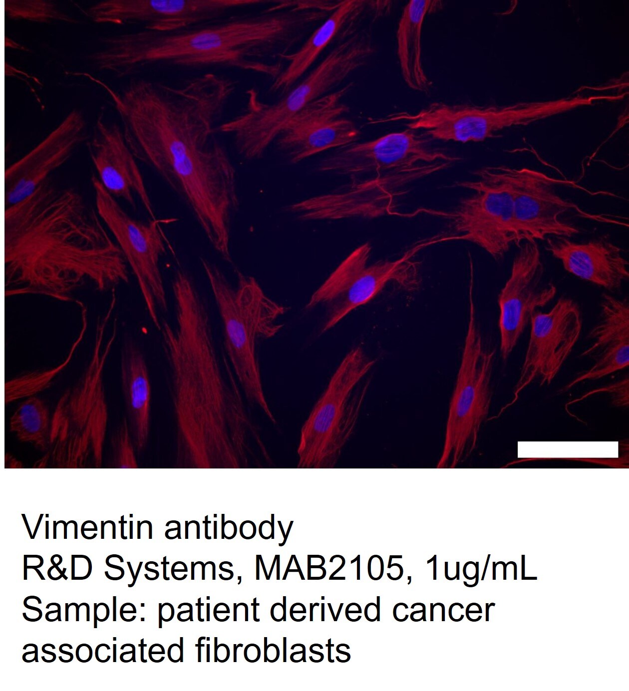
Application: Immunocytochemistry/ImmunofluorescenceSample Tested: Cancer associated FibroblastSpecies: HumanVerified Customer | Posted 09/01/2023Human cancer associated fibroblast cells were stained with Vimentin antibody to confirm identity.

-
Application: Immunocytochemistry/ImmunofluorescenceSample Tested: Epithelial cellsSpecies: MouseVerified Customer | Posted 06/19/2022
-
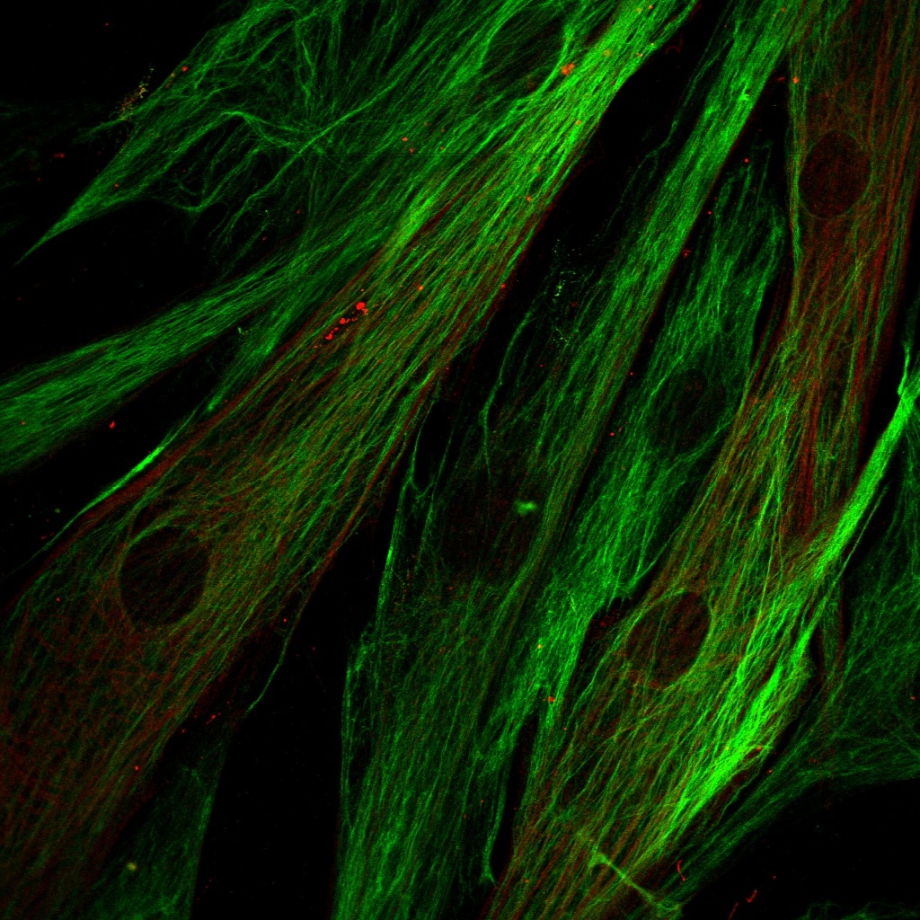
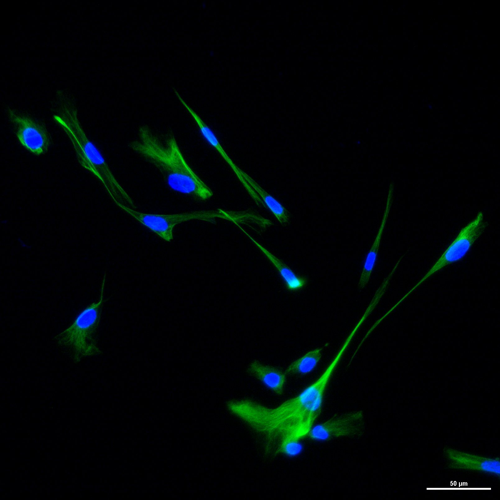
Application: Immunocytochemistry/ImmunofluorescenceSample Tested: Human fibroblastSpecies: HumanVerified Customer | Posted 06/15/2021Vimentin expression in human fibroblasts in culture. Vimentin (green) was used as a fibroblast marker in miofibroblast differentiation assays (a-SMA in red, as a miofibroblast marker).
-
Application: Immunocytochemistry/ImmunofluorescenceSample Tested: Mouse fibroblast cell lineSpecies: MouseVerified Customer | Posted 02/26/2021
-
Application: ImmunohistochemistrySample Tested: Skin tissueSpecies: Rhesus MacaqueVerified Customer | Posted 06/23/2020
-
Application: Western BlotSample Tested: Cancer cell lysatesSpecies: HumanVerified Customer | Posted 11/14/2019Testing several cell lines for mesenchymal characteristics, including vimentin expression, by western blot.
-
Application: Immunohistochemistry-FrozenSample Tested: Cultured Human KeratinocytesSpecies: HumanVerified Customer | Posted 02/22/2019
There are no reviews that match your criteria.
Protocols
Find general support by application which include: protocols, troubleshooting, illustrated assays, videos and webinars.
- 7-Amino Actinomycin D (7-AAD) Cell Viability Flow Cytometry Protocol
- Antigen Retrieval Protocol (PIER)
- Antigen Retrieval for Frozen Sections Protocol
- Appropriate Fixation of IHC/ICC Samples
- Cellular Response to Hypoxia Protocols
- Chromogenic IHC Staining of Formalin-Fixed Paraffin-Embedded (FFPE) Tissue Protocol
- Chromogenic Immunohistochemistry Staining of Frozen Tissue
- ClariTSA™ Fluorophore Kits
- Detection & Visualization of Antibody Binding
- Extracellular Membrane Flow Cytometry Protocol
- Flow Cytometry Protocol for Cell Surface Markers
- Flow Cytometry Protocol for Staining Membrane Associated Proteins
- Flow Cytometry Staining Protocols
- Flow Cytometry Troubleshooting Guide
- Fluorescent IHC Staining of Frozen Tissue Protocol
- Graphic Protocol for Heat-induced Epitope Retrieval
- Graphic Protocol for the Preparation and Fluorescent IHC Staining of Frozen Tissue Sections
- Graphic Protocol for the Preparation and Fluorescent IHC Staining of Paraffin-embedded Tissue Sections
- Graphic Protocol for the Preparation of Gelatin-coated Slides for Histological Tissue Sections
- ICC Cell Smear Protocol for Suspension Cells
- ICC Immunocytochemistry Protocol Videos
- ICC for Adherent Cells
- IHC Sample Preparation (Frozen sections vs Paraffin)
- Immunocytochemistry (ICC) Protocol
- Immunocytochemistry Troubleshooting
- Immunofluorescence of Organoids Embedded in Cultrex Basement Membrane Extract
- Immunofluorescent IHC Staining of Formalin-Fixed Paraffin-Embedded (FFPE) Tissue Protocol
- Immunohistochemistry (IHC) and Immunocytochemistry (ICC) Protocols
- Immunohistochemistry Frozen Troubleshooting
- Immunohistochemistry Paraffin Troubleshooting
- Intracellular Flow Cytometry Protocol Using Alcohol (Methanol)
- Intracellular Flow Cytometry Protocol Using Detergents
- Intracellular Nuclear Staining Flow Cytometry Protocol Using Detergents
- Intracellular Staining Flow Cytometry Protocol Using Alcohol Permeabilization
- Intracellular Staining Flow Cytometry Protocol Using Detergents to Permeabilize Cells
- Preparing Samples for IHC/ICC Experiments
- Preventing Non-Specific Staining (Non-Specific Binding)
- Primary Antibody Selection & Optimization
- Propidium Iodide Cell Viability Flow Cytometry Protocol
- Protocol for Heat-Induced Epitope Retrieval (HIER)
- Protocol for Liperfluo
- Protocol for Making a 4% Formaldehyde Solution in PBS
- Protocol for VisUCyte™ HRP Polymer Detection Reagent
- Protocol for the Characterization of Human Th22 Cells
- Protocol for the Characterization of Human Th9 Cells
- Protocol for the Fluorescent ICC Staining of Cell Smears - Graphic
- Protocol for the Fluorescent ICC Staining of Cultured Cells on Coverslips - Graphic
- Protocol for the Preparation & Fixation of Cells on Coverslips
- Protocol for the Preparation and Chromogenic IHC Staining of Frozen Tissue Sections
- Protocol for the Preparation and Chromogenic IHC Staining of Frozen Tissue Sections - Graphic
- Protocol for the Preparation and Chromogenic IHC Staining of Paraffin-embedded Tissue Sections
- Protocol for the Preparation and Chromogenic IHC Staining of Paraffin-embedded Tissue Sections - Graphic
- Protocol for the Preparation and Fluorescent ICC Staining of Cells on Coverslips
- Protocol for the Preparation and Fluorescent ICC Staining of Non-adherent Cells
- Protocol for the Preparation and Fluorescent ICC Staining of Stem Cells on Coverslips
- Protocol for the Preparation and Fluorescent IHC Staining of Frozen Tissue Sections
- Protocol for the Preparation and Fluorescent IHC Staining of Paraffin-embedded Tissue Sections
- Protocol for the Preparation of Gelatin-coated Slides for Histological Tissue Sections
- Protocol for the Preparation of a Cell Smear for Non-adherent Cell ICC - Graphic
- Protocol: Annexin V and PI Staining by Flow Cytometry
- Protocol: Annexin V and PI Staining for Apoptosis by Flow Cytometry
- R&D Systems Quality Control Western Blot Protocol
- TUNEL and Active Caspase-3 Detection by IHC/ICC Protocol
- The Importance of IHC/ICC Controls
- Troubleshooting Guide: Fluorokine Flow Cytometry Kits
- Troubleshooting Guide: Immunohistochemistry
- Troubleshooting Guide: Western Blot Figures
- Western Blot Conditions
- Western Blot Protocol
- Western Blot Protocol for Cell Lysates
- Western Blot Troubleshooting
- Western Blot Troubleshooting Guide
- View all Protocols, Troubleshooting, Illustrated assays and Webinars

