
Key Product Details
Validated by
Biological Validation
Species Reactivity
Validated:
Human, Mouse, Rat
Cited:
Human, Mouse, Rat, Transgenic Mouse
Applications
Validated:
Immunohistochemistry, Western Blot, Simple Western
Cited:
Western Blot, Simple Western, Western Blot Control
Label
Unconjugated
Antibody Source
Monoclonal Mouse IgG2A Clone # 728526
Loading...
Product Specifications
Immunogen
E. coli-derived recombinant human Vinculin
Lys1020-Gln1134
Accession # P18206
Lys1020-Gln1134
Accession # P18206
Specificity
Detects human Vinculin in direct ELISAs and Western blots.
Clonality
Monoclonal
Host
Mouse
Isotype
IgG2A
Scientific Data Images for Vinculin Antibody (728526)
Detection of Human Vinculin by Western Blot.
Western blot shows lysates of HeLa human cervical epithelial carcinoma cell line and HepG2 human hepatocellular carcinoma cell line. PVDF membrane was probed with 2 µg/mL of Mouse Anti-Human Vinculin Monoclonal Antibody (Catalog # MAB6896) followed by HRP-conjugated Anti-Mouse IgG Secondary Antibody (Catalog # HAF007). A specific band was detected for Vinculin at approximately 124 kDa (as indicated). This experiment was conducted under reducing conditions and using Immunoblot Buffer Group 1.
Vinculin in Human Uterus.
Vinculin was detected in immersion fixed paraffin-embedded sections of human uterus using Mouse Anti-Human Vinculin Monoclonal Antibody (Catalog # MAB6896) at 15 µg/mL overnight at 4 °C. Before incubation with the primary antibody, tissue was subjected to heat-induced epitope retrieval using Antigen Retrieval Reagent-Basic (Catalog # CTS013). Tissue was stained using the Anti-Mouse HRP-DAB Cell & Tissue Staining Kit (brown; Catalog # CTS002) and counterstained with hematoxylin (blue). Specific staining was localized to smooth muscle cells. View our protocol for Chromogenic IHC Staining of Paraffin-embedded Tissue Sections.
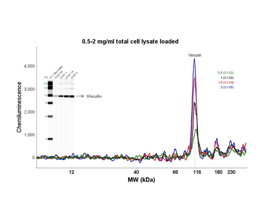
Detection of Human Vinculin by Simple WesternTM.
Simple Western lane view shows lysates of HeLa human cervical epithelial carcinoma cell line and PC‑3 human prostate cancer cell line, loaded at 0.2 mg/mL. A specific band was detected for Vinculin at approximately 122 kDa (as indicated) using 20 µg/mL of Mouse Anti-Human/Mouse/Rat Vinculin Monoclonal Antibody (Catalog # MAB6896). This experiment was conducted under reducing conditions and using the 12-230 kDa separation system.
Detection of Human Vinculin by Western Blot
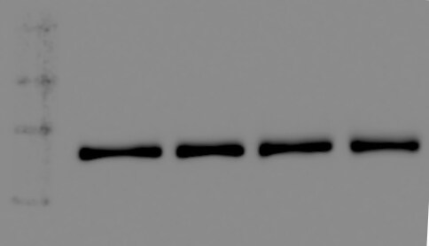
Cr and Ni exposure increases tau levels and phosphorylation in iNeurons and RA-differentiated SH-SY5Y cells. (a,b) iPSCs carrying the R406W tau mutation and isogenic controls were differentiated into iNeurons and treated with Cr (5 µM) and Ni (800 µM) for 72 hours. The levels of total (a) and phospho-tauSer396/404 (PHF-1) (b) tau were measured by western blot. GAPDH and vinculin were used as loading controls. Images show representative immunoblots comparing tau and phospho-tau levels in control and mutant iNeurons before and after Cr and Ni treatment. Plots represents the average ± SEM of 3 independent experiments. (c,d) RA-differentiated SH-SY5Y cells were treated with Cr (2.5 µM) and Nickel (200 µM) for 24 hours before protein extraction. Images show representative immunoblots comparing the levels total tau (c) and phospho-tauSer396/404 (PHF-1) tau (d) before and after heavy metals treatment. GAPDH and vinculin were used as loading control. Plots represent the average ± SEM of 4 independent experiments. Statistical significance was determined by two‐ANOVA followed by Bonferroni’s test for multiple comparisons or one-way ANOVA using GraphPad Prism 6. p-values comparing untreated and heavy metal treated cells are included in the graphs. Statistical significance was considered when p-value ≤ 0.05. Image collected and cropped by CiteAb from the following publication (https://pubmed.ncbi.nlm.nih.gov/31953414), licensed under a CC-BY license. Not internally tested by R&D Systems.
Detection of Human Vinculin by Western Blot
Cr and Ni exposure increases tau levels and phosphorylation in iNeurons and RA-differentiated SH-SY5Y cells. (a,b) iPSCs carrying the R406W tau mutation and isogenic controls were differentiated into iNeurons and treated with Cr (5 µM) and Ni (800 µM) for 72 hours. The levels of total (a) and phospho-tauSer396/404 (PHF-1) (b) tau were measured by western blot. GAPDH and vinculin were used as loading controls. Images show representative immunoblots comparing tau and phospho-tau levels in control and mutant iNeurons before and after Cr and Ni treatment. Plots represents the average ± SEM of 3 independent experiments. (c,d) RA-differentiated SH-SY5Y cells were treated with Cr (2.5 µM) and Nickel (200 µM) for 24 hours before protein extraction. Images show representative immunoblots comparing the levels total tau (c) and phospho-tauSer396/404 (PHF-1) tau (d) before and after heavy metals treatment. GAPDH and vinculin were used as loading control. Plots represent the average ± SEM of 4 independent experiments. Statistical significance was determined by two‐ANOVA followed by Bonferroni’s test for multiple comparisons or one-way ANOVA using GraphPad Prism 6. p-values comparing untreated and heavy metal treated cells are included in the graphs. Statistical significance was considered when p-value ≤ 0.05. Image collected and cropped by CiteAb from the following publication (https://pubmed.ncbi.nlm.nih.gov/31953414), licensed under a CC-BY license. Not internally tested by R&D Systems.
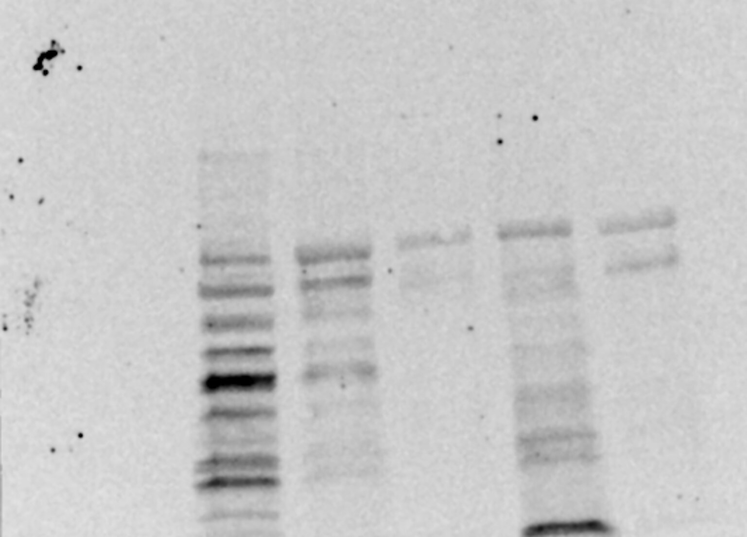
Detection of Human Vinculin by Western Blot
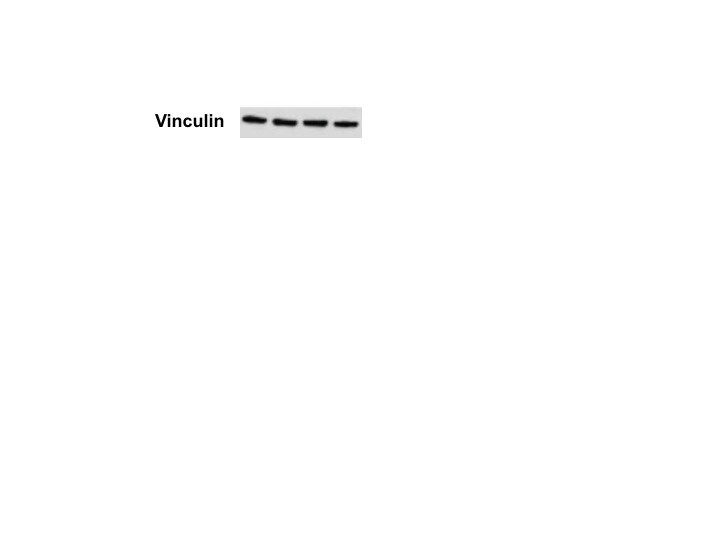
Cr and Ni exposure induces apoptotic cell death in SH-SY5Y cells. Non-differentiated and RA-differentiated SH-SY5Y cells were treated with Cr (2.5 µM) and Ni (200 µM) for 24 hours. Whole cell lysates were collected in order to analyze by western blot the levels of cleaved caspase-3 protein (a), the ratio of anti-apoptotic protein Bcl2 to pro-apoptotic protein Bax (b) and the cleavage and activation of caspase-9 (c). Vinculin and GAPDH were used as loading controls. (a) Representative immunoblot showing the presence of the 17/19KD fragment of caspase-3 after Cr and Ni exposure. (b) Representative immunoblots showing the levels of Bcl2 and Bax proteins after heavy metal exposure. The activation of the intrinsic/mitochondrial apoptosis pathway was assessed by the decrease of the ratio of Bcl2 to full length Bax and the presence of a cleavage form of Bax protein. The plot represent the average ± SEM of 3 independent experiments. Statistical significance was determined by one-way analysis of variance (anova) followed by Bonferroni’s test for multiple comparisons using GraphPad Prism 6. p-values comparing untreated and heavy metal treated cells are included. Statistical significance was considered when p-value ≤ 0.05. (c) Immunoblot showing the presence of the fragment (43KD) of active caspase-9 after Cr and Ni exposure to confirm the activation of the intrinsic/mitochondrial pathway. All experiments were performed in triplicates. Image collected and cropped by CiteAb from the following publication (https://pubmed.ncbi.nlm.nih.gov/31953414), licensed under a CC-BY license. Not internally tested by R&D Systems. [Unconjugated] [MAB6896] -")
Simple Western: Vinculin Antibody (728526) [Unconjugated] [MAB6896] -
Simple Western: Vinculin Antibody (728526) [Unconjugated] [MAB6896] - Western blot analysis for expression levels of mTOR and its downstream factors in the DRG on PID 21. Phosphorylated expressions of mTOR complex 1 and 2 were decreased in the shmTOR group, and its downstream effectors were downregulated as well. Data are expressed as mean +- SEM, * p < 0.05, ** p < 0.01, **** p < 0.0001. One-way ANOVA with Tukey's multiple comparisons test. Image collected and cropped by CiteAb from the following publication (https://pubmed.ncbi.nlm.nih.gov/37958901), licensed under a CC-BY license. Not internally tested by R&D Systems.Applications for Vinculin Antibody (728526)
Application
Recommended Usage
Immunohistochemistry
8-25 µg/mL
Sample: Immersion fixed paraffin-embedded sections of human uterus
Sample: Immersion fixed paraffin-embedded sections of human uterus
Simple Western
20 µg/mL
Sample: HeLa human cervical epithelial carcinoma cell line and PC‑3 human prostate cancer cell line
Sample: HeLa human cervical epithelial carcinoma cell line and PC‑3 human prostate cancer cell line
Western Blot
2 µg/mL
Sample: HeLa human cervical epithelial carcinoma cell line and HepG2 human hepatocellular carcinoma cell line
Sample: HeLa human cervical epithelial carcinoma cell line and HepG2 human hepatocellular carcinoma cell line
Reviewed Applications
Read 10 reviews rated 4.1 using MAB6896 in the following applications:
Formulation, Preparation, and Storage
Purification
Protein A or G purified from hybridoma culture supernatant
Reconstitution
Sterile PBS to a final concentration of 0.5 mg/mL. For liquid material, refer to CoA for concentration.
Loading...
Formulation
Lyophilized from a 0.2 μm filtered solution in PBS with Trehalose. *Small pack size (SP) is supplied either lyophilized or as a 0.2 µm filtered solution in PBS.
Shipping
Lyophilized product is shipped at ambient temperature. Liquid small pack size (-SP) is shipped with polar packs. Upon receipt, store immediately at the temperature recommended below.
Stability & Storage
Use a manual defrost freezer and avoid repeated freeze-thaw cycles.
- 12 months from date of receipt, -20 to -70 °C as supplied.
- 1 month, 2 to 8 °C under sterile conditions after reconstitution.
- 6 months, -20 to -70 °C under sterile conditions after reconstitution.
Calculators
Background: Vinculin
Long Name
Metavinculin
Alternate Names
MVCL, VCL
Gene Symbol
VCL
UniProt
Additional Vinculin Products
Product Documents for Vinculin Antibody (728526)
Certificate of Analysis
To download a Certificate of Analysis, please enter a lot or batch number in the search box below.
Note: Certificate of Analysis not available for kit components.
Product Specific Notices for Vinculin Antibody (728526)
For research use only
Related Research Areas
Citations for Vinculin Antibody (728526)
 Powered by Bioz
Customer Reviews for Vinculin Antibody (728526) (10)
4.1 out of 5
10 Customer Ratings
Have you used Vinculin Antibody (728526)?
Submit a review and receive an Amazon gift card!
$25/€18/£15/$25CAN/¥2500 Yen for a review with an image
$10/€7/£6/$10CAN/¥1110 Yen for a review without an image
Submit a review
Customer Images
Showing
1
-
5 of
10 reviews
Showing All
Filter By:
-
Application: Western BlotSample Tested: Tumor cell lyastesSpecies: HumanVerified Customer | Posted 10/14/2025We used vinculin as a loading control for western blots of treated cell lines. This vinculin antibody demonstrated a significant amount of unanticipated staining, which we believe is nonspecific since we did not see this issue with a vinculin antibody purchased from another supplier.
 Bio-Techne ResponseThank you for reviewing our product. We are sorry to hear that this product did not perform as expected. We have been in touch with the customer to resolve this issue according to our Product Guarantee and to the customer’s satisfaction. To reduce extra bands in your Western blot, we recommend adjusting the primary antibody concentration, % milk or BSA in the solution, and NaCl in the blotting buffer. You can find helpful troubleshooting tips here
 -
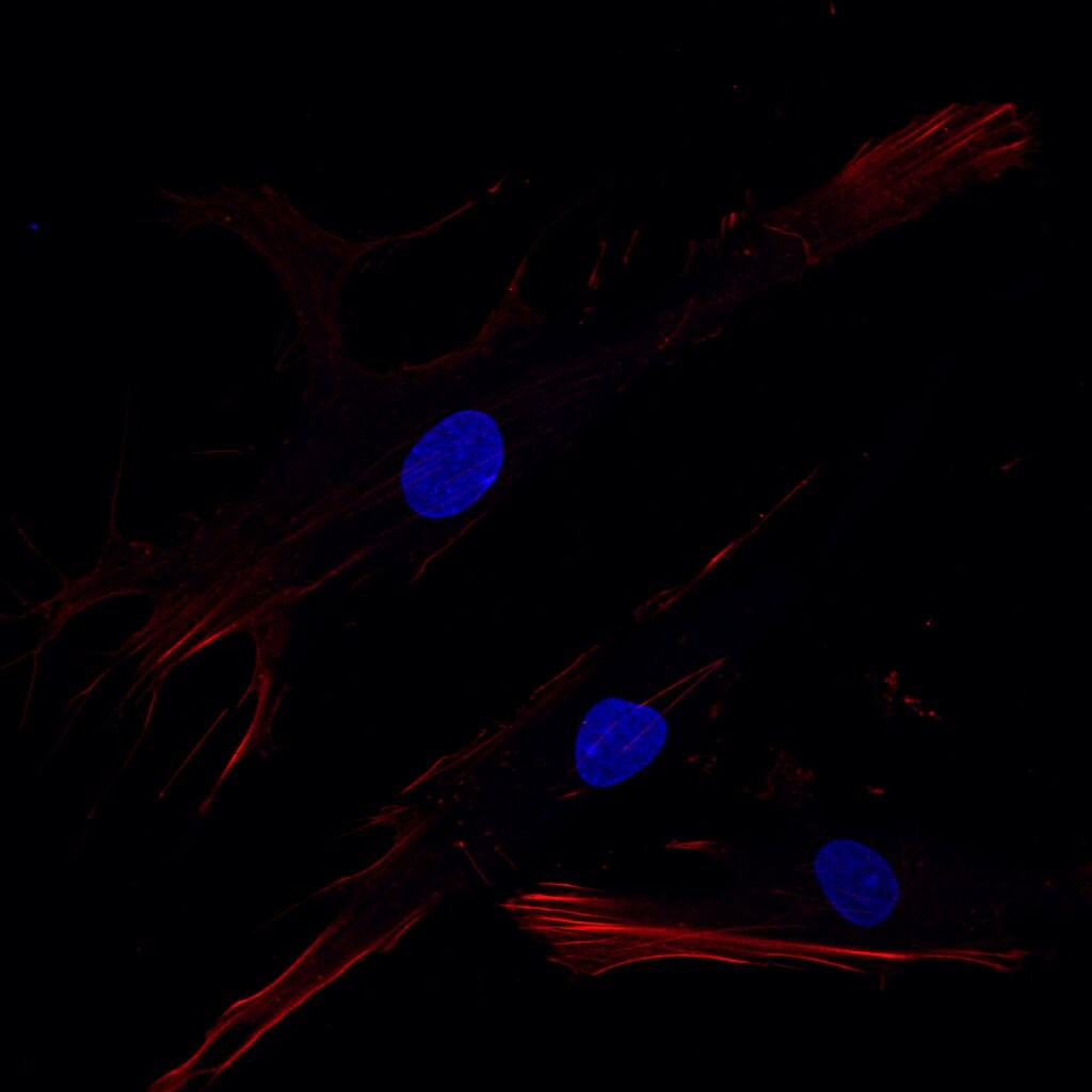
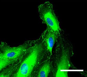
Application: Immunocytochemistry/ImmunofluorescenceSample Tested: Human fibroblastSpecies: HumanVerified Customer | Posted 11/23/2023Paraformaldehyde-fixed fibroblast stained with vinculin (red), and counterstained with DAPI (blue)
-
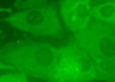
Application: Immunocytochemistry/ImmunofluorescenceSample Tested: Human osteoblast-like cells (MG63)Species: HumanVerified Customer | Posted 08/27/2021
-
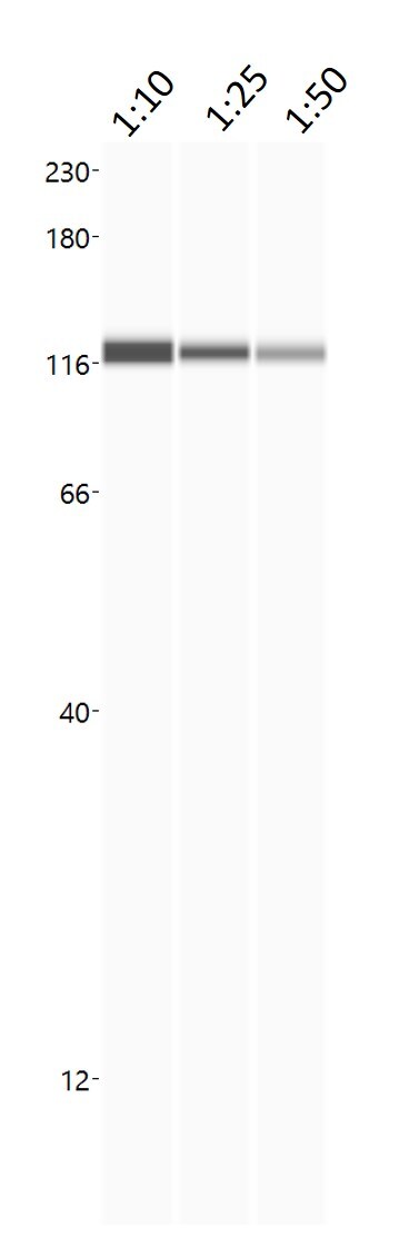
Application: Simple WesternSample Tested: HCT116 cell line lysateSpecies: HumanVerified Customer | Posted 08/27/2020Vinculin Ab titration on HCT116 cell lysateAb titration on HCT116 cell lysate
-
Application: Western BlotSample Tested: IPS2 induced pluripotent stem cellsSpecies: MouseVerified Customer | Posted 01/25/2018
-
Application: Immunocytochemistry/ImmunofluorescenceSample Tested: HMVEC human microvascular endothelial cellsSpecies: HumanVerified Customer | Posted 12/28/2017
-
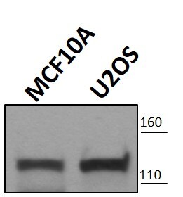
Application: Western BlotSample Tested: MCF 10A human breast epithelial cell line and U2OS human osteosarcoma cell lineSpecies: HumanVerified Customer | Posted 12/07/2017
-
Application: Western BlotSample Tested: Kidney tissueSpecies: MouseVerified Customer | Posted 08/23/2017- 1:3000 dilution of sample size stock - used milk for block and Ab dilution - 10ug of total protein loaded in each well
-
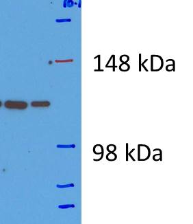
Application: Western BlotSample Tested: Hela whole cell lysateSpecies: HumanVerified Customer | Posted 10/16/2015Hela cells lysate 25 ug protein loaded on SDS gel (6%) used Tris Glycine for electrophoresis. Major bands ~124 kDa
-
Application: Simple WesternSample Tested: CaCo2 whole cell lysateSpecies: MouseVerified Customer | Posted 09/23/20150.5-1.5 mg/ml of total cell lysate (CaCo2 intestinal cells)
There are no reviews that match your criteria.
Protocols
Find general support by application which include: protocols, troubleshooting, illustrated assays, videos and webinars.
- Antigen Retrieval Protocol (PIER)
- Antigen Retrieval for Frozen Sections Protocol
- Appropriate Fixation of IHC/ICC Samples
- Cellular Response to Hypoxia Protocols
- Chromogenic IHC Staining of Formalin-Fixed Paraffin-Embedded (FFPE) Tissue Protocol
- Chromogenic Immunohistochemistry Staining of Frozen Tissue
- ClariTSA™ Fluorophore Kits
- Detection & Visualization of Antibody Binding
- Fluorescent IHC Staining of Frozen Tissue Protocol
- Graphic Protocol for Heat-induced Epitope Retrieval
- Graphic Protocol for the Preparation and Fluorescent IHC Staining of Frozen Tissue Sections
- Graphic Protocol for the Preparation and Fluorescent IHC Staining of Paraffin-embedded Tissue Sections
- Graphic Protocol for the Preparation of Gelatin-coated Slides for Histological Tissue Sections
- IHC Sample Preparation (Frozen sections vs Paraffin)
- Immunofluorescent IHC Staining of Formalin-Fixed Paraffin-Embedded (FFPE) Tissue Protocol
- Immunohistochemistry (IHC) and Immunocytochemistry (ICC) Protocols
- Immunohistochemistry Frozen Troubleshooting
- Immunohistochemistry Paraffin Troubleshooting
- Preparing Samples for IHC/ICC Experiments
- Preventing Non-Specific Staining (Non-Specific Binding)
- Primary Antibody Selection & Optimization
- Protocol for Heat-Induced Epitope Retrieval (HIER)
- Protocol for Making a 4% Formaldehyde Solution in PBS
- Protocol for VisUCyte™ HRP Polymer Detection Reagent
- Protocol for the Preparation & Fixation of Cells on Coverslips
- Protocol for the Preparation and Chromogenic IHC Staining of Frozen Tissue Sections
- Protocol for the Preparation and Chromogenic IHC Staining of Frozen Tissue Sections - Graphic
- Protocol for the Preparation and Chromogenic IHC Staining of Paraffin-embedded Tissue Sections
- Protocol for the Preparation and Chromogenic IHC Staining of Paraffin-embedded Tissue Sections - Graphic
- Protocol for the Preparation and Fluorescent IHC Staining of Frozen Tissue Sections
- Protocol for the Preparation and Fluorescent IHC Staining of Paraffin-embedded Tissue Sections
- Protocol for the Preparation of Gelatin-coated Slides for Histological Tissue Sections
- R&D Systems Quality Control Western Blot Protocol
- TUNEL and Active Caspase-3 Detection by IHC/ICC Protocol
- The Importance of IHC/ICC Controls
- Troubleshooting Guide: Immunohistochemistry
- Troubleshooting Guide: Western Blot Figures
- Western Blot Conditions
- Western Blot Protocol
- Western Blot Protocol for Cell Lysates
- Western Blot Troubleshooting
- Western Blot Troubleshooting Guide
- View all Protocols, Troubleshooting, Illustrated assays and Webinars
Loading...