Smads are a family of intracellular proteins that transmit transforming growth factor beta (TGF-beta ) superfamily signals from the cell surface to the nucleus. The Smad family is divided into three subclasses: receptor regulated Smads, (Smads 1, 2, 3, 5 and 8); the common partner, (Smad4); and the inhibitory Smads, (Smads 6 and 7). The binding of TGF-beta or activin to their cognate receptor induces phosphorylation of Smads 2 and 3. The activated Smads associate with the common-mediator subunit, Smad4, and the heteromeric complex translocates into the nucleus to initiate transcription. Smad3, also known as Mothers Against Decapentaplegic homolog 3 (MADH3), shares 83% amino acid identity with Smad2, also known as Mothers Against Decapentaplegic homolog 2 (MADH2). Human Smad2 has 99% identity to mouse and rat Smad2. Human Smad3 has 99% identity to mouse and rat Smad3.

Key Product Details
Validated by
Biological Validation
Species Reactivity
Validated:
Human, Mouse
Cited:
Human, Mouse, Rat, Frog - Xenopus (African Clawed Frog)
Applications
Validated:
Immunohistochemistry, Western Blot, Immunocytochemistry, Simple Western, Chromatin Immunoprecipitation (ChIP)
Cited:
Immunohistochemistry, Western Blot, Flow Cytometry, Immunocytochemistry, Simple Western, Immunoprecipitation, Chromatin Immunoprecipitation (ChIP)
Label
Unconjugated
Antibody Source
Polyclonal Goat IgG
Loading...
Product Specifications
Immunogen
E. coli-derived recombinant human Smad3
Ser2-Ala230
Accession # P84022
Ser2-Ala230
Accession # P84022
Specificity
Detects human, mouse, and rat Smad2/3 in Western blots. Predicted to detect rat based on sequence homology.
Clonality
Polyclonal
Host
Goat
Isotype
IgG
Scientific Data Images for Smad2/3 Antibody
Detection of Human/Mouse Smad2/3 by Western Blot.
Western blot shows lysates of A549 human lung carcinoma cell line, Jurkat human acute T cell leukemia cell line, HEK293 human embryonic kidney cell line, and C2C12 mouse myoblast cell line. PVDF membrane was probed with 0.5 µg/mL Goat Anti-Human/Mouse Smad2/3 Antigen Affinity-purified Polyclonal Antibody (Catalog # AF3797) followed by HRP-conjugated Anti-Goat IgG Secondary Antibody (Catalog # HAF109). For additional reference, recombinant human Smad2 and Smad3 were included. Specific bands for Smad2 were detected at approximately 64 and 58 kDa (as indicated). This experiment was conducted under reducing conditions and using Immunoblot Buffer Group 2.
Detection of Smad2/3-regulated Genes by Chromatin Immunoprecipitation.
Jurkat human acute T cell leukemia cell line treated with 50 ng/mL PMA and 200 ng/mL calcium ionomycin for 30 minutes was fixed using formaldehyde, resuspended in lysis buffer, and sonicated to shear chromatin. Smad2/3/DNA complexes were immunoprecipitated using 5 µg Goat Anti-Human/Mouse Smad2/3 Antigen Affinity-purified Polyclonal Antibody (Catalog # AF3797) or control antibody (Catalog # AB-108-C) for 15 minutes in an ultrasonic bath, followed by Biotinylated Anti-Goat IgG Secondary Antibody (Catalog # BAF109). Immunocomplexes were captured using 50 µL of MagCellect Streptavidin Ferrofluid (Catalog # MAG999) and DNA was purified using chelating resin solution. Thec-mycpromoter was detected by standard PCR.
Smad2/3 in MCF‑7 Human Cell Line.
Smad2/3 was detected in immersion fixed MCF-7 human breast cancer cell line induced (upper panel) or non-induced (lower panel) to undergo epithelial-mesenchymal transition (EMT) using Goat Anti-Human/Mouse Smad2/3 Antigen Affinity-purified Polyclonal Antibody (Catalog # AF3797) at 10 µg/mL for 3 hours at room temperature. Cells were stained using the NorthernLights™ 557-conjugated Anti-Goat IgG Secondary Antibody (red; Catalog # NL001) and counterstained with DAPI (blue). Specific staining was localized to cytoplasm and, in EMT-induced cells, nuclei. View our protocol for Fluorescent ICC Staining of Cells on Coverslips.
Detection of Human Smad2/3 by Simple WesternTM.
Simple Western lane view shows lysates of A549 human lung carcinoma cell line, COLO 205 human colorectal adenocarcinoma cell line, and HT-29 human colon adenocarcinoma cell line, loaded at 0.2 mg/mL. A specific band was detected for Smad2/3 at approximately 64 kDa (as indicated) using 25 µg/mL of Goat Anti-Human/Mouse Smad2/3 Antigen Affinity-purified Polyclonal Antibody (Catalog # AF3797) followed by 1:50 dilution of HRP-conjugated Anti-Goat IgG Secondary Antibody (Catalog # HAF109). This experiment was conducted under reducing conditions and using the 12-230 kDa separation system..")
Smad2/3 in Human Brain (Cortex).
Smad2/3 was detected in immersion fixed paraffin-embedded sections of human brain (cortex) using Goat Anti-Human/Mouse Smad2/3 Antigen Affinity-purified Polyclonal Antibody (Catalog # AF3797) at 3 µg/mL for 1 hour at room temperature followed by incubation with the Anti-Goat IgG VisUCyte™ HRP Polymer Antibody (VC004). Before incubation with the primary antibody, tissue was subjected to heat-induced epitope retrieval using Antigen Retrieval Reagent-Basic (CTS013). Tissue was stained using DAB (brown) and counterstained with hematoxylin (blue). Specific staining was localized to cytoplasm in neurons. Staining was performed using our protocol for IHC Staining with VisUCyte HRP Polymer Detection Reagents..")
Smad2/3 in Human Brain (Cerebellum).
Smad2/3 was detected in immersion fixed paraffin-embedded sections of human brain (cerebellum) using Goat Anti-Human/Mouse Smad2/3 Antigen Affinity-purified Polyclonal Antibody (Catalog # AF3797) at 10 µg/mL for 1 hour at room temperature followed by incubation with the Anti-Goat IgG VisUCyte™ HRP Polymer Antibody (VC004). Before incubation with the primary antibody, tissue was subjected to heat-induced epitope retrieval using Antigen Retrieval Reagent-Basic (CTS013). Tissue was stained using DAB (brown) and counterstained with hematoxylin (blue). Specific staining was localized to cytoplasm in Purkinje neurons. Staining was performed using our protocol for IHC Staining with VisUCyte HRP Polymer Detection Reagents.Applications for Smad2/3 Antibody
Application
Recommended Usage
Chromatin Immunoprecipitation (ChIP)
5 µg/5 x 106 cells
Sample: PMA and calcium ionomycin treated Jurkat human acute T cell leukemia cell line chromatin, c-myc promoter detected by standard PCR
Sample: PMA and calcium ionomycin treated Jurkat human acute T cell leukemia cell line chromatin, c-myc promoter detected by standard PCR
Immunocytochemistry
5-15 µg/mL
Sample: Immersion fixed MCF‑7 human breast cancer cell line non-induced or induced to undergo epithelial-mesenchymal transition
Sample: Immersion fixed MCF‑7 human breast cancer cell line non-induced or induced to undergo epithelial-mesenchymal transition
Immunohistochemistry
3-15 µg/mL
Sample: Immersion fixed paraffin-embedded sections of human brain (cortex and cerebellum)
Sample: Immersion fixed paraffin-embedded sections of human brain (cortex and cerebellum)
Simple Western
25 µg/mL
Sample: A549 human lung carcinoma cell line, COLO 205 human colorectal adenocarcinoma cell line, and HT‑29 human colon adenocarcinoma cell line
Sample: A549 human lung carcinoma cell line, COLO 205 human colorectal adenocarcinoma cell line, and HT‑29 human colon adenocarcinoma cell line
Western Blot
0.5 µg/mL
Sample: A549 human lung carcinoma cell line, Jurkat human acute T cell leukemia cell line, HEK293 human embryonic kidney cell line, and C2C12 mouse myoblast cell line
Sample: A549 human lung carcinoma cell line, Jurkat human acute T cell leukemia cell line, HEK293 human embryonic kidney cell line, and C2C12 mouse myoblast cell line
Reviewed Applications
Read 4 reviews rated 4.8 using AF3797 in the following applications:
Formulation, Preparation, and Storage
Purification
Antigen Affinity-purified
Reconstitution
Reconstitute at 0.2 mg/mL in sterile PBS. For liquid material, refer to CoA for concentration.
Loading...
Formulation
Lyophilized from a 0.2 μm filtered solution in PBS with Trehalose. *Small pack size (SP) is supplied either lyophilized or as a 0.2 µm filtered solution in PBS.
Shipping
Lyophilized product is shipped at ambient temperature. Liquid small pack size (-SP) is shipped with polar packs. Upon receipt, store immediately at the temperature recommended below.
Stability & Storage
Use a manual defrost freezer and avoid repeated freeze-thaw cycles.
- 12 months from date of receipt, -20 to -70 °C as supplied.
- 1 month, 2 to 8 °C under sterile conditions after reconstitution.
- 6 months, -20 to -70 °C under sterile conditions after reconstitution.
Calculators
Background: Smad2/3
Long Name
Mothers Against DPP Homolog 2/3
Alternate Names
hMAD-2, hSMAD2, JV18, JV18-1SMAD, mothers against DPP homolog 2 (Drosophila), MAD homolog 2, Mad protein homolog, Mad, mothers against decapentaplegic homolog 2, MADH2MGC34440, MADR2MGC22139, Mad-related protein 2, mother against DPP homolog 2, mothers against decapentaplegic homolog 2, Mothers against DPP homolog 2, Sma- and Mad-related protein 2, SMAD 2, SMAD family member 2MAD, mothers against decapentaplegic homolog 2 (Drosophila), SMAD, mothers against DPP homolog 2, Smad2
UniProt
Additional Smad2/3 Products
Product Documents for Smad2/3 Antibody
Certificate of Analysis
To download a Certificate of Analysis, please enter a lot or batch number in the search box below.
Note: Certificate of Analysis not available for kit components.
Product Specific Notices for Smad2/3 Antibody
For research use only
Citations for Smad2/3 Antibody
 Powered by Bioz
Customer Reviews for Smad2/3 Antibody (4)
4.8 out of 5
4 Customer Ratings
Have you used Smad2/3 Antibody?
Submit a review and receive an Amazon gift card!
$25/€18/£15/$25CAN/¥2500 Yen for a review with an image
$10/€7/£6/$10CAN/¥1110 Yen for a review without an image
Submit a review
Customer Images
Showing
1
-
4 of
4 reviews
Showing All
Filter By:
-
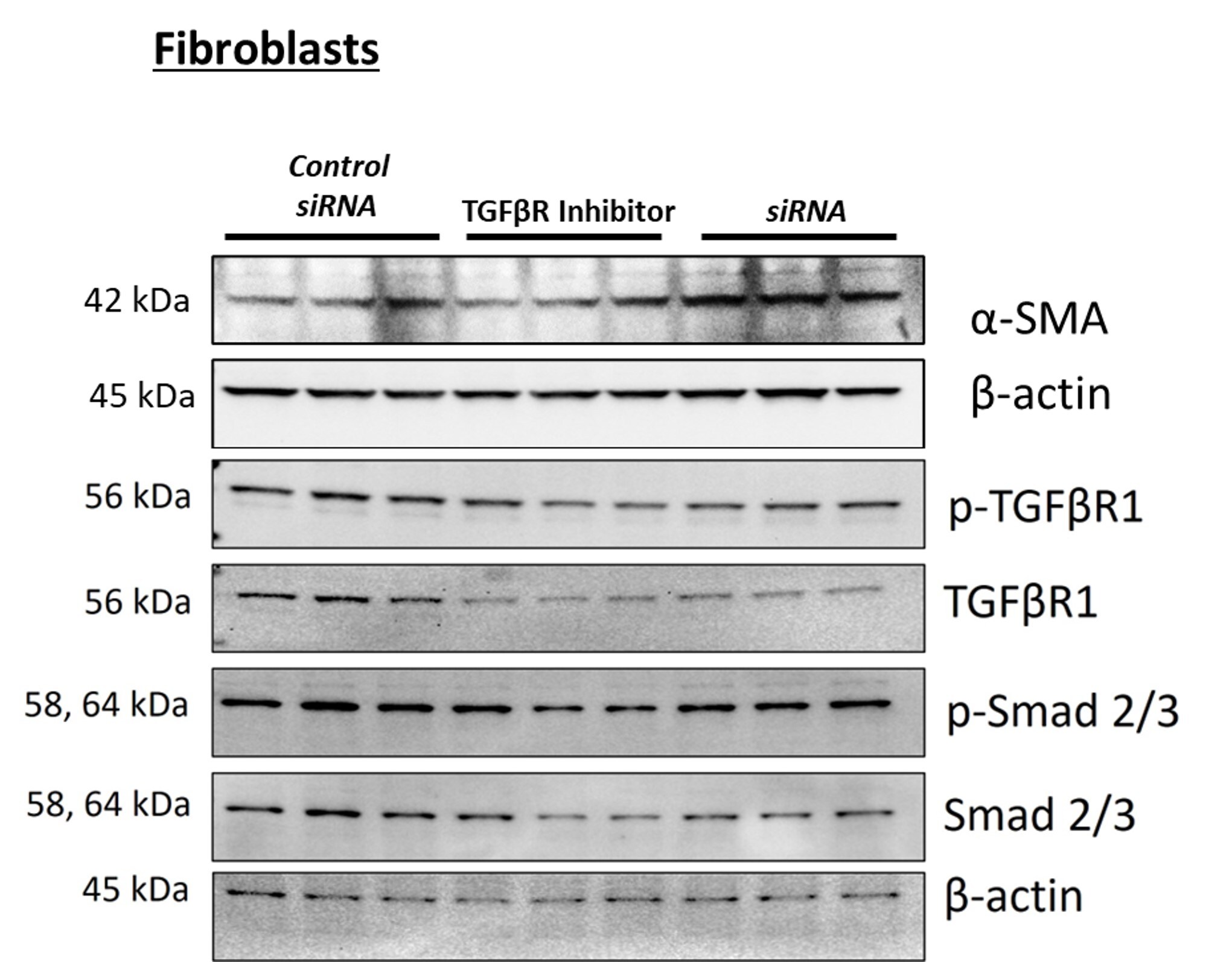
Application: Western BlotSample Tested: MOSUE CARDIAC fibroblastsSpecies: MouseVerified Customer | Posted 12/20/2024Mouse cardiac fibroblast assessed for Smad2/3 expression

-
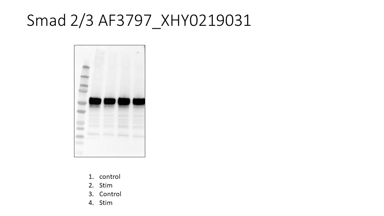
Application: Western BlotSample Tested: Mv1Lu mink lung epithelial cell lineSpecies: MinkVerified Customer | Posted 07/08/2020
-
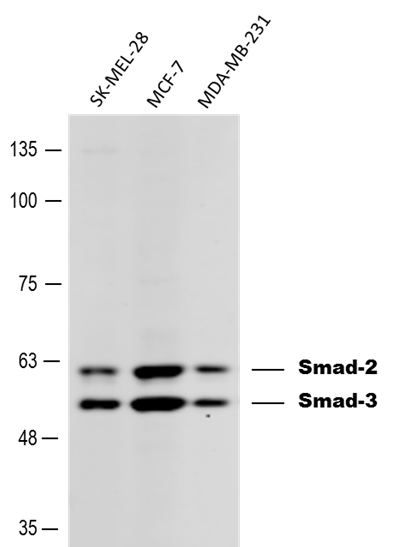
Application: Western BlotSample Tested: Human cell lineSpecies: HumanVerified Customer | Posted 03/14/2017Detection of Human Smad 2/3 in melanoma and mammary tumor cell lines. Dilution: 1:500 in PBS with 5% BSA. Secondary Ab: anti-Goat IgG 1:5,000.
-
Application: Western BlotSample Tested: See PMID 22868230Species: MouseVerified Customer | Posted 01/08/2015
There are no reviews that match your criteria.
Protocols
Find general support by application which include: protocols, troubleshooting, illustrated assays, videos and webinars.
- Antigen Retrieval Protocol (PIER)
- Antigen Retrieval for Frozen Sections Protocol
- Appropriate Fixation of IHC/ICC Samples
- Cellular Response to Hypoxia Protocols
- ChIP Protocol Video
- Chromatin Immunoprecipitation (ChIP) Protocol
- Chromatin Immunoprecipitation Protocol
- Chromogenic IHC Staining of Formalin-Fixed Paraffin-Embedded (FFPE) Tissue Protocol
- Chromogenic Immunohistochemistry Staining of Frozen Tissue
- ClariTSA™ Fluorophore Kits
- Detection & Visualization of Antibody Binding
- Fluorescent IHC Staining of Frozen Tissue Protocol
- Graphic Protocol for Heat-induced Epitope Retrieval
- Graphic Protocol for the Preparation and Fluorescent IHC Staining of Frozen Tissue Sections
- Graphic Protocol for the Preparation and Fluorescent IHC Staining of Paraffin-embedded Tissue Sections
- Graphic Protocol for the Preparation of Gelatin-coated Slides for Histological Tissue Sections
- ICC Cell Smear Protocol for Suspension Cells
- ICC Immunocytochemistry Protocol Videos
- ICC for Adherent Cells
- IHC Sample Preparation (Frozen sections vs Paraffin)
- Immunocytochemistry (ICC) Protocol
- Immunocytochemistry Troubleshooting
- Immunofluorescence of Organoids Embedded in Cultrex Basement Membrane Extract
- Immunofluorescent IHC Staining of Formalin-Fixed Paraffin-Embedded (FFPE) Tissue Protocol
- Immunohistochemistry (IHC) and Immunocytochemistry (ICC) Protocols
- Immunohistochemistry Frozen Troubleshooting
- Immunohistochemistry Paraffin Troubleshooting
- Preparing Samples for IHC/ICC Experiments
- Preventing Non-Specific Staining (Non-Specific Binding)
- Primary Antibody Selection & Optimization
- Protocol for Heat-Induced Epitope Retrieval (HIER)
- Protocol for Making a 4% Formaldehyde Solution in PBS
- Protocol for VisUCyte™ HRP Polymer Detection Reagent
- Protocol for the Fluorescent ICC Staining of Cell Smears - Graphic
- Protocol for the Fluorescent ICC Staining of Cultured Cells on Coverslips - Graphic
- Protocol for the Preparation & Fixation of Cells on Coverslips
- Protocol for the Preparation and Chromogenic IHC Staining of Frozen Tissue Sections
- Protocol for the Preparation and Chromogenic IHC Staining of Frozen Tissue Sections - Graphic
- Protocol for the Preparation and Chromogenic IHC Staining of Paraffin-embedded Tissue Sections
- Protocol for the Preparation and Chromogenic IHC Staining of Paraffin-embedded Tissue Sections - Graphic
- Protocol for the Preparation and Fluorescent ICC Staining of Cells on Coverslips
- Protocol for the Preparation and Fluorescent ICC Staining of Non-adherent Cells
- Protocol for the Preparation and Fluorescent ICC Staining of Stem Cells on Coverslips
- Protocol for the Preparation and Fluorescent IHC Staining of Frozen Tissue Sections
- Protocol for the Preparation and Fluorescent IHC Staining of Paraffin-embedded Tissue Sections
- Protocol for the Preparation of Gelatin-coated Slides for Histological Tissue Sections
- Protocol for the Preparation of a Cell Smear for Non-adherent Cell ICC - Graphic
- R&D Systems Quality Control Western Blot Protocol
- TUNEL and Active Caspase-3 Detection by IHC/ICC Protocol
- The Importance of IHC/ICC Controls
- Troubleshooting Guide: Immunohistochemistry
- Troubleshooting Guide: Western Blot Figures
- Western Blot Conditions
- Western Blot Protocol
- Western Blot Protocol for Cell Lysates
- Western Blot Troubleshooting
- Western Blot Troubleshooting Guide
- View all Protocols, Troubleshooting, Illustrated assays and Webinars
Loading...