
Key Product Details
Validated by
Knockout/Knockdown
Species Reactivity
Validated:
Human, Mouse
Cited:
Human, Mouse
Applications
Validated:
Knockout Validated, Western Blot, Simple Western, Immunoprecipitation
Cited:
Western Blot
Label
Unconjugated
Antibody Source
Polyclonal Goat IgG
Loading...
Product Specifications
Immunogen
E. coli-derived recombinant human STAT1
Specificity
Detects human and mouse STAT1 p91 in Western blots. In Western blots, less than 1% cross-reactivity with human or mouse STAT1 p84 is observed. Additionally, no cross-reactivity with the other STATs has been detected by immunoprecipitation or Western blotting.
Clonality
Polyclonal
Host
Goat
Isotype
IgG
Scientific Data Images for STAT1 p91 Antibody
Detection of Human and Mouse STAT1 p91 by Western Blot.
Western blot shows lysates of Daudi human Burkitt's lymphoma cell line, HeLa human cervical epithelial carcinoma cell line, and NIH-3T3 mouse embryonic fibroblast cell line. PVDF Membrane was probed with 1 µg/mL of Goat Anti-Human/Mouse STAT1 p91 Antigen Affinity-purified Polyclonal Antibody (Catalog # PAF-ST1) followed by HRP-conjugated Anti-Goat IgG Secondary Antibody (Catalog # HAF109). A specific band was detected for STAT1 p91 at approximately 91 kDa (as indicated). This experiment was conducted under reducing conditions and using Immunoblot Buffer Group 1.
Immunoprecipitation of Human STAT1.
STAT1 p91 was immunoprecipitated from 200 µg of cell lysate of HeLa human cervical epithelial carcinoma cell line following incubation with 3 µg Goat Anti-Human/Mouse STAT1 p91 Antigen Affinity-purified Polyclonal Antibody (Catalog # PAF-ST1) overnight at 4 °C. STAT1 p91-antibody complexes were absorbed using Protein G sepharose (Invitrogen, Catalog # 10-1242). Immunoprecipitated STAT1 p91 was detected by Western blot using 1 µg/mL Human STAT1 Monoclonal Antibody (Catalog # MAB14901). View our recommended buffer recipes for immunoprecipitation.
Western Blot Shows Human STAT1 Specificity by Using Knockout Cell Line.
Western blot shows lysates of HeLa human cervical epithelial carcinoma parental cell line and STAT1 knockout HeLa cell line (KO). PVDF membrane was probed with 0.25 µg/mL of Goat Anti-Human/Mouse STAT1 p91 Antigen Affinity-purified Polyclonal Antibody (Catalog # PAF-ST1) followed by HRP-conjugated Anti-Goat IgG Secondary Antibody (Catalog # HAF017). A specific band was detected for STAT1 at approximately 90 kDa (as indicated) in the parental HeLa cell line, but is not detectable in knockout HeLa cell line. GAPDH (Catalog # AF5718) is shown as a loading control. This experiment was conducted under reducing conditions and using Immunoblot Buffer Group 1.
Western Blot Shows Human STAT1 Specificity by Using Knockout Cell Line.
Western blot shows lysates of HeLa human cervical epithelial carcinoma parental cell line, STAT1 knockout (KO) HeLa cell line, STAT2 KO HeLa cell line, STAT3 KO HeLa cell line, STAT5a KO HeLa cell line, STAT5b KO HeLa cell line, and STAT6 KO HeLa cell line. PVDF membrane was probed with 0.25 µg/mL of Goat Anti-Human/Mouse STAT1 p91 Antigen Affinity-purified Polyclonal Antibody (Catalog # PAF-ST1) followed by HRP-conjugated Anti-Goat IgG Secondary Antibody (Catalog # HAF017). A specific band was detected for STAT1 at approximately 90 kDa (as indicated) in the parental HeLa cell line, but is not detectable in knockout HeLa cell line. GAPDH (Catalog # AF5718) is shown as a loading control. This experiment was conducted under reducing conditions and using Immunoblot Buffer Group 1.
Detection of Human STAT1 by Simple WesternTM.
Simple Western shows lysates of Daudi human Burkitt's lymphoma cell line, loaded at 0.5 mg/ml. A specific band was detected for STAT1 at approximately 84 kDa (as indicated) using 10 µg/mL of Goat Anti-Human/Mouse STAT1 p91 Antigen Affinity-purified Polyclonal Antibody (Catalog # PAF-ST1). This experiment was conducted under reducing conditions and using the 12-230kDa separation system.Applications for STAT1 p91 Antibody
Application
Recommended Usage
Immunoprecipitation
3 µg/106 cells
Sample: HeLa human cervical epithelial carcinoma cell line, see our available Western blot detection antibodies
Sample: HeLa human cervical epithelial carcinoma cell line, see our available Western blot detection antibodies
Knockout Validated
STAT1
is specifically detected in HeLa human cervical epithelial carcinoma parental cell line but is not detectable in
STAT1 knockout HeLa cell line.
Simple Western
10 µg/mL
Sample: Daudi human Burkitt's lymphoma cell line
Sample: Daudi human Burkitt's lymphoma cell line
Western Blot
0.25-1 µg/mL
Sample: Daudi human Burkitt's lymphoma cell line, HeLa human cervical epithelial carcinoma cell line, and NIH‑3T3 mouse embryonic fibroblast cell line
Sample: Daudi human Burkitt's lymphoma cell line, HeLa human cervical epithelial carcinoma cell line, and NIH‑3T3 mouse embryonic fibroblast cell line
Reviewed Applications
Read 1 review rated 5 using PAF-ST1 in the following applications:
Formulation, Preparation, and Storage
Purification
Antigen Affinity-purified
Reconstitution
Sterile PBS to a final concentration of 0.2 mg/mL.
Loading...
Formulation
Lyophilized from a 0.2 μm filtered solution in PBS with Trehalose.
Shipping
The product is shipped at ambient temperature. Upon receipt, store it immediately at the temperature recommended below.
Stability & Storage
Use a manual defrost freezer and avoid repeated freeze-thaw cycles.
- 12 months from date of receipt, -20 to -70 °C as supplied.
- 1 month, 2 to 8 °C under sterile conditions after reconstitution.
- 6 months, -20 to -70 °C under sterile conditions after reconstitution.
Calculators
Background: STAT1
Long Name
Signal Transducer and Activator of Transcription 1
Alternate Names
CANDF7, ISGF-3, STAT91
Gene Symbol
STAT1
Additional STAT1 Products
Product Documents for STAT1 p91 Antibody
Certificate of Analysis
To download a Certificate of Analysis, please enter a lot or batch number in the search box below.
Note: Certificate of Analysis not available for kit components.
Product Specific Notices for STAT1 p91 Antibody
For research use only
Citations for STAT1 p91 Antibody
 Powered by Bioz
Customer Reviews for STAT1 p91 Antibody (1)
5 out of 5
1 Customer Rating
Have you used STAT1 p91 Antibody?
Submit a review and receive an Amazon gift card!
$25/€18/£15/$25CAN/¥2500 Yen for a review with an image
$10/€7/£6/$10CAN/¥1110 Yen for a review without an image
Submit a review
Customer Images
Showing
1
-
1 of
1 review
Showing All
Filter By:
-
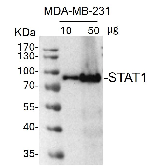
Application: Western BlotSample Tested: human MDAMB-231 whole cell lysateSpecies: HumanVerified Customer | Posted 11/28/2024Western Blot: whole cell lysates from MDA-MB-231 cells were loaded with10 or 50 ug/lane. 10% SDS-PAGE. STAT1 Antibody (PAF-ST1) was used for primary antibody: 1:1000, 4℃, overnight.

There are no reviews that match your criteria.
Protocols
Find general support by application which include: protocols, troubleshooting, illustrated assays, videos and webinars.
- Cellular Response to Hypoxia Protocols
- Immunoprecipitation Protocol
- R&D Systems Quality Control Western Blot Protocol
- Troubleshooting Guide: Western Blot Figures
- Western Blot Conditions
- Western Blot Protocol
- Western Blot Protocol for Cell Lysates
- Western Blot Troubleshooting
- Western Blot Troubleshooting Guide
- View all Protocols, Troubleshooting, Illustrated assays and Webinars


