STAT family proteins mediate cytokine signaling by acting as signal transducers in the cytoplasm and transcription activators in the nucleus. STAT5a and STAT5b are encoded by separate genes and share 93% amino acid identity.

Key Product Details
Validated by
Biological Validation
Species Reactivity
Validated:
Human, Mouse
Cited:
Human, Mouse
Applications
Validated:
Western Blot, Immunocytochemistry, Simple Western
Cited:
Immunohistochemistry, Western Blot, Immunocytochemistry
Label
Unconjugated
Antibody Source
Monoclonal Mouse IgG3 Clone # 251619
Loading...
Product Specifications
Immunogen
Human STAT5a synthetic peptide
SLDSRLSPPAGLFTSARGSLS
Accession # NP_003143
SLDSRLSPPAGLFTSARGSLS
Accession # NP_003143
Specificity
Detects human and mouse STAT5a. Does not cross-react with STAT5b.
Clonality
Monoclonal
Host
Mouse
Isotype
IgG3
Scientific Data Images for STAT5a Antibody (251619)
Detection of Human and Mouse STAT5a by Western Blot.
Western blot shows lysates of K562 human chronic myelogenous leukemia cell line and M1 mouse myeloid leukemia cell line. PVDF membrane was probed with 2 µg/mL of Mouse Anti-Human/Mouse STAT5a Monoclonal Antibody (Catalog # MAB2174) followed by HRP-conjugated Anti-Mouse IgG Secondary Antibody (Catalog # HAF007). A specific band was detected for STAT5a at approximately 91 kDa (as indicated). This experiment was conducted under reducing conditions and using Immunoblot Buffer Group 1.
STAT5a in K562 Human Cell Line.
STAT5a was detected in immersion fixed K562 human chronic myelogenous leukemia cell line using Mouse Anti-Human/Mouse STAT5a Monoclonal Antibody (Catalog # MAB2174) at 25 µg/mL for 3 hours at room temperature. Cells were stained using the NorthernLights™ 557-conjugated Anti-Mouse IgG Secondary Antibody (red; Catalog # NL007) and counterstained with DAPI (blue). Specific staining was localized to cytoplasm. View our protocol for Fluorescent ICC Staining of Non-adherent Cells.
Detection of Human STAT5a by Simple WesternTM.
Left: Simple Western lane view shows lysates of K562 human chronic myelogenous leukemia cell line, loaded at 1 mg/ml. A specific band was detected for STAT5a at approximately 91 kDa (as indicated) using both 2 µg/ml and 10 µg/ml of Mouse Anti-Human/Mouse STAT5a Monoclonal Antibody (Catalog # MAB2174) followed by HRP-conjugated Goat Anti-Mouse Secondary Antibody (Catalog # 042-205). This experiment was conducted under reducing conditions and using the 12-230kDa separation system. Right: Simple Western electropherogram showing the same Mouse Anti-Human/Mouse STAT5a Monoclonal Antibody (Catalog # MAB2174) tested at 2 µg/ml (blue line) and 10 µg/ml (green line) in the K562 human chronic myelogenous leukemia cell line.
Detection of Mouse STAT5A by Western Blot
Effects of inhibition of IGF-IR on IGF-IR, BCR-ABL and downstream target proteins in CML cell lines. (A) PPP induces concentration-dependent decrease in IGF-IR tyrosine kinase activity in K562 and KBM-5 cell lines. In contrast, PPP fails to cause similar effect in BCR-ABL tyrosine kinase activity. The results represent the means ± S.D. of three consistent experiments. *: P < 0.01 and †: P < 0.001 compared with control untreated cells. (B) Western blotting and co-immunoprecipitation studies confirm that PPP decreases the tyrosine phosphorylation of IGF-IR in a concentration-dependent fashion (results shown are representative and were obtained from the KBM-5 cell line). The basal levels of IGF-IR did not change after treatment with PPP. The phosphorylation level of BCR-ABL remains unchanged after treatment with PPP. The decrease in pIGF-IR is associated with down-regulation of pAkt and pSTAT5, two oncogenic proteins in CML. Changes are not seen in Akt and STAT5. Also, PPP induces changes consistent with apoptotic cell death including down-regulation of Bcl-2, Bcl-XL and caspase-3. Moreover, treatment with PPP induces up-regulation of cyclin B1 and down-regulation of cyclin E and pCdc2, whereas the levels of Cdc2 and p16 remain unchanged. Overall, the changes in the cell cycle regulatory proteins are consistent with G2/M-phase cell cycle arrest. beta -Actin shows equal loading of the proteins. (C) IGF-IR siRNA decreases IGF-IR levels in the KBM-5 cell line and this decrease is associated with down-regulation of pAkt and pSTAT5. Basal levels of these two proteins remain unchanged. IGF-IR siRNA did not induce alterations in BCR-ABL or pBCR-ABL protein. beta -Actin confirms equal loading of the proteins. Image collected and cropped by CiteAb from the following publication (https://pubmed.ncbi.nlm.nih.gov/19508387), licensed under a CC-BY license. Not internally tested by R&D Systems.Applications for STAT5a Antibody (251619)
Application
Recommended Usage
Immunocytochemistry
8-25 µg/mL
Sample: Immersion fixed human peripheral blood mononuclear cells and K562 human chronic myelogenous leukemia cell line
Sample: Immersion fixed human peripheral blood mononuclear cells and K562 human chronic myelogenous leukemia cell line
Simple Western
2-10 µg/mL
Sample: K562 human chronic myelogenous leukemia cell line
Sample: K562 human chronic myelogenous leukemia cell line
Western Blot
2 µg/mL
Sample: K562 human chronic myelogenous leukemia cell line and M1 mouse myeloid leukemia cell line
Sample: K562 human chronic myelogenous leukemia cell line and M1 mouse myeloid leukemia cell line
Reviewed Applications
Read 1 review rated 3 using MAB2174 in the following applications:
Formulation, Preparation, and Storage
Purification
Protein A or G purified from hybridoma culture supernatant
Reconstitution
Reconstitute at 0.5 mg/mL in sterile PBS. For liquid material, refer to CoA for concentration.
Loading...
Formulation
Lyophilized from a 0.2 μm filtered solution in PBS with Trehalose. See Certificate of Analysis for details.
*Small pack size (-SP) is supplied either lyophilized or as a 0.2 µm filtered solution in PBS.
*Small pack size (-SP) is supplied either lyophilized or as a 0.2 µm filtered solution in PBS.
Shipping
Lyophilized product is shipped at ambient temperature. Liquid small pack size (-SP) is shipped with polar packs. Upon receipt, store immediately at the temperature recommended below.
Stability & Storage
Use a manual defrost freezer and avoid repeated freeze-thaw cycles.
- 12 months from date of receipt, -20 to -70 °C as supplied.
- 1 month, 2 to 8 °C under sterile conditions after reconstitution.
- 6 months, -20 to -70 °C under sterile conditions after reconstitution.
Calculators
Background: STAT5a
Long Name
Signal Transducer and Activator of Transcription 5
Alternate Names
MGF, signal transducer and activator of transcription 5A, STAT5
Gene Symbol
STAT5A
UniProt
Additional STAT5a Products
Product Documents for STAT5a Antibody (251619)
Certificate of Analysis
To download a Certificate of Analysis, please enter a lot or batch number in the search box below.
Note: Certificate of Analysis not available for kit components.
Product Specific Notices for STAT5a Antibody (251619)
For research use only
Citations for STAT5a Antibody (251619)
 Powered by Bioz
Customer Reviews for STAT5a Antibody (251619) (1)
3 out of 5
1 Customer Rating
Have you used STAT5a Antibody (251619)?
Submit a review and receive an Amazon gift card!
$25/€18/£15/$25CAN/¥2500 Yen for a review with an image
$10/€7/£6/$10CAN/¥1110 Yen for a review without an image
Submit a review
Customer Images
Showing
1
-
1 of
1 review
Showing All
Filter By:
-
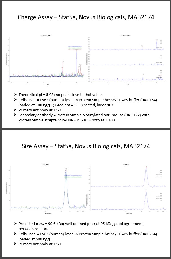
Application: Simple WesternSample Tested: k562 cellsSpecies: HumanVerified Customer | Posted 11/28/2016Theoretical pI = 5.98; no peak close to that value Cells used = K562 (human) lysed in Protein Simple bicine/CHAPS buffer (040-764) loaded at 100 ng/µL; Gradient = 5 – 8 nested, ladder# 3 Primary antibody at 1:50 Secondary antibody = Protein Simple biotinylated anti-mouse (041-127) with Protein Simple streptavidin-HRP (041-106) both at 1:100 Predicted m.w. = 90.6 kDa; well defined peak at 95 kDa, good agreement between replicates Cells used = K562 (human) lysed in Protein Simple bicine/CHAPS buffer (040-764) loaded at 500 ng/µL Primary antibody at 1:50

There are no reviews that match your criteria.
Protocols
Find general support by application which include: protocols, troubleshooting, illustrated assays, videos and webinars.
- Appropriate Fixation of IHC/ICC Samples
- Cellular Response to Hypoxia Protocols
- ClariTSA™ Fluorophore Kits
- Detection & Visualization of Antibody Binding
- ICC Cell Smear Protocol for Suspension Cells
- ICC Immunocytochemistry Protocol Videos
- ICC for Adherent Cells
- Immunocytochemistry (ICC) Protocol
- Immunocytochemistry Troubleshooting
- Immunofluorescence of Organoids Embedded in Cultrex Basement Membrane Extract
- Immunohistochemistry (IHC) and Immunocytochemistry (ICC) Protocols
- Preparing Samples for IHC/ICC Experiments
- Preventing Non-Specific Staining (Non-Specific Binding)
- Primary Antibody Selection & Optimization
- Protocol for VisUCyte™ HRP Polymer Detection Reagent
- Protocol for the Fluorescent ICC Staining of Cell Smears - Graphic
- Protocol for the Fluorescent ICC Staining of Cultured Cells on Coverslips - Graphic
- Protocol for the Preparation and Fluorescent ICC Staining of Cells on Coverslips
- Protocol for the Preparation and Fluorescent ICC Staining of Non-adherent Cells
- Protocol for the Preparation and Fluorescent ICC Staining of Stem Cells on Coverslips
- Protocol for the Preparation of a Cell Smear for Non-adherent Cell ICC - Graphic
- R&D Systems Quality Control Western Blot Protocol
- TUNEL and Active Caspase-3 Detection by IHC/ICC Protocol
- The Importance of IHC/ICC Controls
- Troubleshooting Guide: Western Blot Figures
- Western Blot Conditions
- Western Blot Protocol
- Western Blot Protocol for Cell Lysates
- Western Blot Troubleshooting
- Western Blot Troubleshooting Guide
- View all Protocols, Troubleshooting, Illustrated assays and Webinars


