NLGN3 (Neuroligin 3; also gliotactin homolog) is a 110-114 kDa member of the type-B carboxyesterase/lipase family of proteins. It is a neuronal transmembrane protein that forms Ca++-dependent intercellular junctions with short beta -neurexin isoforms. This seems to contribute to both glutamatergic and GABAergic synapse formation. Mutations in NLGN3 are associated with a reduction in protein expression and the occurrence of autism. Mature human NLGN3 is an 811 amino acid (aa) type I transmembrane protein. It contains a 672 aa extracellular domain (ECD) (aa 38-709), plus a 118 aa cytoplasmic region. The ECD possesses a nonfunctional carboxyesterase domain (aa 41-625). Multiple splice variants exist. There is a deletion of aa 153-172 that may also be accompanied by an alternative start site at Met118, and a deletion of aa 153-192 that may also be accompanied by a five
Human Neuroligin 3/NLGN3 Antibody (566209)
R&D Systems | Catalog # MAB6088

Key Product Details
Species Reactivity
Validated:
Human
Cited:
Mouse
Applications
Validated:
Immunohistochemistry, Immunocytochemistry
Cited:
Immunohistochemistry
Label
Unconjugated
Antibody Source
Monoclonal Mouse IgG2B Clone # 566209
Loading...
Product Specifications
Immunogen
Mouse myeloma cell line NS0-derived recombinant human Neuroligin 3
Gln38-Leu709
Accession # NP_061850
Gln38-Leu709
Accession # NP_061850
Specificity
Detects human Neuroligin 3 in direct ELISAs. In this format, 100% cross-reactivity with recombinant human (rh) Neuroligin 3 variant 2 and recombinant rat Neuroligin 3 variant 2 and no cross-reactivity with rhNeuroligin 1 variant 2, rhNeuroligin 2, rhNeuroligin 4, recombinant rat Neuroligin 1, 1 variant 2, 2 variant 2, or 3 variant 4 is observed.
Clonality
Monoclonal
Host
Mouse
Isotype
IgG2B
Scientific Data Images for Human Neuroligin 3/NLGN3 Antibody (566209)
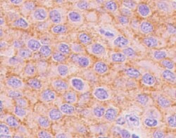
Neuroligin 3/NLGN3 in Human Brain.
Neuroligin 3/NLGN3 was detected in immersion fixed paraffin-embedded sections of human brain (cerebellum) using Human Neuroligin 3/NLGN3 Monoclonal Antibody (Catalog # MAB6088) at 15 µg/mL overnight at 4 °C. Tissue was stained using the Anti-Mouse HRP-DAB Cell & Tissue Staining Kit (brown; Catalog # CTS002) and counterstained with hematoxylin (blue). Lower panel shows a lack of labeling if primary antibodies are omitted and tissue is stained only with secondary antibody followed by incubation with detection reagents. View our protocol for Chromogenic IHC Staining of Paraffin-embedded Tissue Sections.Applications for Human Neuroligin 3/NLGN3 Antibody (566209)
Application
Recommended Usage
Immunocytochemistry
8-25 µg/mL
Sample: Immersion-fixed 7-day differentiated rat cortical stem cells
Sample: Immersion-fixed 7-day differentiated rat cortical stem cells
Immunohistochemistry
8-25 µg/mL
Sample: Immersion fixed paraffin-embedded sections of human brain (cerebellum)
Sample: Immersion fixed paraffin-embedded sections of human brain (cerebellum)
Reviewed Applications
Read 1 review rated 5 using MAB6088 in the following applications:
Formulation, Preparation, and Storage
Purification
Protein A or G purified from hybridoma culture supernatant
Reconstitution
Reconstitute at 0.5 mg/mL in sterile PBS. For liquid material, refer to CoA for concentration.
Loading...
Formulation
Lyophilized from a 0.2 μm filtered solution in PBS with Trehalose. *Small pack size (SP) is supplied either lyophilized or as a 0.2 µm filtered solution in PBS.
Shipping
Lyophilized product is shipped at ambient temperature. Liquid small pack size (-SP) is shipped with polar packs. Upon receipt, store immediately at the temperature recommended below.
Stability & Storage
Use a manual defrost freezer and avoid repeated freeze-thaw cycles.
- 12 months from date of receipt, -20 to -70 °C as supplied.
- 1 month, 2 to 8 °C under sterile conditions after reconstitution.
- 6 months, -20 to -70 °C under sterile conditions after reconstitution.
Calculators
Background: Neuroligin 3/NLGN3
Alternate Names
ASPGX1, AUTSX1, HNL3, NL3, NLGN3
Entrez Gene IDs
54413 (Human)
Gene Symbol
NLGN3
UniProt
Additional Neuroligin 3/NLGN3 Products
Product Documents for Human Neuroligin 3/NLGN3 Antibody (566209)
Certificate of Analysis
To download a Certificate of Analysis, please enter a lot or batch number in the search box below.
Note: Certificate of Analysis not available for kit components.
Product Specific Notices for Human Neuroligin 3/NLGN3 Antibody (566209)
For research use only
Citations for Human Neuroligin 3/NLGN3 Antibody (566209)
 Powered by Bioz
Customer Reviews for Human Neuroligin 3/NLGN3 Antibody (566209) (1)
5 out of 5
1 Customer Rating
Have you used Human Neuroligin 3/NLGN3 Antibody (566209)?
Submit a review and receive an Amazon gift card!
$25/€18/£15/$25CAN/¥2500 Yen for a review with an image
$10/€7/£6/$10CAN/¥1110 Yen for a review without an image
Submit a review
Customer Images
Showing
1
-
1 of
1 review
Showing All
Filter By:
-
Application: ImmunohistochemistrySample Tested: Brain tissueSpecies: HumanVerified Customer | Posted 05/22/2022

There are no reviews that match your criteria.
Protocols
Find general support by application which include: protocols, troubleshooting, illustrated assays, videos and webinars.
- Antigen Retrieval Protocol (PIER)
- Antigen Retrieval for Frozen Sections Protocol
- Appropriate Fixation of IHC/ICC Samples
- Cellular Response to Hypoxia Protocols
- Chromogenic IHC Staining of Formalin-Fixed Paraffin-Embedded (FFPE) Tissue Protocol
- Chromogenic Immunohistochemistry Staining of Frozen Tissue
- ClariTSA™ Fluorophore Kits
- Detection & Visualization of Antibody Binding
- Fluorescent IHC Staining of Frozen Tissue Protocol
- Graphic Protocol for Heat-induced Epitope Retrieval
- Graphic Protocol for the Preparation and Fluorescent IHC Staining of Frozen Tissue Sections
- Graphic Protocol for the Preparation and Fluorescent IHC Staining of Paraffin-embedded Tissue Sections
- Graphic Protocol for the Preparation of Gelatin-coated Slides for Histological Tissue Sections
- ICC Cell Smear Protocol for Suspension Cells
- ICC Immunocytochemistry Protocol Videos
- ICC for Adherent Cells
- IHC Sample Preparation (Frozen sections vs Paraffin)
- Immunocytochemistry (ICC) Protocol
- Immunocytochemistry Troubleshooting
- Immunofluorescence of Organoids Embedded in Cultrex Basement Membrane Extract
- Immunofluorescent IHC Staining of Formalin-Fixed Paraffin-Embedded (FFPE) Tissue Protocol
- Immunohistochemistry (IHC) and Immunocytochemistry (ICC) Protocols
- Immunohistochemistry Frozen Troubleshooting
- Immunohistochemistry Paraffin Troubleshooting
- Preparing Samples for IHC/ICC Experiments
- Preventing Non-Specific Staining (Non-Specific Binding)
- Primary Antibody Selection & Optimization
- Protocol for Heat-Induced Epitope Retrieval (HIER)
- Protocol for Making a 4% Formaldehyde Solution in PBS
- Protocol for VisUCyte™ HRP Polymer Detection Reagent
- Protocol for the Fluorescent ICC Staining of Cell Smears - Graphic
- Protocol for the Fluorescent ICC Staining of Cultured Cells on Coverslips - Graphic
- Protocol for the Preparation & Fixation of Cells on Coverslips
- Protocol for the Preparation and Chromogenic IHC Staining of Frozen Tissue Sections
- Protocol for the Preparation and Chromogenic IHC Staining of Frozen Tissue Sections - Graphic
- Protocol for the Preparation and Chromogenic IHC Staining of Paraffin-embedded Tissue Sections
- Protocol for the Preparation and Chromogenic IHC Staining of Paraffin-embedded Tissue Sections - Graphic
- Protocol for the Preparation and Fluorescent ICC Staining of Cells on Coverslips
- Protocol for the Preparation and Fluorescent ICC Staining of Non-adherent Cells
- Protocol for the Preparation and Fluorescent ICC Staining of Stem Cells on Coverslips
- Protocol for the Preparation and Fluorescent IHC Staining of Frozen Tissue Sections
- Protocol for the Preparation and Fluorescent IHC Staining of Paraffin-embedded Tissue Sections
- Protocol for the Preparation of Gelatin-coated Slides for Histological Tissue Sections
- Protocol for the Preparation of a Cell Smear for Non-adherent Cell ICC - Graphic
- TUNEL and Active Caspase-3 Detection by IHC/ICC Protocol
- The Importance of IHC/ICC Controls
- Troubleshooting Guide: Immunohistochemistry
- View all Protocols, Troubleshooting, Illustrated assays and Webinars
Loading...