NADPH Oxidase 4/NOX4, also known as Renal NADPH Oxidase/RENOX and Kidney Oxidase 1/KOX1, is a 66 to 75 kDa member of the NOX family of NADPH oxidases. NOX family members are a major source of reactive oxygen species (ROS), including hydrogen peroxide and superoxide, and are critical mediators of redox signaling. NOX4 is strongly expressed in the kidney, especially in the proximal convoluted tubule cells of the renal cortex, and may function as an oxygen sensor that regulates the synthesis of erythropoietin. Expression is also high in endothelial cells, and because aortas from deficient mice exhibit increased inflammation, hypertrophy and endothelial dysfunction, NOX4 may function to protect the vasculature during ischemic or inflammatory stress. Human NOX4 is 578 amino acids (aa) in length, with several additional smaller isoforms identified. Over aa 450-578, human NOX4 has 92% sequence identity with mouse NOX4, and 91% identity with rat NOX4.

Key Product Details
Species Reactivity
Validated:
Human
Cited:
Human
Applications
Validated:
Immunohistochemistry, Immunocytochemistry
Cited:
IF/IHC
Label
Unconjugated
Antibody Source
Monoclonal Mouse IgG2A Clone # 996715
Loading...
Product Specifications
Immunogen
E. coli-derived recombinant human NOX4
Lys450-Ser578
Accession # Q9NPH5
Lys450-Ser578
Accession # Q9NPH5
Specificity
Detects human NOX4 in direct ELISAs.
Clonality
Monoclonal
Host
Mouse
Isotype
IgG2A
Scientific Data Images for Human Nox4 Antibody (996715)
Nox4 in HUVECs.
Nox4 was detected in immersion fixed human umbilical vein endothelial cells (HUVECs; left panel) using Mouse Anti-Human Nox4 Monoclonal Antibody (Catalog # MAB8158) at 8 µg/mL for 3 hours at room temperature. Cells were stained using the NorthernLights™ 557-conjugated Anti-Mouse IgG Secondary Antibody (red; Catalog # NL007) and counterstained with DAPI (blue). Specific staining was localized to cytoplasm and nuclei. MCF-7 human breast cancer cell line is shown as negative staining (right panel). View our protocol for Fluorescent ICC Staining of Cells on Coverslips.
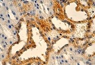
Nox4 in Human Kidney.
Nox4 was detected in immersion fixed paraffin-embedded sections of human kidney using Mouse Anti-Human Nox4 Monoclonal Antibody (Catalog # MAB8158) at 1.7 µg/mL for 1 hour at room temperature followed by incubation with the Anti-Mouse IgG VisUCyte™ HRP Polymer Antibody (Catalog # VC001). Before incubation with the primary antibody, tissue was subjected to heat-induced epitope retrieval using Antigen Retrieval Reagent-Basic (Catalog # CTS013). Tissue was stained using DAB (brown) and counterstained with hematoxylin (blue). Specific staining was localized to cytoplasm and nuclei. View our protocol for IHC Staining with VisUCyte HRP Polymer Detection Reagents.Applications for Human Nox4 Antibody (996715)
Application
Recommended Usage
Immunocytochemistry
5-25 µg/mL
Sample: Immersion fixed human umbilical vein endothelial cells (HUVECs)
Sample: Immersion fixed human umbilical vein endothelial cells (HUVECs)
Immunohistochemistry
1-25 µg/mL
Sample: Immersion fixed paraffin-embedded sections of human kidney
Sample: Immersion fixed paraffin-embedded sections of human kidney
Reviewed Applications
Read 1 review rated 5 using MAB8158 in the following applications:
Formulation, Preparation, and Storage
Purification
Protein A or G purified from hybridoma culture supernatant
Reconstitution
Reconstitute at 0.5 mg/mL in sterile PBS. For liquid material, refer to CoA for concentration.
Loading...
Formulation
Lyophilized from a 0.2 μm filtered solution in PBS with Trehalose. *Small pack size (SP) is supplied either lyophilized or as a 0.2 µm filtered solution in PBS.
Shipping
Lyophilized product is shipped at ambient temperature. Liquid small pack size (-SP) is shipped with polar packs. Upon receipt, store immediately at the temperature recommended below.
Stability & Storage
Use a manual defrost freezer and avoid repeated freeze-thaw cycles.
- 12 months from date of receipt, -20 to -70 °C as supplied.
- 1 month, 2 to 8 °C under sterile conditions after reconstitution.
- 6 months, -20 to -70 °C under sterile conditions after reconstitution.
Calculators
Background: Nox4
Long Name
NADPH Oxidase 4
Alternate Names
KOX-1, RENOX
Gene Symbol
NOX4
UniProt
Additional Nox4 Products
Product Documents for Human Nox4 Antibody (996715)
Certificate of Analysis
To download a Certificate of Analysis, please enter a lot or batch number in the search box below.
Note: Certificate of Analysis not available for kit components.
Product Specific Notices for Human Nox4 Antibody (996715)
For research use only
Related Research Areas
Citations for Human Nox4 Antibody (996715)
 Powered by Bioz
Customer Reviews for Human Nox4 Antibody (996715) (1)
5 out of 5
1 Customer Rating
Have you used Human Nox4 Antibody (996715)?
Submit a review and receive an Amazon gift card!
$25/€18/£15/$25CAN/¥2500 Yen for a review with an image
$10/€7/£6/$10CAN/¥1110 Yen for a review without an image
Submit a review
Customer Images
Showing
1
-
1 of
1 review
Showing All
Filter By:
-
Application: ImmunohistochemistrySample Tested: Kidney tissueSpecies: HumanVerified Customer | Posted 02/03/2022

There are no reviews that match your criteria.
Protocols
Find general support by application which include: protocols, troubleshooting, illustrated assays, videos and webinars.
- Antigen Retrieval Protocol (PIER)
- Antigen Retrieval for Frozen Sections Protocol
- Appropriate Fixation of IHC/ICC Samples
- Cellular Response to Hypoxia Protocols
- Chromogenic IHC Staining of Formalin-Fixed Paraffin-Embedded (FFPE) Tissue Protocol
- Chromogenic Immunohistochemistry Staining of Frozen Tissue
- ClariTSA™ Fluorophore Kits
- Detection & Visualization of Antibody Binding
- Fluorescent IHC Staining of Frozen Tissue Protocol
- Graphic Protocol for Heat-induced Epitope Retrieval
- Graphic Protocol for the Preparation and Fluorescent IHC Staining of Frozen Tissue Sections
- Graphic Protocol for the Preparation and Fluorescent IHC Staining of Paraffin-embedded Tissue Sections
- Graphic Protocol for the Preparation of Gelatin-coated Slides for Histological Tissue Sections
- ICC Cell Smear Protocol for Suspension Cells
- ICC Immunocytochemistry Protocol Videos
- ICC for Adherent Cells
- IHC Sample Preparation (Frozen sections vs Paraffin)
- Immunocytochemistry (ICC) Protocol
- Immunocytochemistry Troubleshooting
- Immunofluorescence of Organoids Embedded in Cultrex Basement Membrane Extract
- Immunofluorescent IHC Staining of Formalin-Fixed Paraffin-Embedded (FFPE) Tissue Protocol
- Immunohistochemistry (IHC) and Immunocytochemistry (ICC) Protocols
- Immunohistochemistry Frozen Troubleshooting
- Immunohistochemistry Paraffin Troubleshooting
- Preparing Samples for IHC/ICC Experiments
- Preventing Non-Specific Staining (Non-Specific Binding)
- Primary Antibody Selection & Optimization
- Protocol for Heat-Induced Epitope Retrieval (HIER)
- Protocol for Making a 4% Formaldehyde Solution in PBS
- Protocol for VisUCyte™ HRP Polymer Detection Reagent
- Protocol for the Fluorescent ICC Staining of Cell Smears - Graphic
- Protocol for the Fluorescent ICC Staining of Cultured Cells on Coverslips - Graphic
- Protocol for the Preparation & Fixation of Cells on Coverslips
- Protocol for the Preparation and Chromogenic IHC Staining of Frozen Tissue Sections
- Protocol for the Preparation and Chromogenic IHC Staining of Frozen Tissue Sections - Graphic
- Protocol for the Preparation and Chromogenic IHC Staining of Paraffin-embedded Tissue Sections
- Protocol for the Preparation and Chromogenic IHC Staining of Paraffin-embedded Tissue Sections - Graphic
- Protocol for the Preparation and Fluorescent ICC Staining of Cells on Coverslips
- Protocol for the Preparation and Fluorescent ICC Staining of Non-adherent Cells
- Protocol for the Preparation and Fluorescent ICC Staining of Stem Cells on Coverslips
- Protocol for the Preparation and Fluorescent IHC Staining of Frozen Tissue Sections
- Protocol for the Preparation and Fluorescent IHC Staining of Paraffin-embedded Tissue Sections
- Protocol for the Preparation of Gelatin-coated Slides for Histological Tissue Sections
- Protocol for the Preparation of a Cell Smear for Non-adherent Cell ICC - Graphic
- TUNEL and Active Caspase-3 Detection by IHC/ICC Protocol
- The Importance of IHC/ICC Controls
- Troubleshooting Guide: Immunohistochemistry
- View all Protocols, Troubleshooting, Illustrated assays and Webinars

