Placental (P) - Cadherin (PCAD) is a member of the Cadherin family of cell adhesion molecules. Cadherins are calcium-dependent transmembrane proteins, which bind to one another in a homophilic manner. On their cytoplasmic side, they associate with the three catenins, alpha, beta, and gamma (plakoglobin). This association links the cadherin protein to the cytoskeleton. Without association with the catenins, the cadherins are non-adhesive. Cadherins play a role in development, specifically in tissue formation. They may also help to maintain tissue architecture in the adult. P-Cadherin is a classical cadherin molecule. Classical cadherins consist of a large extracellular domain which contains DXD and DXNDN repeats responsible for mediating calcium-dependent adhesion, a single-pass transmembrane domain, and a short carboxy-terminal cytoplasmic domain responsible for interacting with the catenins. Human P-Cadherin is an 829 amino acid (aa) protein with a 26 aa signal sequence and an 803 aa propeptide. The mature protein begins at aa 108 and has a 548 aa extracellular region, a 23 aa transmembrane region, and a 151 aa cytoplasmic region. The human and mouse mature PCAD proteins share 87% homology.
Human P-Cadherin Antibody (104805)
R&D Systems | Catalog # MAB861

Key Product Details
Validated by
Species Reactivity
Validated:
Cited:
Applications
Validated:
Cited:
Label
Antibody Source
Product Specifications
Immunogen
Asp108-Gly654
Accession # CAA45177
Specificity
Clonality
Host
Isotype
Scientific Data Images for Human P-Cadherin Antibody (104805)
Detection of Human P‑Cadherin by Western Blot.
Western blot shows lysate of ZR-75 human breast cancer cell line. PVDF membrane was probed with 1 µg/mL of Mouse Anti-Human P-Cadherin Monoclonal Antibody (Catalog # MAB861) followed by HRP-conjugated Anti-Mouse IgG Secondary Antibody (Catalog # HAF018). A specific band was detected for P-Cadherin at approximately 120 kDa (as indicated). This experiment was conducted under reducing conditions and using Immunoblot Buffer Group 1.
Detection of P-Cadherin in A431 Human Cell Line by Flow Cytometry.
A431 human carcinoma cell line was stained with Mouse Anti-Human P-Cadherin Monoclonal Antibody (Catalog # MAB861, filled histogram) or isotype control antibody (Catalog # MAB002, open histogram), followed by Allophycocyanin-conjugated Anti-Mouse IgG F(ab')2 Secondary Antibody (Catalog # F0101B). Cells were stained in a buffer containing Ca2+ and Mg2+. View our protocol for Staining Membrane-associated Proteins.
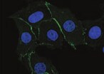
P‑Cadherin in A431 Human Cell Line.
P-Cadherin was detected in immersion fixed A431 human epithelial carcinoma cell line using Mouse Anti-Human P-Cadherin Monoclonal Antibody (Catalog # MAB861) at 10 µg/mL for 3 hours at room temperature. Cells were stained using the NorthernLights™ 557-conjugated Anti-Mouse IgG Secondary Antibody (red; Catalog # NL007) and counterstained with DAPI(blue). Specific staining was localized to the cell surface. View our protocol for Fluorescent ICC Staining of Cells on Coverslips.
Detection of Human P‑Cadherin by Simple WesternTM.
Simple Western lane view shows lysates of ZR‑75 human breast cancer cell line and A431 human epithelial carcinoma cell line, loaded at 0.2 mg/mL. A specific band was detected for P‑Cadherin at approximately 135 kDa (as indicated) using 10 µg/mL of Mouse Anti-Human P‑Cadherin Monoclonal Antibody (Catalog # MAB861). This experiment was conducted under reducing conditions and using the 12-230 kDa separation system.
Western Blot Shows Human P‑Cadherin Specificity by Using Knockout Cell Line.
Western blot shows lysates of A431 human epithelial carcinoma parental cell line and P-Cadherin knockout A431 cell line (KO). PVDF membrane was probed with 2 µg/mL of Mouse Anti-Human P-Cadherin Monoclonal Antibody (Catalog # MAB861) followed by HRP-conjugated Anti-Mouse IgG Secondary Antibody (Catalog # HAF018). A specific band was detected for P-Cadherin at approximately 150 kDa (as indicated) in the parental A431 cell line, but is not detectable in knockout A431 cell line. GAPDH (Catalog # MAB5718) is shown as a loading control. This experiment was conducted under reducing conditions and using Immunoblot Buffer Group 1.
P-Cadherin Specificity is Shown by Flow Cytometry in Knockout Cell Line.
P-Cadherin knockout A431 human epithelial carcinoma cell line was stained with Mouse Anti-Human P-Cadherin Monoclonal Antibody (Catalog # MAB861, filled histogram) or isotype control antibody (Catalog # MAB002, open histogram) followed by anti-Mouse IgG PE-conjugated Secondary Antibody (Catalog # F0102B). No staining in the P-Cadherin knockout A431 cell line was observed. Cells were stained in a buffer containing Ca2+ and Mg2+. View our protocol for Staining Membrane-associated Proteins.
P‑Cadherin Specificity is Shown by Immunocytochemistry in Knockout Cell Line.
P-Cadherin was detected in immersion fixed A431 human epithelial carcinoma cell line, wildtype (left panel) but is not detected in P-Cadherin knockout (right panel), using Mouse Anti-Human P-Cadherin Monoclonal Antibody (Catalog # MAB861) at 1 µg/mL for 3 hours at room temperature. Cells were stained using the NorthernLights™ 557-conjugated Anti-Mouse IgG Secondary Antibody (red; Catalog # NL007) and counterstained with DAPI (blue). Specific staining was localized to plasma membrane in wildtype cells. View our protocol for Fluorescent ICC Staining of Cells on Coverslips.
Human P-Cadherin ELISA Standard Curve
Recombinant Human P‑Cadherin Fc Chimera (Catalog # 861-PC) was serially diluted and captured by Mouse Anti-Human P‑Cadherin Monoclonal Antibody (Catalog # MAB861) coated on a Clear Polystyrene Microplate (Catalog # DY990). Goat Anti-Human P‑Cadherin Antigen Affinity-purified Polyclonal Antibody (Catalog # AF861) was biotinylated and incubated with the protein captured on the plate. Detection of the standard curve was achieved by incubating Streptavidin-HRP (Catalog # DY998)Applications for Human P-Cadherin Antibody (104805)
Competitive ELISA
CyTOF-ready
Flow Cytometry
Sample: A431 human epithelial carcinoma cell line stained in buffer containing Ca2+ and Mg2+
Immunocytochemistry
Sample: Immersion fixed A431 human epithelial carcinoma cell line, wildtype and knockout
Knockout Validated
Simple Western
Sample: ZR‑75 human breast cancer cell line and A431 human epithelial carcinoma cell line
Western Blot
Sample: ZR‑75 human breast cancer cell line
Human P-Cadherin Sandwich Immunoassay
Reviewed Applications
Read 2 reviews rated 4.5 using MAB861 in the following applications:
Flow Cytometry Panel Builder
Bio-Techne Knows Flow Cytometry
Save time and reduce costly mistakes by quickly finding compatible reagents using the Panel Builder Tool.
Advanced Features
- Spectra Viewer - Custom analysis of spectra from multiple fluorochromes
- Spillover Popups - Visualize the spectra of individual fluorochromes
- Antigen Density Selector - Match fluorochrome brightness with antigen density

Formulation, Preparation, and Storage
Purification
Reconstitution
Reconstitute at 0.5 mg/mL in sterile PBS. For liquid material, refer to CoA for concentration.
Formulation
Shipping
Stability & Storage
- 12 months from date of receipt, -20 to -70 °C as supplied.
- 1 month, 2 to 8 °C under sterile conditions after reconstitution.
- 6 months, -20 to -70 °C under sterile conditions after reconstitution.
Calculators
Background: P-Cadherin
References
- Shimoyama, Y. et al. (1989) J. Cell Biol. 109:1787.
- Bussemakers, M.J.G. et al. (1993) Mol. Biol. Reports 17:123.
- Overduin, M. et al. (1995) Science 267:386.
- Takeichi, M. (1991) Science 251:1451.
- Nose, A. et al. (1987) EMBO J. 6:3655.
Long Name
Alternate Names
Gene Symbol
UniProt
Additional P-Cadherin Products
Product Documents for Human P-Cadherin Antibody (104805)
Certificate of Analysis
To download a Certificate of Analysis, please enter a lot or batch number in the search box below.
Note: Certificate of Analysis not available for kit components.
Product Specific Notices for Human P-Cadherin Antibody (104805)
For research use only
Related Research Areas
Citations for Human P-Cadherin Antibody (104805)
 Powered by Bioz
Customer Reviews for Human P-Cadherin Antibody (104805) (2)
Have you used Human P-Cadherin Antibody (104805)?
Submit a review and receive an Amazon gift card!
$25/€18/£15/$25CAN/¥2500 Yen for a review with an image
$10/€7/£6/$10CAN/¥1110 Yen for a review without an image
Submit a review
Customer Images
-
Application: Immunocytochemistry/ImmunofluorescenceSample Tested: Breast cancer cellsSpecies: HumanVerified Customer | Posted 10/12/2021

-
Application: Flow CytometrySample Tested: Cancer CellsSpecies: HumanVerified Customer | Posted 04/26/2016
There are no reviews that match your criteria.
Protocols
Find general support by application which include: protocols, troubleshooting, illustrated assays, videos and webinars.
- 7-Amino Actinomycin D (7-AAD) Cell Viability Flow Cytometry Protocol
- Appropriate Fixation of IHC/ICC Samples
- Cellular Response to Hypoxia Protocols
- ClariTSA™ Fluorophore Kits
- Detection & Visualization of Antibody Binding
- Extracellular Membrane Flow Cytometry Protocol
- Flow Cytometry Protocol for Cell Surface Markers
- Flow Cytometry Protocol for Staining Membrane Associated Proteins
- Flow Cytometry Staining Protocols
- Flow Cytometry Troubleshooting Guide
- ICC Cell Smear Protocol for Suspension Cells
- ICC Immunocytochemistry Protocol Videos
- ICC for Adherent Cells
- Immunocytochemistry (ICC) Protocol
- Immunocytochemistry Troubleshooting
- Immunofluorescence of Organoids Embedded in Cultrex Basement Membrane Extract
- Immunohistochemistry (IHC) and Immunocytochemistry (ICC) Protocols
- Intracellular Flow Cytometry Protocol Using Alcohol (Methanol)
- Intracellular Flow Cytometry Protocol Using Detergents
- Intracellular Nuclear Staining Flow Cytometry Protocol Using Detergents
- Intracellular Staining Flow Cytometry Protocol Using Alcohol Permeabilization
- Intracellular Staining Flow Cytometry Protocol Using Detergents to Permeabilize Cells
- Preparing Samples for IHC/ICC Experiments
- Preventing Non-Specific Staining (Non-Specific Binding)
- Primary Antibody Selection & Optimization
- Propidium Iodide Cell Viability Flow Cytometry Protocol
- Protocol for Liperfluo
- Protocol for VisUCyte™ HRP Polymer Detection Reagent
- Protocol for the Characterization of Human Th22 Cells
- Protocol for the Characterization of Human Th9 Cells
- Protocol for the Fluorescent ICC Staining of Cell Smears - Graphic
- Protocol for the Fluorescent ICC Staining of Cultured Cells on Coverslips - Graphic
- Protocol for the Preparation and Fluorescent ICC Staining of Cells on Coverslips
- Protocol for the Preparation and Fluorescent ICC Staining of Non-adherent Cells
- Protocol for the Preparation and Fluorescent ICC Staining of Stem Cells on Coverslips
- Protocol for the Preparation of a Cell Smear for Non-adherent Cell ICC - Graphic
- Protocol: Annexin V and PI Staining by Flow Cytometry
- Protocol: Annexin V and PI Staining for Apoptosis by Flow Cytometry
- R&D Systems Quality Control Western Blot Protocol
- TUNEL and Active Caspase-3 Detection by IHC/ICC Protocol
- The Importance of IHC/ICC Controls
- Troubleshooting Guide: Fluorokine Flow Cytometry Kits
- Troubleshooting Guide: Western Blot Figures
- Western Blot Conditions
- Western Blot Protocol
- Western Blot Protocol for Cell Lysates
- Western Blot Troubleshooting
- Western Blot Troubleshooting Guide
- View all Protocols, Troubleshooting, Illustrated assays and Webinars
FAQs for Human P-Cadherin Antibody (104805)
-
Q: Why does the staining protocol with this Cadherin antibody use buffers containing Ca2+ and Mg2+?
A: The staining protocol with this and other Cadherin antibodies uses buffer containing Ca2+ and Mg2+ because Cadherin function is Calcium-dependent.