Tumor Protein 63 (p63), also named TP73L, TP63, p51, p40 or KET, is a p53 homolog. It is one of several proteins that are produced from a single gene using two promoters and alternative splicing of the primary RNA transcript. p63 is highly expressed in embryonic ectoderm and in the nuclei of basal regenerative cells of many epithelial tissues in the adult. p63 is suggested to play a role in development, epithelial cell maintenance and tumorigenesis (1‑3).

Key Product Details
Species Reactivity
Validated:
Human
Cited:
Human, Mouse, Transgenic Mouse, Xenograft
Applications
Validated:
Immunohistochemistry, Western Blot, Immunocytochemistry
Cited:
Immunohistochemistry, Immunocytochemistry, Chromatin Immunoprecipitation (ChIP)
Label
Unconjugated
Antibody Source
Polyclonal Goat IgG
Loading...
Product Specifications
Immunogen
E. coli-derived recombinant human p63/TP73L
Met40-Cys339
Accession # Q9H3D4
Met40-Cys339
Accession # Q9H3D4
Specificity
Detects human p63/TP73L in direct ELISAs and Western blots.
Clonality
Polyclonal
Host
Goat
Isotype
IgG
Scientific Data Images for Human p63/TP73L Antibody
p63/TP73L in Human Breast.
p63/TP73L was detected in immersion fixed paraffin-embedded sections of human breast using Goat Anti-Human p63/TP73L Antigen Affinity-purified Polyclonal Antibody (Catalog # AF1916) at 15 µg/mL overnight at 4 °C. Tissue was stained using the Anti-Goat HRP-DAB Cell & Tissue Staining Kit (brown; Catalog # CTS008) and counterstained with hematoxylin (blue). Specific staining was localized to nuclei. View our protocol for Chromogenic IHC Staining of Paraffin-embedded Tissue Sections.
p63/TP73L in SCC-25 Human Cell Line.
p63/TP73L was detected in immersion fixed SCC-25 human tongue carcinoma cell line using Goat Anti-Human p63/TP73L Antigen Affinity-purified Polyclonal Antibody (Catalog # AF1916) at 10 µg/mL for 3 hours at room temperature. Cells were stained using the NorthernLights™ 557-conjugated Anti-Goat IgG Secondary Antibody (yellow; Catalog # NL001) and counterstained with DAPI (blue). Specific staining was localized to nuclei. View our protocol for Fluorescent ICC Staining of Cells on Coverslips.
Detection of Mouse p63/TP73L by Immunocytochemistry/Immunofluorescence
Analysis of p73 and p63 co-expression in human and murine skin.(A) Scatter plot of TP63 versus TP73 RNA-seq expression [units = transcripts per million (TPM)] by human tissue type (n = 37) from the Human Protein Atlas (172 total samples) [30]. Mean expression (TPM + 0.1) for each tissue is plotted on a log2 scale with a LOESS smooth local regression line (gray). Correlation between TP63 and TP73 was quantified using Spearman’s rank correlation coefficient (rs). (B) Representative micrographs of H&E and immunofluorescence (IF) staining on serial human (top) and mouse (bottom) skin sections; DAPI (blue), p63 alpha (green), and p73 (red). Regions of the skin in micrographs are labeled as: interfollicular epidermis (IFE), hair follicle (HF), outer root sheath (ORS), HF bulge (Bu), hair bulb (HB), sebaceous gland (SG), hair shaft (HS), and arrector pili muscle (APM). Scale bars represent 200 μm for human and 50 μm for murine tissue. See also S1 and S2 Figs. Image collected and cropped by CiteAb from the following publication (https://pubmed.ncbi.nlm.nih.gov/31216312), licensed under a CC-BY license. Not internally tested by R&D Systems.
Detection of Human p63/TP73L by Western Blot
Analysis of delta Np73 in epidermal programming using an induced basal keratinocyte (iKC) model system.(A) Immunoblot of KLF4, p63 alpha, and p73 protein expression in neonatal human dermal fibroblast (HDFn) cells infected with lentivirus encoding delta Np73 isoforms ( delta Np73 alpha and delta Np73 beta ) or empty vector control in combination with KLF4 and delta Np63 alpha. Cells were grown for 3 days and protein was harvested for immunoblot analysis. (B) Bar graphs of RNA expression for the indicated keratinocyte genes in HDFn cells infected in (A). Cells were grown for 3 days and RNA was harvested for qRT-PCR analysis. Expression data are represented as the fold increase relative to control. The mean of three replicates is shown with error bars representing SEM. *p-value < 0.05, **p-value < 0.01, ***p-value < 0.001. (C) Principal component analysis (PCA) plot of RNA-seq from HDFn cells infected with lentivirus encoding delta Np73 beta or empty vector control in combination with KLF4 and delta Np63 alpha. Cells were grown for 6 days and RNA was harvested for RNA-seq analysis. The percentage of variance contributed by each PC is listed in parentheses. (D and E) Tables listing the enriched Genome Ontology (GO) categories and pathways among the top 250 genes contributing to PC1 from (C). (F) Heatmap with the expression of a set of 44 genes that underlie the enrichment of GO categories from (D). Genes are annotated based on known roles in iKC-related processes (gray box) and the presence of a p63/p73 ChIP-seq peak within 50 kb of its TSS in multiple basal cell types (brown box). See also S5 and S6 Figs and S2–S9 Tables. Image collected and cropped by CiteAb from the following publication (https://pubmed.ncbi.nlm.nih.gov/31216312), licensed under a CC-BY license. Not internally tested by R&D Systems.
Immunofluorescent Staining of Adult stem cell-derived Lung Organoids.
Adult stem cells isolated from human lung biopsy tissue were cultured following the steps detailed in the human lung organoid culture protocol. Lung organoids were stained with a (A) Goat Anti-Human p63/TP73L Polyclonal Antibody (Catalog # AF1916; red) and a rabbit anti-human cytokeratin 5 (KRT5) monoclonal antibody (green) to visualize basal cells; a (B) Hamster Anti-Mouse Podoplanin (PDPN) Monoclonal Antibody (Novus Biologicals, Catalog # NB600-1015; green) to visualize alveolar type I cells and a Goat Anti-Human p63/TP73L Polyclonal Antibody (Catalog # AF1916; red) to visualize basal cells; and a (C, D) Mouse Anti-MUC5AC Monoclonal Antibody (Novus Biologicals, Catalog # NBP2-15196; green) to visualize goblet cells and a Mouse Anti-Human/Mouse/Rat SOX2 Monoclonal Antibody (Catalog # MAB2018; red). All samples were counterstained with DAPI (Catalog # 5748; blue).Applications for Human p63/TP73L Antibody
Application
Recommended Usage
Immunocytochemistry
5-15 µg/mL
Sample: Immersion fixed FaDu human hypopharyngeal carcinoma cell line and SCC-25 human tongue carcinoma cell line
Sample: Immersion fixed FaDu human hypopharyngeal carcinoma cell line and SCC-25 human tongue carcinoma cell line
Immunohistochemistry
5-15 µg/mL
Sample: Immersion fixed paraffin-embedded sections of human breast
Sample: Immersion fixed paraffin-embedded sections of human breast
Western Blot
0.1 µg/mL
Sample: Recombinant Human p63/TP73L
Sample: Recombinant Human p63/TP73L
Reviewed Applications
Read 4 reviews rated 4.3 using AF1916 in the following applications:
Formulation, Preparation, and Storage
Purification
Antigen Affinity-purified
Reconstitution
Reconstitute at 0.2 mg/mL in sterile PBS. For liquid material, refer to CoA for concentration.
Loading...
Formulation
Lyophilized from a 0.2 μm filtered solution in PBS with Trehalose. *Small pack size (SP) is supplied either lyophilized or as a 0.2 µm filtered solution in PBS.
Shipping
Lyophilized product is shipped at ambient temperature. Liquid small pack size (-SP) is shipped with polar packs. Upon receipt, store immediately at the temperature recommended below.
Stability & Storage
Use a manual defrost freezer and avoid repeated freeze-thaw cycles.
- 12 months from date of receipt, -20 to -70 °C as supplied.
- 1 month, 2 to 8 °C under sterile conditions after reconstitution.
- 6 months, -20 to -70 °C under sterile conditions after reconstitution.
Calculators
Background: p63/TP73L
References
- Harms, K. et al. (2004), Cell Mol. Life Sci. 61(7):822.
- Koster, M.I. and D.R. Roop, J. Dermatol. Sci. 34(1):3.
- Benard, J. et al. (2003) Hum. Mutat. 21(3):182.
Long Name
Tumor Protein 63
Alternate Names
EEC3, KET, OFC8, p40, p51, p73L, SHFM4, TP63, TP73L
Entrez Gene IDs
8626 (Human)
Gene Symbol
TP63
UniProt
Additional p63/TP73L Products
Product Documents for Human p63/TP73L Antibody
Certificate of Analysis
To download a Certificate of Analysis, please enter a lot or batch number in the search box below.
Note: Certificate of Analysis not available for kit components.
Product Specific Notices for Human p63/TP73L Antibody
For research use only
Related Research Areas
Citations for Human p63/TP73L Antibody
 Powered by Bioz
Customer Reviews for Human p63/TP73L Antibody (4)
4.3 out of 5
4 Customer Ratings
Have you used Human p63/TP73L Antibody?
Submit a review and receive an Amazon gift card!
$25/€18/£15/$25CAN/¥2500 Yen for a review with an image
$10/€7/£6/$10CAN/¥1110 Yen for a review without an image
Submit a review
Customer Images
Showing
1
-
4 of
4 reviews
Showing All
Filter By:
-
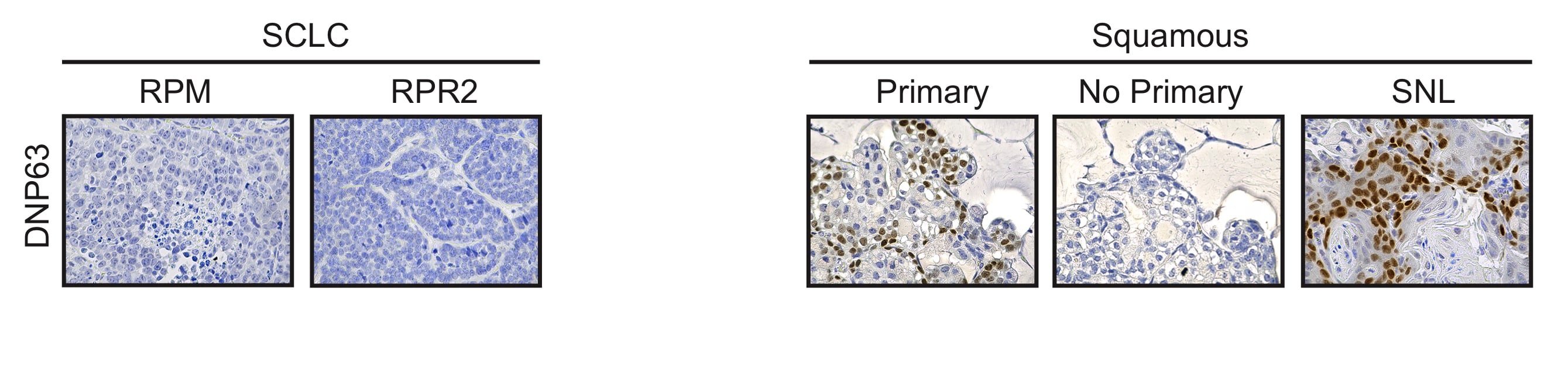
Application: Immunohistochemistry-ParaffinSample Tested: Mouse lung paraffin embedded sections and mouse lung tissueSpecies: MouseVerified Customer | Posted 09/23/2020p63/TP73L was detected in paraffin-embedded mouse tissue from Lkb1fl/fl; Ptenfl/fl and Sox2LSL/LSL ;Nkx2-1fl/fl; Lkb1fl/fl (SNL) mice, which present with lung squamous cell carcinoma (LSCC) tumors.p63/TP73L was detected in paraffin-embedded mouse tissue from Lkb1fl/fl; Ptenfl/fl and Sox2LSL/LSL ;Nkx2-1fl/fl; Lkb1fl/fl (SNL) mice, which present with lung squamous cell carcinoma (LSCC) tumors, using the Goat Anti-Human p63/TP73L Antigen Affinity-purified Polyclonal Antibody (Catalog # AF1916) overnight at 4℃ in blocking buffer at a dilution of 1/400. Citrate buffer pH 6.0 was used for high temperature antigen retrieval for 15 minutes in a pressure cooker. Tissue was stained using a HRP-conjugated secondary antibody and DAB staining and counterstained with hematoxylin (blue). P63 positivity was seen in the basal cells found in the trachea of mice and in LSCC tumors but not in small cell lung cancer (SCLC) tumors from Rb1/p53/Myc and Rb1/p53/Rbl2 mice or in mucinous adenocarcinoma tumors found in SNL mice.

-
Application: Western BlotSample Tested: 293T human embryonic kidney cell lineSpecies: HumanVerified Customer | Posted 12/07/2017
-
Application: Western BlotSample Tested: Mammary gland tissueSpecies: HumanVerified Customer | Posted 05/16/2017
-
Application: Immunocytochemistry/ImmunofluorescenceSample Tested: Prostate cancerSpecies: HumanVerified Customer | Posted 12/15/2016p63 is prostate stem cell marker. i have isolated stem cells from BPH patients and stained with p63 marker. I found excellent results with DAB kit and also p63 antibody performed superb job.
There are no reviews that match your criteria.
Protocols
Find general support by application which include: protocols, troubleshooting, illustrated assays, videos and webinars.
- Antigen Retrieval Protocol (PIER)
- Antigen Retrieval for Frozen Sections Protocol
- Appropriate Fixation of IHC/ICC Samples
- Cellular Response to Hypoxia Protocols
- Chromogenic IHC Staining of Formalin-Fixed Paraffin-Embedded (FFPE) Tissue Protocol
- Chromogenic Immunohistochemistry Staining of Frozen Tissue
- ClariTSA™ Fluorophore Kits
- Detection & Visualization of Antibody Binding
- Fluorescent IHC Staining of Frozen Tissue Protocol
- Graphic Protocol for Heat-induced Epitope Retrieval
- Graphic Protocol for the Preparation and Fluorescent IHC Staining of Frozen Tissue Sections
- Graphic Protocol for the Preparation and Fluorescent IHC Staining of Paraffin-embedded Tissue Sections
- Graphic Protocol for the Preparation of Gelatin-coated Slides for Histological Tissue Sections
- ICC Cell Smear Protocol for Suspension Cells
- ICC Immunocytochemistry Protocol Videos
- ICC for Adherent Cells
- IHC Sample Preparation (Frozen sections vs Paraffin)
- Immunocytochemistry (ICC) Protocol
- Immunocytochemistry Troubleshooting
- Immunofluorescence of Organoids Embedded in Cultrex Basement Membrane Extract
- Immunofluorescent IHC Staining of Formalin-Fixed Paraffin-Embedded (FFPE) Tissue Protocol
- Immunohistochemistry (IHC) and Immunocytochemistry (ICC) Protocols
- Immunohistochemistry Frozen Troubleshooting
- Immunohistochemistry Paraffin Troubleshooting
- Preparing Samples for IHC/ICC Experiments
- Preventing Non-Specific Staining (Non-Specific Binding)
- Primary Antibody Selection & Optimization
- Protocol for Heat-Induced Epitope Retrieval (HIER)
- Protocol for Making a 4% Formaldehyde Solution in PBS
- Protocol for VisUCyte™ HRP Polymer Detection Reagent
- Protocol for the Fluorescent ICC Staining of Cell Smears - Graphic
- Protocol for the Fluorescent ICC Staining of Cultured Cells on Coverslips - Graphic
- Protocol for the Preparation & Fixation of Cells on Coverslips
- Protocol for the Preparation and Chromogenic IHC Staining of Frozen Tissue Sections
- Protocol for the Preparation and Chromogenic IHC Staining of Frozen Tissue Sections - Graphic
- Protocol for the Preparation and Chromogenic IHC Staining of Paraffin-embedded Tissue Sections
- Protocol for the Preparation and Chromogenic IHC Staining of Paraffin-embedded Tissue Sections - Graphic
- Protocol for the Preparation and Fluorescent ICC Staining of Cells on Coverslips
- Protocol for the Preparation and Fluorescent ICC Staining of Non-adherent Cells
- Protocol for the Preparation and Fluorescent ICC Staining of Stem Cells on Coverslips
- Protocol for the Preparation and Fluorescent IHC Staining of Frozen Tissue Sections
- Protocol for the Preparation and Fluorescent IHC Staining of Paraffin-embedded Tissue Sections
- Protocol for the Preparation of Gelatin-coated Slides for Histological Tissue Sections
- Protocol for the Preparation of a Cell Smear for Non-adherent Cell ICC - Graphic
- R&D Systems Quality Control Western Blot Protocol
- TUNEL and Active Caspase-3 Detection by IHC/ICC Protocol
- The Importance of IHC/ICC Controls
- Troubleshooting Guide: Immunohistochemistry
- Troubleshooting Guide: Western Blot Figures
- Western Blot Conditions
- Western Blot Protocol
- Western Blot Protocol for Cell Lysates
- Western Blot Troubleshooting
- Western Blot Troubleshooting Guide
- View all Protocols, Troubleshooting, Illustrated assays and Webinars
Loading...
Associated Pathways
