Human PD-L1/B7-H1 Antibody (130021)
R&D Systems | Catalog # MAB1561

Key Product Details
Validated by
Biological Validation
Species Reactivity
Validated:
Human
Cited:
Human, Mouse
Applications
Validated:
Immunohistochemistry, Flow Cytometry, Dual RNAscope ISH-IHC Compatible, CyTOF-ready
Cited:
Immunohistochemistry, Immunohistochemistry-Paraffin, Western Blot, Flow Cytometry, Immunofluorescence, Immunocytochemistry, ELISA Detection, ELISA Development (Capture)
Label
Unconjugated
Antibody Source
Monoclonal Mouse IgG1 Clone # 130021
Loading...
Product Specifications
Immunogen
Mouse myeloma cell line NS0-derived recombinant human PD-L1/B7-H1
Phe19-Thr239
Accession # Q9NZQ7
Phe19-Thr239
Accession # Q9NZQ7
Specificity
Detects human PD-L1/B7-H1 in direct ELISAs. In direct ELISAs, no cross-reactivity with recombinant human (rh) B7-1, -2, -H2, -H3, -H3b,-H4, rhPD-L2, recombinant mouse B7-H1, recombinant rat (rr) B7-1, or rrB7-2 is observed.
Clonality
Monoclonal
Host
Mouse
Isotype
IgG1
Scientific Data Images for Human PD-L1/B7-H1 Antibody (130021)
Detection of PD-L1/B7-H1 in Jurkat Human Cell Line by Flow Cytometry.
Jurkat human acute T cell leukemia cell line was stained with Mouse Anti-Human PD-L1/B7-H1 Monoclonal Antibody (Catalog # MAB1561, filled histogram) or isotype control antibody (Catalog # MAB002, open histogram), followed by Phycoerythrin-conjugated Anti-Mouse IgG F(ab')2Secondary Antibody (Catalog # F0102B).View our protocol for Staining Membrane-associated Proteins.
Detection of PD-L1/B7-H1 in MDA-MB-231 Human Cell Line by Flow Cytometry.
MDA-MB-231 human breast adenocarcinoma cell line was stained with Mouse Anti-Human PD-L1/B7-H1 Monoclonal Antibody (Catalog # MAB1561, filled histogram) or isotype control antibody (Catalog # MAB002, open histogram), followed by Phycoerythrin-conjugated Anti-Mouse IgG F(ab')2 Secondary Antibody (Catalog # F0102B). Adherent cells were prepared by either manual scraping or with TrypLE Express treatment with similar results. View our protocol for Staining Membrane-associated Proteins.
PD-L1/B7-H1 in Human Colon Cancer.
PD-L1/B7-H1 was detected in formalin fixed paraffin-embedded sections of human colon cancer using Mouse Anti-Human PD-L1/B7-H1 Monoclonal Antibody (Catalog # MAB1561) at 15 µg/mL overnight at 4 °C. Tissue was stained using the Anti-Mouse HRP-DAB Cell & Tissue Staining Kit (brown; Catalog # CTS002) and counterstained with hematoxylin (blue). Specific staining was observed in the cytoplasm. View our protocol for Chromogenic IHC Staining of Paraffin-embedded Tissue Sections.
Detection of PD-L1/B7-H1 in Human Colon Cancer.
Formalin-fixed paraffin-embedded tissue sections of human colon cancer were probed for PDL1 mRNA (ACD RNAScope Probe, catalog # 600861; Fast Red chromogen, ACD catalog # 322360). Adjacent tissue section was processed for immunohistochemistry using mouse anti-human PDL1 monoclonal antibody (R&D Systems catalog # MAB1561) at 5ug/mL with overnight incubation at 4 degrees Celsius followed by incubation with anti-mouse IgG VisUCyte HRP Polymer Antibody (Catalog # VC001) and DAB chromogen (yellow-brown). Tissue was counterstained with hematoxylin (blue). Specific staining was localized to lymphocytes.
Detection of PD-L1/B7-H1 by Western Blot
Chemotherapeutics induce PD-L1 expression in NPC cells and PD-1 expression in NK cells via upregulation of NF-kappa B. NPC cells a or NK cells c were incubated with the NF-kappa B inhibitor BMS-345541 for 1 h before incubation with chemotherapeutics. NPC cells were transfected with NF-kappa B siRNA b or NK cells with NF-kappa B siRNA d for 16 h and then incubated with chemotherapeutics. Expression of NF-kappa B, PD-L1 and PD-1 was analyzed by immunoblot Image collected and cropped by CiteAb from the following open publication (https://pubmed.ncbi.nlm.nih.gov/32737537), licensed under a CC-BY license. Not internally tested by R&D Systems.
Detection of PD-L1/B7-H1 by Western Blot
Chemotherapeutics induce PD-L1 expression in NPC cells and PD-1 expression in NK cells via upregulation of NF-kappa B. NPC cells a or NK cells c were incubated with the NF-kappa B inhibitor BMS-345541 for 1 h before incubation with chemotherapeutics. NPC cells were transfected with NF-kappa B siRNA b or NK cells with NF-kappa B siRNA d for 16 h and then incubated with chemotherapeutics. Expression of NF-kappa B, PD-L1 and PD-1 was analyzed by immunoblot Image collected and cropped by CiteAb from the following open publication (https://pubmed.ncbi.nlm.nih.gov/32737537), licensed under a CC-BY license. Not internally tested by R&D Systems.Applications for Human PD-L1/B7-H1 Antibody (130021)
Application
Recommended Usage
CyTOF-ready
Ready to be labeled using established conjugation methods. No BSA or other carrier proteins that could interfere with conjugation.
Dual RNAscope ISH-IHC Compatible
3-25 µg/mL
Sample: Immersion fixed paraffin-embedded sections of human colon cancer
Sample: Immersion fixed paraffin-embedded sections of human colon cancer
Flow Cytometry
0.25 µg/106 cells
Sample: Jurkat human acute T cell leukemia cell line or MDA-MB-231 human breast adenocarcinoma cell line
Sample: Jurkat human acute T cell leukemia cell line or MDA-MB-231 human breast adenocarcinoma cell line
Immunohistochemistry
8-25 µg/mL
Sample: Immersion fixed paraffin-embedded sections of human colon
Sample: Immersion fixed paraffin-embedded sections of human colon
Reviewed Applications
Read 5 reviews rated 4.8 using MAB1561 in the following applications:
Flow Cytometry Panel Builder
Bio-Techne Knows Flow Cytometry
Save time and reduce costly mistakes by quickly finding compatible reagents using the Panel Builder Tool.
Advanced Features
- Spectra Viewer - Custom analysis of spectra from multiple fluorochromes
- Spillover Popups - Visualize the spectra of individual fluorochromes
- Antigen Density Selector - Match fluorochrome brightness with antigen density

Formulation, Preparation, and Storage
Purification
Protein A or G purified from hybridoma culture supernatant
Reconstitution
Reconstitute at 0.5 mg/mL in sterile PBS. For liquid material, refer to CoA for concentration.
Loading...
Formulation
Lyophilized from a 0.2 μm filtered solution in PBS with Trehalose. See Certificate of Analysis for details.
*Small pack size (-SP) is supplied either lyophilized or as a 0.2 µm filtered solution in PBS.
*Small pack size (-SP) is supplied either lyophilized or as a 0.2 µm filtered solution in PBS.
Shipping
Lyophilized product is shipped at ambient temperature. Liquid small pack size (-SP) is shipped with polar packs. Upon receipt, store immediately at the temperature recommended below.
Stability & Storage
Use a manual defrost freezer and avoid repeated freeze-thaw cycles.
- 12 months from date of receipt, -20 to -70 °C as supplied.
- 1 month, 2 to 8 °C under sterile conditions after reconstitution.
- 6 months, -20 to -70 °C under sterile conditions after reconstitution.
Calculators
Background: PD-L1/B7-H1
References
- Nishimura, H. and T. Honjo (2001) Trends Immunol. 22:265.
- Freeman, G.J. et al. (2000) J. Exp. Med. 192:1027.
- Latchman, Y. et al. (2001) Nat. Immunol. 2:261.
Long Name
Programmed Death Ligand 1
Alternate Names
B7-H1, B7H1, CD274, PDCD1L1, PDCD1LG1, PDL1
Entrez Gene IDs
Gene Symbol
CD274
UniProt
Additional PD-L1/B7-H1 Products
Product Documents for Human PD-L1/B7-H1 Antibody (130021)
Certificate of Analysis
To download a Certificate of Analysis, please enter a lot or batch number in the search box below.
Note: Certificate of Analysis not available for kit components.
Product Specific Notices for Human PD-L1/B7-H1 Antibody (130021)
For research use only
Citations for Human PD-L1/B7-H1 Antibody (130021)
 Powered by Bioz
Customer Reviews for Human PD-L1/B7-H1 Antibody (130021) (5)
4.8 out of 5
5 Customer Ratings
Have you used Human PD-L1/B7-H1 Antibody (130021)?
Submit a review and receive an Amazon gift card!
$25/€18/£15/$25CAN/¥2500 Yen for a review with an image
$10/€7/£6/$10CAN/¥1110 Yen for a review without an image
Submit a review
Customer Images
Showing
1
-
5 of
5 reviews
Showing All
Filter By:
-
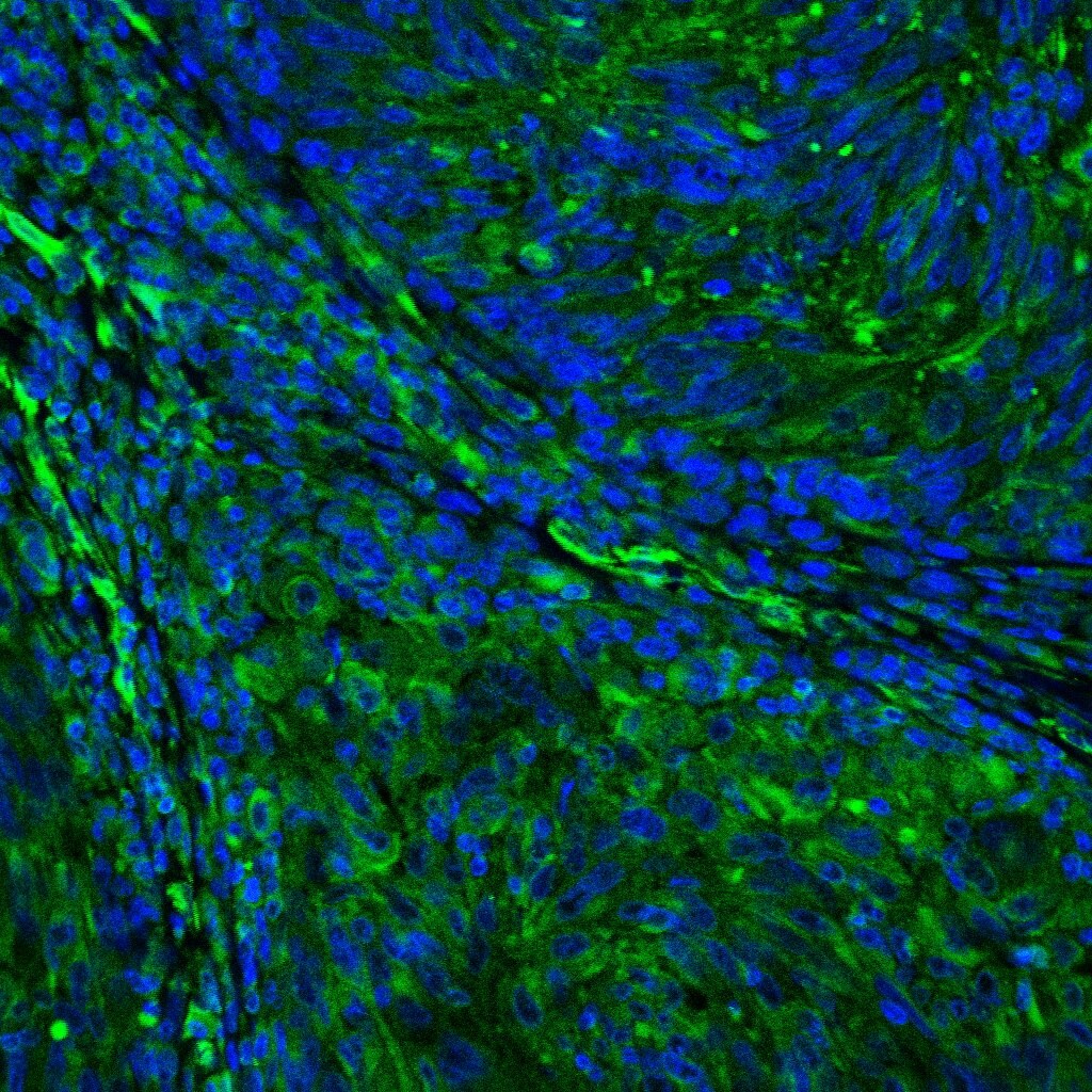
Application: Immunocytochemistry/ImmunofluorescenceSample Tested: Melanoma tissueSpecies: HumanVerified Customer | Posted 11/04/2020

-
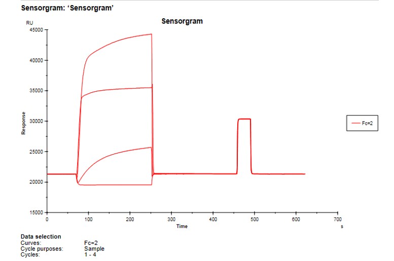
Application: SPR ASSAYSample Tested: MCF7Species: HumanVerified Customer | Posted 12/16/2017
-
Application: ImmunohistochemistrySample Tested: Colon cancer tissueSpecies: HumanVerified Customer | Posted 12/11/2017
-
Application: SPRSample Tested: Human recombinant test antibodySpecies: MouseVerified Customer | Posted 12/04/2017
-
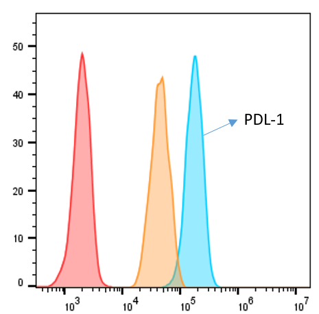
Application: Flow CytometrySample Tested: MDA-MB-231 human breast cancer cell lineSpecies: HumanVerified Customer | Posted 11/20/2017
There are no reviews that match your criteria.
Protocols
Find general support by application which include: protocols, troubleshooting, illustrated assays, videos and webinars.
- 7-Amino Actinomycin D (7-AAD) Cell Viability Flow Cytometry Protocol
- Antigen Retrieval Protocol (PIER)
- Antigen Retrieval for Frozen Sections Protocol
- Appropriate Fixation of IHC/ICC Samples
- Cellular Response to Hypoxia Protocols
- Chromogenic IHC Staining of Formalin-Fixed Paraffin-Embedded (FFPE) Tissue Protocol
- Chromogenic Immunohistochemistry Staining of Frozen Tissue
- ClariTSA™ Fluorophore Kits
- Detection & Visualization of Antibody Binding
- Extracellular Membrane Flow Cytometry Protocol
- Flow Cytometry Protocol for Cell Surface Markers
- Flow Cytometry Protocol for Staining Membrane Associated Proteins
- Flow Cytometry Staining Protocols
- Flow Cytometry Troubleshooting Guide
- Fluorescent IHC Staining of Frozen Tissue Protocol
- Graphic Protocol for Heat-induced Epitope Retrieval
- Graphic Protocol for the Preparation and Fluorescent IHC Staining of Frozen Tissue Sections
- Graphic Protocol for the Preparation and Fluorescent IHC Staining of Paraffin-embedded Tissue Sections
- Graphic Protocol for the Preparation of Gelatin-coated Slides for Histological Tissue Sections
- IHC Sample Preparation (Frozen sections vs Paraffin)
- ISH-IHC Protocol for Chromogenic Detection on Formalin Fixed Paraffin Embedded (FFPE) Tissue
- Immunofluorescent IHC Staining of Formalin-Fixed Paraffin-Embedded (FFPE) Tissue Protocol
- Immunohistochemistry (IHC) and Immunocytochemistry (ICC) Protocols
- Immunohistochemistry Frozen Troubleshooting
- Immunohistochemistry Paraffin Troubleshooting
- Intracellular Flow Cytometry Protocol Using Alcohol (Methanol)
- Intracellular Flow Cytometry Protocol Using Detergents
- Intracellular Nuclear Staining Flow Cytometry Protocol Using Detergents
- Intracellular Staining Flow Cytometry Protocol Using Alcohol Permeabilization
- Intracellular Staining Flow Cytometry Protocol Using Detergents to Permeabilize Cells
- Preparing Samples for IHC/ICC Experiments
- Preventing Non-Specific Staining (Non-Specific Binding)
- Primary Antibody Selection & Optimization
- Propidium Iodide Cell Viability Flow Cytometry Protocol
- Protocol for Heat-Induced Epitope Retrieval (HIER)
- Protocol for Liperfluo
- Protocol for Making a 4% Formaldehyde Solution in PBS
- Protocol for VisUCyte™ HRP Polymer Detection Reagent
- Protocol for the Characterization of Human Th22 Cells
- Protocol for the Characterization of Human Th9 Cells
- Protocol for the Preparation & Fixation of Cells on Coverslips
- Protocol for the Preparation and Chromogenic IHC Staining of Frozen Tissue Sections
- Protocol for the Preparation and Chromogenic IHC Staining of Frozen Tissue Sections - Graphic
- Protocol for the Preparation and Chromogenic IHC Staining of Paraffin-embedded Tissue Sections
- Protocol for the Preparation and Chromogenic IHC Staining of Paraffin-embedded Tissue Sections - Graphic
- Protocol for the Preparation and Fluorescent IHC Staining of Frozen Tissue Sections
- Protocol for the Preparation and Fluorescent IHC Staining of Paraffin-embedded Tissue Sections
- Protocol for the Preparation of Gelatin-coated Slides for Histological Tissue Sections
- Protocol: Annexin V and PI Staining by Flow Cytometry
- Protocol: Annexin V and PI Staining for Apoptosis by Flow Cytometry
- TUNEL and Active Caspase-3 Detection by IHC/ICC Protocol
- The Importance of IHC/ICC Controls
- Troubleshooting Guide: Fluorokine Flow Cytometry Kits
- Troubleshooting Guide: Immunohistochemistry
- View all Protocols, Troubleshooting, Illustrated assays and Webinars
Loading...