PGC1 alpha (PPAR-gamma Coactivator 1 alpha; also LEM6) is a 97-120 kDa member of the PGC-1 family of proteins. It is expressed in select cell types, including brown adipocytes, skeletal muscle and hepatocytes. PGC1 alpha participates in both RNA processing and transcriptional coactivation in conjunction with multiple nuclear hormone receptors such as PPAR gamma, RAR and TR. Human PCG1 alpha is 798 amino acids (aa) in length. It contains an LxxLL nuclear receptor binding motif (aa 144-148), one PPAR-gamma interaction domain (aa 293-339), two NLSs and an RNA binding/processing region (aa 566-710). PGC1 alpha activity is regulated by phosphorylation. AMPK is known to phosphorylate Thr178 and Ser539, promoting cotranscriptional activity. Conversely, Akt-mediated phosphorylation at Ser571 is reported to downregulate PGC1 alpha activity. This latter effect is achieved by an initial Ser571 phosphorylation, followed by GCN5 binding and subsequent PCG1 alpha acetylation that promotes PGC-1 alpha dissociation from target gene promoters.
Human PGC1 alpha Antibody (474915)
R&D Systems | Catalog # MAB10784

Key Product Details
Species Reactivity
Human
Applications
Immunohistochemistry, Western Blot, Immunocytochemistry
Label
Unconjugated
Antibody Source
Monoclonal Mouse IgG1 Clone # 474915
Loading...
Product Specifications
Immunogen
E. coli-derived human PGC1 alpha
Glu11-Ile280
Accession # Q9UBK2
Glu11-Ile280
Accession # Q9UBK2
Specificity
Detects human PGC1 alpha in direct ELISAs.
Clonality
Monoclonal
Host
Mouse
Isotype
IgG1
Scientific Data Images for Human PGC1 alpha Antibody (474915)
Detection of Human PGC1 alpha by Western Blot.
Western blot shows lysates of Jurkat human acute T cell leukemia cell line. PVDF membrane was probed with 2 µg/mL of Mouse Anti-Human PGC1 alpha Monoclonal Antibody (Catalog # MAB10784) followed by HRP-conjugated Anti-Mouse IgG Secondary Antibody (HAF018). A specific band was detected for PGC1 alpha at approximately 145 kDa (as indicated). This experiment was conducted under reducing conditions and using Western Blot Buffer Group 1.
PGC1 alpha in A431 Human Cell Line.
PGC1 alpha was detected in immersion fixed A431 human epithelial carcinoma cell line using Mouse Anti-Human PGC1 alpha Monoclonal Antibody (Catalog # MAB10784) at 8 µg/mL for 3 hours at room temperature. Cells were stained using the NorthernLights™ 557-conjugated Anti-Mouse IgG Secondary Antibody (red; NL007) and counterstained with DAPI (blue). Specific staining was localized to cell nuclei. Staining was performed using our protocol for Fluorescent ICC Staining of Non-adherent Cells.
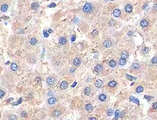
PGC1 alpha in Human Liver Cancer.
PGC1 alpha was detected in immersion fixed paraffin-embedded sections of human liver cancer tissue using Mouse Anti-Human PGC1 alpha Monoclonal Antibody (Catalog # MAB10784) at 5 µg/mL for 1 hour at room temperature followed by incubation with the Anti-Mouse IgG VisUCyte™ HRP Polymer Antibody (VC001). Before incubation with the primary antibody, tissue was subjected to heat-induced epitope retrieval using Antigen Retrieval Reagent-Basic (CTS013). Tissue was stained using DAB (brown) and counterstained with hematoxylin (blue). Specific staining was localized to nuclei in hepatocytes. Staining was performed using our protocol for IHC Staining with VisUCyte HRP Polymer Detection Reagents.Applications for Human PGC1 alpha Antibody (474915)
Application
Recommended Usage
Immunocytochemistry
8-25 µg/mL
Sample: Immersion fixed A431 human epithelial carcinoma cell line
Sample: Immersion fixed A431 human epithelial carcinoma cell line
Immunohistochemistry
5-25 µg/mL
Sample: Immersion fixed paraffin-embedded sections of human liver cancer
Sample: Immersion fixed paraffin-embedded sections of human liver cancer
Western Blot
2 µg/mL
Sample: Jurkat human acute T cell leukemia cell line
Sample: Jurkat human acute T cell leukemia cell line
Reviewed Applications
Read 1 review rated 5 using MAB10784 in the following applications:
Formulation, Preparation, and Storage
Purification
Protein A or G purified from hybridoma culture supernatant
Reconstitution
Reconstitute at 0.5 mg/mL in sterile PBS. For liquid material, refer to CoA for concentration.
Loading...
Formulation
Lyophilized from a 0.2 μm filtered solution in PBS with Trehalose. *Small pack size (SP) is supplied either lyophilized or as a 0.2 µm filtered solution in PBS.
Shipping
Lyophilized product is shipped at ambient temperature. Liquid small pack size (-SP) is shipped with polar packs. Upon receipt, store immediately at the temperature recommended below.
Stability & Storage
Use a manual defrost freezer and avoid repeated freeze-thaw cycles.
- 12 months from date of receipt, -20 to -70 °C as supplied.
- 1 month, 2 to 8 °C under sterile conditions after reconstitution.
- 6 months, -20 to -70 °C under sterile conditions after reconstitution.
Calculators
Background: PGC1 alpha
Long Name
Peroxisome Proliferator-activated Receptor gamma, Coactivator 1 alpha
Alternate Names
LEM6, PPARGC1A
Gene Symbol
PPARGC1A
UniProt
Additional PGC1 alpha Products
Product Documents for Human PGC1 alpha Antibody (474915)
Certificate of Analysis
To download a Certificate of Analysis, please enter a lot or batch number in the search box below.
Note: Certificate of Analysis not available for kit components.
Product Specific Notices for Human PGC1 alpha Antibody (474915)
For research use only
Citations for Human PGC1 alpha Antibody (474915)
 Powered by Bioz
Customer Reviews for Human PGC1 alpha Antibody (474915) (1)
5 out of 5
1 Customer Rating
Have you used Human PGC1 alpha Antibody (474915)?
Submit a review and receive an Amazon gift card!
$25/€18/£15/$25CAN/¥2500 Yen for a review with an image
$10/€7/£6/$10CAN/¥1110 Yen for a review without an image
Submit a review
Customer Images
Showing
1
-
1 of
1 review
Showing All
Filter By:
-
Application: ImmunohistochemistrySample Tested: Liver cancer tissueSpecies: HumanVerified Customer | Posted 06/19/2022

There are no reviews that match your criteria.
Protocols
Find general support by application which include: protocols, troubleshooting, illustrated assays, videos and webinars.
- Antigen Retrieval Protocol (PIER)
- Antigen Retrieval for Frozen Sections Protocol
- Appropriate Fixation of IHC/ICC Samples
- Cellular Response to Hypoxia Protocols
- Chromogenic IHC Staining of Formalin-Fixed Paraffin-Embedded (FFPE) Tissue Protocol
- Chromogenic Immunohistochemistry Staining of Frozen Tissue
- ClariTSA™ Fluorophore Kits
- Detection & Visualization of Antibody Binding
- Fluorescent IHC Staining of Frozen Tissue Protocol
- Graphic Protocol for Heat-induced Epitope Retrieval
- Graphic Protocol for the Preparation and Fluorescent IHC Staining of Frozen Tissue Sections
- Graphic Protocol for the Preparation and Fluorescent IHC Staining of Paraffin-embedded Tissue Sections
- Graphic Protocol for the Preparation of Gelatin-coated Slides for Histological Tissue Sections
- ICC Cell Smear Protocol for Suspension Cells
- ICC Immunocytochemistry Protocol Videos
- ICC for Adherent Cells
- IHC Sample Preparation (Frozen sections vs Paraffin)
- Immunocytochemistry (ICC) Protocol
- Immunocytochemistry Troubleshooting
- Immunofluorescence of Organoids Embedded in Cultrex Basement Membrane Extract
- Immunofluorescent IHC Staining of Formalin-Fixed Paraffin-Embedded (FFPE) Tissue Protocol
- Immunohistochemistry (IHC) and Immunocytochemistry (ICC) Protocols
- Immunohistochemistry Frozen Troubleshooting
- Immunohistochemistry Paraffin Troubleshooting
- Preparing Samples for IHC/ICC Experiments
- Preventing Non-Specific Staining (Non-Specific Binding)
- Primary Antibody Selection & Optimization
- Protocol for Heat-Induced Epitope Retrieval (HIER)
- Protocol for Making a 4% Formaldehyde Solution in PBS
- Protocol for VisUCyte™ HRP Polymer Detection Reagent
- Protocol for the Fluorescent ICC Staining of Cell Smears - Graphic
- Protocol for the Fluorescent ICC Staining of Cultured Cells on Coverslips - Graphic
- Protocol for the Preparation & Fixation of Cells on Coverslips
- Protocol for the Preparation and Chromogenic IHC Staining of Frozen Tissue Sections
- Protocol for the Preparation and Chromogenic IHC Staining of Frozen Tissue Sections - Graphic
- Protocol for the Preparation and Chromogenic IHC Staining of Paraffin-embedded Tissue Sections
- Protocol for the Preparation and Chromogenic IHC Staining of Paraffin-embedded Tissue Sections - Graphic
- Protocol for the Preparation and Fluorescent ICC Staining of Cells on Coverslips
- Protocol for the Preparation and Fluorescent ICC Staining of Non-adherent Cells
- Protocol for the Preparation and Fluorescent ICC Staining of Stem Cells on Coverslips
- Protocol for the Preparation and Fluorescent IHC Staining of Frozen Tissue Sections
- Protocol for the Preparation and Fluorescent IHC Staining of Paraffin-embedded Tissue Sections
- Protocol for the Preparation of Gelatin-coated Slides for Histological Tissue Sections
- Protocol for the Preparation of a Cell Smear for Non-adherent Cell ICC - Graphic
- R&D Systems Quality Control Western Blot Protocol
- TUNEL and Active Caspase-3 Detection by IHC/ICC Protocol
- The Importance of IHC/ICC Controls
- Troubleshooting Guide: Immunohistochemistry
- Troubleshooting Guide: Western Blot Figures
- Western Blot Conditions
- Western Blot Protocol
- Western Blot Protocol for Cell Lysates
- Western Blot Troubleshooting
- Western Blot Troubleshooting Guide
- View all Protocols, Troubleshooting, Illustrated assays and Webinars
Loading...
Associated Pathways
