CXC chemokine ligand 16 (CXCL16) is a type I membrane protein containing a non-ELR motif-containing CXC chemokine domain in its extracellular region. Together with Fractalkine (CX3CL1), CXCL16 constitute the only two transmembrane chemokines within the superfamily. The gene for human CXCL16 predicts a 273 amino acid (aa) residue precursor protein with a putative signal peptide, a CXC chemokine domain, a mucin-like spacer region, a transmembrane domain and a cytoplasmic domain with a potential tyrosine phosphorylation and SH2 protein-binding site. Mouse and human CXCL16 share 70% aa sequence similarity within their chemokine domains and 49% overall aa sequence identity. By northern blot analysis, CXCL16 expression is detected in various human organs except for brain, bone marrow, skeletal muscle or colon. By flow cytometry, CXCL16 has been detected on the surface CD19+ B cells, CD14+ monocytes/macrophages, and CD11c+ splenic and lymph node dendritic cells. Functional CXCL16 can be shed from the cell surface as an approximately 35 kDa soluble protein. The functional receptor for CXCL16 has been identified as CXCR6 (also known as Bonzo, STRL33 or TYMSTR), a receptor previously shown to be a co-receptor for HIV entry. CXCL16 has also been independently cloned and named SRPSOX (scavenger receptor that binds phosphatidylserine and oxidized lipoprotein). It was shown to be a specific receptor for OxLDL but not LDL or acetyl-LDL.

Key Product Details
Species Reactivity
Validated:
Human, Primate
Cited:
Human, Mouse
Applications
Validated:
Immunohistochemistry, Western Blot, ELISA Capture (Matched Antibody Pair), Neutralization, Flow Cytometry, CyTOF-ready
Cited:
Immunohistochemistry, Immunohistochemistry-Paraffin, Immunohistochemistry-Frozen, Western Blot, Neutralization, Flow Cytometry, Immunocytochemistry
Label
Unconjugated
Antibody Source
Polyclonal Goat IgG
Loading...
Product Specifications
Immunogen
E. coli-derived recombinant human CXCL16
Asn49-Pro137
Accession # NP_071342
Asn49-Pro137
Accession # NP_071342
Specificity
Detects human CXCL16 in ELISAs and Western blots. In ELISAs, less than 0.05% cross-reactivity with recombinant mouse (rm) CXCL16, rm6Ckine, recombinant human (rh) BLC, rhCTACK, rhIL-8, rhPF4, and rhLymphotactin is observed.
Clonality
Polyclonal
Host
Goat
Isotype
IgG
Endotoxin Level
<0.10 EU per 1 μg of the antibody by the LAL method.
Scientific Data Images for CXCL16 Antibody
Chemotaxis Induced by CXCL16 and Neutralization by Human CXCL16 Antibody.
Recombinant Human CXCL16 Chemokine Domain (976-CX) induces chemotaxis in BaF3 mouse pro-B cell line transfected with mouse CXCR6 in a dose-dependent manner (orange line). Chemotaxis elicited by Recombinant Human CXCL16 Chemokine Domain (20 ng/mL) is neutralized (green line) by increasing concentrations of Goat Anti-Human/Primate CXCL16 Antigen Affinity-purified Polyclonal Antibody (Catalog # AF976). The ND50 is typically 0.05-0.25 µg/mL.
CXCL16 in Human Lymph Node.
CXCL16 was detected in immersion fixed paraffin-embedded sections of human lymph node using Goat Anti-Human/Primate CXCL16 Antigen Affinity-purified Polyclonal Antibody (Catalog # AF976) at 10 µg/mL overnight at 4 °C. Before incubation with the primary antibody tissue was subjected to heat-induced epitope retrieval using Antigen Retrieval Reagent-Basic (CTS013). Tissue was stained using the Anti-Goat HRP-DAB Cell & Tissue Staining Kit (brown; CTS008) and counterstained with hematoxylin (blue). View our protocol for Chromogenic IHC Staining of Paraffin-embedded Tissue Sections.
CXCL16 in Human Lymphoma.
CXCL16 was detected in immersion fixed paraffin-embedded sections of human lymphoma using Goat Anti-Human/Primate CXCL16 Antigen Affinity-purified Polyclonal Antibody (Catalog # AF976) at 15 µg/mL overnight at 4 °C. Tissue was stained using the Anti-Goat HRP-DAB Cell & Tissue Staining Kit (brown; CTS008) and counterstained with hematoxylin (blue). Lower panel shows a lack of labeling if primary antibodies are omitted and tissue is stained only with secondary antibody followed by incubation with detection reagents. View our protocol for Chromogenic IHC Staining of Paraffin-embedded Tissue Sections.
Detection of CXCL16 in Human PBMCs by Flow Cytometry.
Human PBMCs were stained with Goat Anti-Human CXCL16 Antigen Affinity-purified Polyclonal Antibody (AF976, filled histogram) or Goat IgG Isotype Control (AB-108-C, open histogram), followed by Phycoerythrin-conjugated Anti-Goat IgG Secondary Antibody (F0107). View our protocol for Staining Membrane-associated Proteins.
Human CXCL16 ELISA Standard Curve
Recombinant Human CXCL16 Extracellular Domain (1164-CX) was serially diluted and captured by Goat Anti-Human/Primate CXCL16 Antigen Affinity-purified Polyclonal Antibody (Catalog # AF976) coated on a Clear Polystyrene Microplate (DY990). Goat Anti-Human/Primate CXCL16 Antigen Affinity-purified Polyclonal Antibody (AF976) was biotinylated and incubated with the protein captured on the plate. Detection of the standard curve was achieved by incubating Streptavidin-HRP (DY998).
Human CXCL16 ELISA Standard Curve
Recombinant Human CXCL16 Extracellular Domain (1164-CX) was serially diluted and captured by Goat Anti-Human/Primate CXCL16 Antigen Affinity-purified Polyclonal Antibody (AF976) coated on a Clear Polystyrene Microplate (DY990). Goat Anti-Human/Primate CXCL16 Antigen Affinity-purified Polyclonal Antibody (AF976) was biotinylated and incubated with the protein captured on the plate. Detection of the standard curve was achieved by incubating Streptavidin-HRP (DY998)Applications for CXCL16 Antibody
Application
Recommended Usage
CyTOF-ready
Ready to be labeled using established conjugation methods. No BSA or other carrier proteins that could interfere with conjugation.
Flow Cytometry
0.25 µg/106 cells
Sample: Human PBMCs.
Sample: Human PBMCs.
Immunohistochemistry
5-15 µg/mL
Sample: Immersion fixed paraffin-embedded sections of human lymph node subjected to Antigen Retrieval Reagent-Basic (Catalog # CTS013) and human lymphoma
Sample: Immersion fixed paraffin-embedded sections of human lymph node subjected to Antigen Retrieval Reagent-Basic (Catalog # CTS013) and human lymphoma
Western Blot
0.1 µg/mL
Sample: Recombinant Human CXCL16 Chemokine Domain (Catalog # 976-CX)
Sample: Recombinant Human CXCL16 Chemokine Domain (Catalog # 976-CX)
Neutralization
Measured by its ability to neutralize CXCL16-induced chemotaxis in BaF3 mouse pro‑B cell line transfected with mouse CXCR6. Matloubian, M. et al. (2000) Nat. Immunol.The Neutralization Dose (ND50) is typically
0.05-0.25 μg/mL in the presence of 20 ng/mL Recombinant Human CXCL16 Chemokine Domain.
0.05-0.25 μg/mL in the presence of 20 ng/mL Recombinant Human CXCL16 Chemokine Domain.
Human/Primate CXCL16 Sandwich Immunoassay
Please Note: Optimal dilutions of this antibody should be experimentally determined.
Reviewed Applications
Read 4 reviews rated 4.5 using AF976 in the following applications:
Flow Cytometry Panel Builder
Bio-Techne Knows Flow Cytometry
Save time and reduce costly mistakes by quickly finding compatible reagents using the Panel Builder Tool.
Advanced Features
- Spectra Viewer - Custom analysis of spectra from multiple fluorochromes
- Spillover Popups - Visualize the spectra of individual fluorochromes
- Antigen Density Selector - Match fluorochrome brightness with antigen density

Formulation, Preparation, and Storage
Purification
Antigen Affinity-purified
Reconstitution
Reconstitute at 0.2 mg/mL in sterile PBS. For liquid material, refer to CoA for concentration.
Loading...
Formulation
Lyophilized from a 0.2 μm filtered solution in PBS with Trehalose. See Certificate of Analysis for details.
*Small pack size (-SP) is supplied either lyophilized or as a 0.2 µm filtered solution in PBS.
*Small pack size (-SP) is supplied either lyophilized or as a 0.2 µm filtered solution in PBS.
Shipping
Lyophilized product is shipped at ambient temperature. Liquid small pack size (-SP) is shipped with polar packs. Upon receipt, store immediately at the temperature recommended below.
Stability & Storage
Use a manual defrost freezer and avoid repeated freeze-thaw cycles.
- 12 months from date of receipt, -20 to -70 °C as supplied.
- 1 month, 2 to 8 °C under sterile conditions after reconstitution.
- 6 months, -20 to -70 °C under sterile conditions after reconstitution.
Calculators
Background: CXCL16
References
- Matloubian, M. et al. (2000) Nature Immun. 1:298.
- Shimaoka, T. et al. (2000) J. Biol. Chem. 275:40663.
- Wilbanks, A. et al. (2001) J. Immun. 166:5145.
Alternate Names
CXCL16
Gene Symbol
CXCL16
UniProt
Additional CXCL16 Products
Product Documents for CXCL16 Antibody
Certificate of Analysis
To download a Certificate of Analysis, please enter a lot or batch number in the search box below.
Note: Certificate of Analysis not available for kit components.
Product Specific Notices for CXCL16 Antibody
For research use only
Related Research Areas
Citations for CXCL16 Antibody
 Powered by Bioz
Customer Reviews for CXCL16 Antibody (4)
4.5 out of 5
4 Customer Ratings
Have you used CXCL16 Antibody?
Submit a review and receive an Amazon gift card!
$25/€18/£15/$25CAN/¥2500 Yen for a review with an image
$10/€7/£6/$10CAN/¥1110 Yen for a review without an image
Submit a review
Customer Images
Showing
1
-
4 of
4 reviews
Showing All
Filter By:
-
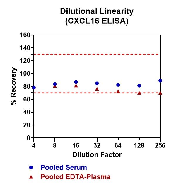
Application: ELISASample Tested: Serum and PlasmaSpecies: HumanVerified Customer | Posted 04/18/2021

-
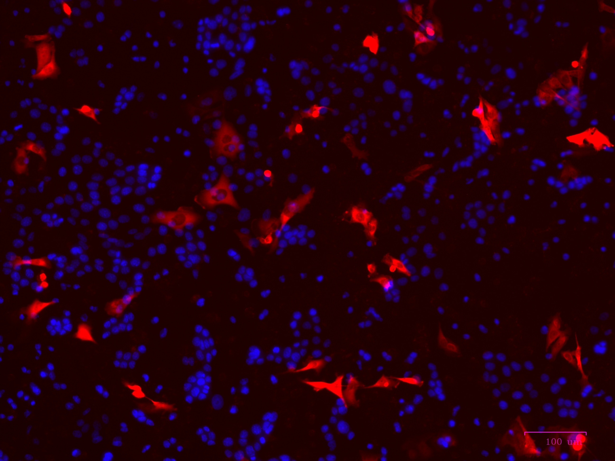
Application: Immunocytochemistry/ImmunofluorescenceSample Tested: OVARIAN CELL LINE and OVCAR5 CELL LINESpecies: HumanVerified Customer | Posted 06/16/2017
-
Application: ImmunofluorescenceSample Tested: See PMID: 23009930Species: HumanVerified Customer | Posted 01/12/2015
-
Application: ELISASample Tested: See PMID 20506383Species: HumanVerified Customer | Posted 01/12/2015
There are no reviews that match your criteria.
Protocols
Find general support by application which include: protocols, troubleshooting, illustrated assays, videos and webinars.
- 7-Amino Actinomycin D (7-AAD) Cell Viability Flow Cytometry Protocol
- Antigen Retrieval Protocol (PIER)
- Antigen Retrieval for Frozen Sections Protocol
- Appropriate Fixation of IHC/ICC Samples
- Cellular Response to Hypoxia Protocols
- Chromogenic IHC Staining of Formalin-Fixed Paraffin-Embedded (FFPE) Tissue Protocol
- Chromogenic Immunohistochemistry Staining of Frozen Tissue
- ClariTSA™ Fluorophore Kits
- Detection & Visualization of Antibody Binding
- Extracellular Membrane Flow Cytometry Protocol
- Flow Cytometry Protocol for Cell Surface Markers
- Flow Cytometry Protocol for Staining Membrane Associated Proteins
- Flow Cytometry Staining Protocols
- Flow Cytometry Troubleshooting Guide
- Fluorescent IHC Staining of Frozen Tissue Protocol
- Graphic Protocol for Heat-induced Epitope Retrieval
- Graphic Protocol for the Preparation and Fluorescent IHC Staining of Frozen Tissue Sections
- Graphic Protocol for the Preparation and Fluorescent IHC Staining of Paraffin-embedded Tissue Sections
- Graphic Protocol for the Preparation of Gelatin-coated Slides for Histological Tissue Sections
- IHC Sample Preparation (Frozen sections vs Paraffin)
- Immunofluorescent IHC Staining of Formalin-Fixed Paraffin-Embedded (FFPE) Tissue Protocol
- Immunohistochemistry (IHC) and Immunocytochemistry (ICC) Protocols
- Immunohistochemistry Frozen Troubleshooting
- Immunohistochemistry Paraffin Troubleshooting
- Intracellular Flow Cytometry Protocol Using Alcohol (Methanol)
- Intracellular Flow Cytometry Protocol Using Detergents
- Intracellular Nuclear Staining Flow Cytometry Protocol Using Detergents
- Intracellular Staining Flow Cytometry Protocol Using Alcohol Permeabilization
- Intracellular Staining Flow Cytometry Protocol Using Detergents to Permeabilize Cells
- Preparing Samples for IHC/ICC Experiments
- Preventing Non-Specific Staining (Non-Specific Binding)
- Primary Antibody Selection & Optimization
- Propidium Iodide Cell Viability Flow Cytometry Protocol
- Protocol for Heat-Induced Epitope Retrieval (HIER)
- Protocol for Liperfluo
- Protocol for Making a 4% Formaldehyde Solution in PBS
- Protocol for VisUCyte™ HRP Polymer Detection Reagent
- Protocol for the Characterization of Human Th22 Cells
- Protocol for the Characterization of Human Th9 Cells
- Protocol for the Preparation & Fixation of Cells on Coverslips
- Protocol for the Preparation and Chromogenic IHC Staining of Frozen Tissue Sections
- Protocol for the Preparation and Chromogenic IHC Staining of Frozen Tissue Sections - Graphic
- Protocol for the Preparation and Chromogenic IHC Staining of Paraffin-embedded Tissue Sections
- Protocol for the Preparation and Chromogenic IHC Staining of Paraffin-embedded Tissue Sections - Graphic
- Protocol for the Preparation and Fluorescent IHC Staining of Frozen Tissue Sections
- Protocol for the Preparation and Fluorescent IHC Staining of Paraffin-embedded Tissue Sections
- Protocol for the Preparation of Gelatin-coated Slides for Histological Tissue Sections
- Protocol: Annexin V and PI Staining by Flow Cytometry
- Protocol: Annexin V and PI Staining for Apoptosis by Flow Cytometry
- R&D Systems Quality Control Western Blot Protocol
- TUNEL and Active Caspase-3 Detection by IHC/ICC Protocol
- The Importance of IHC/ICC Controls
- Troubleshooting Guide: Fluorokine Flow Cytometry Kits
- Troubleshooting Guide: Immunohistochemistry
- Troubleshooting Guide: Western Blot Figures
- Western Blot Conditions
- Western Blot Protocol
- Western Blot Protocol for Cell Lysates
- Western Blot Troubleshooting
- Western Blot Troubleshooting Guide
- View all Protocols, Troubleshooting, Illustrated assays and Webinars
Loading...
Associated Pathways
