
Key Product Details
Species Reactivity
Validated:
Human, Primate
Cited:
Human, Bovine
Applications
Validated:
Immunohistochemistry, ELISA Capture (Matched Antibody Pair)
Cited:
Western Blot, ELISA Capture, ELISA Development
Label
Unconjugated
Antibody Source
Monoclonal Mouse IgG1 Clone # 10827
Loading...
Product Specifications
Immunogen
E. coli-derived recombinant human EGF
Specificity
Detects human and primate EGF in ELISAs. In sandwich ELISAs, no cross-reactivity or interference is observed with recombinant mouse (rm) EGF, bovine FGF acidic, bovine FGF basic, recombinant human (rh) G-CSF, rhGM-CSF, rmGM-CSF, rhLIF, human PDGF, porcine PDGF, rhTGF‑ alpha, hTGF-beta 1, pTGF-beta 1, rhTGF-beta 1, porcine TGF-beta 1.2, porcine TGF-beta 2, rhTNF-alpha, or rhTNF-beta.
Clonality
Monoclonal
Host
Mouse
Isotype
IgG1
Scientific Data Images for EGF Antibody (10827)
EGF in Human Lung Cancer Tissue.
EGF was detected in immersion fixed paraffin-embedded sections of human lung cancer tissue using Mouse Anti-Human/Primate EGF Monoclonal Antibody (Catalog # MAB636) at 15 µg/mL overnight at 4 °C. Tissue was stained using the Anti-Mouse HRP-DAB Cell & Tissue Staining Kit (brown; Catalog # CTS002) and counterstained with hematoxylin (blue). Specific staining was localized to the plasma membrane and cytoplasm. View our protocol for Chromogenic IHC Staining of Paraffin-embedded Tissue Sections.
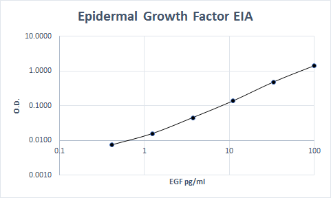
Human EGF ELISA Standard Curve
Recombinant Human EGF (Catalog # 236-EG) was serially diluted and captured by Mouse Anti-Human/Primate EGF Monoclonal Antibody (Catalog # MAB636) coated on a Clear Polystyrene Microplate (Catalog # DY990). Goat Anti-Human EGF Antigen Affinity-purified Polyclonal Antibody (Catalog # AF236) was biotinylated and incubated with the protein captured on the plate. Detection of the standard curve was achieved by incubating Streptavidin-HRP (Catalog # DY998)Applications for EGF Antibody (10827)
Application
Recommended Usage
Immunohistochemistry
8-25 µg/mL
Sample: Immersion fixed paraffin-embedded sections of human lung cancer tissue
Sample: Immersion fixed paraffin-embedded sections of human lung cancer tissue
Human/Primate EGF Sandwich Immunoassay
Please Note: Optimal dilutions of this antibody should be experimentally determined.
Reviewed Applications
Read 2 reviews rated 5 using MAB636 in the following applications:
Formulation, Preparation, and Storage
Purification
Protein A or G purified from hybridoma culture supernatant
Reconstitution
Reconstitute at 0.5 mg/mL in sterile PBS. For liquid material, refer to CoA for concentration.
Loading...
Formulation
Lyophilized from a 0.2 μm filtered solution in PBS with Trehalose. *Small pack size (SP) is supplied either lyophilized or as a 0.2 µm filtered solution in PBS.
Shipping
Lyophilized product is shipped at ambient temperature. Liquid small pack size (-SP) is shipped with polar packs. Upon receipt, store immediately at the temperature recommended below.
Stability & Storage
Use a manual defrost freezer and avoid repeated freeze-thaw cycles.
- 12 months from date of receipt, -20 to -70 °C as supplied.
- 1 month, 2 to 8 °C under sterile conditions after reconstitution.
- 6 months, -20 to -70 °C under sterile conditions after reconstitution.
Calculators
Background: EGF
Long Name
Epidermal Growth Factor
Alternate Names
HOMG4, URG, Urogastrone
Gene Symbol
EGF
Additional EGF Products
Product Documents for EGF Antibody (10827)
Certificate of Analysis
To download a Certificate of Analysis, please enter a lot or batch number in the search box below.
Note: Certificate of Analysis not available for kit components.
Product Specific Notices for EGF Antibody (10827)
For research use only
Related Research Areas
Citations for EGF Antibody (10827)
 Powered by Bioz
Customer Reviews for EGF Antibody (10827) (2)
5 out of 5
2 Customer Ratings
Have you used EGF Antibody (10827)?
Submit a review and receive an Amazon gift card!
$25/€18/£15/$25CAN/¥2500 Yen for a review with an image
$10/€7/£6/$10CAN/¥1110 Yen for a review without an image
Submit a review
Customer Images
Showing
1
-
2 of
2 reviews
Showing All
Filter By:
-
Application: ELISASample Tested: Serum and PlasmaSpecies: HumanVerified Customer | Posted 07/19/2019

-
Application: ELISASample Tested: Serum and PlasmaSpecies: HumanVerified Customer | Posted 11/07/2017This antibody was used to develop a sandwich ELISA for the measurement of EGF in human serum. It worked very well using AF236 as a matched pair.
There are no reviews that match your criteria.
Protocols
Find general support by application which include: protocols, troubleshooting, illustrated assays, videos and webinars.
- Antigen Retrieval Protocol (PIER)
- Antigen Retrieval for Frozen Sections Protocol
- Appropriate Fixation of IHC/ICC Samples
- Cellular Response to Hypoxia Protocols
- Chromogenic IHC Staining of Formalin-Fixed Paraffin-Embedded (FFPE) Tissue Protocol
- Chromogenic Immunohistochemistry Staining of Frozen Tissue
- ClariTSA™ Fluorophore Kits
- Detection & Visualization of Antibody Binding
- Fluorescent IHC Staining of Frozen Tissue Protocol
- Graphic Protocol for Heat-induced Epitope Retrieval
- Graphic Protocol for the Preparation and Fluorescent IHC Staining of Frozen Tissue Sections
- Graphic Protocol for the Preparation and Fluorescent IHC Staining of Paraffin-embedded Tissue Sections
- Graphic Protocol for the Preparation of Gelatin-coated Slides for Histological Tissue Sections
- IHC Sample Preparation (Frozen sections vs Paraffin)
- Immunofluorescent IHC Staining of Formalin-Fixed Paraffin-Embedded (FFPE) Tissue Protocol
- Immunohistochemistry (IHC) and Immunocytochemistry (ICC) Protocols
- Immunohistochemistry Frozen Troubleshooting
- Immunohistochemistry Paraffin Troubleshooting
- Preparing Samples for IHC/ICC Experiments
- Preventing Non-Specific Staining (Non-Specific Binding)
- Primary Antibody Selection & Optimization
- Protocol for Heat-Induced Epitope Retrieval (HIER)
- Protocol for Making a 4% Formaldehyde Solution in PBS
- Protocol for VisUCyte™ HRP Polymer Detection Reagent
- Protocol for the Preparation & Fixation of Cells on Coverslips
- Protocol for the Preparation and Chromogenic IHC Staining of Frozen Tissue Sections
- Protocol for the Preparation and Chromogenic IHC Staining of Frozen Tissue Sections - Graphic
- Protocol for the Preparation and Chromogenic IHC Staining of Paraffin-embedded Tissue Sections
- Protocol for the Preparation and Chromogenic IHC Staining of Paraffin-embedded Tissue Sections - Graphic
- Protocol for the Preparation and Fluorescent IHC Staining of Frozen Tissue Sections
- Protocol for the Preparation and Fluorescent IHC Staining of Paraffin-embedded Tissue Sections
- Protocol for the Preparation of Gelatin-coated Slides for Histological Tissue Sections
- TUNEL and Active Caspase-3 Detection by IHC/ICC Protocol
- The Importance of IHC/ICC Controls
- Troubleshooting Guide: Immunohistochemistry
- View all Protocols, Troubleshooting, Illustrated assays and Webinars


