Protein Tyrosine Phosphatase, Non-receptor type 14 (PTPN14), also called Phosphatase with Ezrin Domain (PEZ) and PTP36, is a 135 kDa cytosolic protein that dephosphorylates tyrosine residues in proteins. PTPN14’s Ezrin domain is homologous to erythrocyte Band 4.1 protein, targeting the phosphatase to cytoskeletal elements such as actin. PTPN14 associates with beta-catenin in adhesion zones in cells that are touching, but is nuclear in isolated, proliferating cells. Loss-of-function mutations in PTPN14 are found in 26% of colorectal cancers, suggesting it is a tumor suppressor.

Key Product Details
Species Reactivity
Validated:
Human
Cited:
Human
Applications
Validated:
Immunohistochemistry, Western Blot
Cited:
Immunohistochemistry, Immunohistochemistry-Paraffin, Western Blot
Label
Unconjugated
Antibody Source
Monoclonal Mouse IgG2B Clone # 448701
Loading...
Product Specifications
Immunogen
E. coli-derived recombinant human PTPN14
Leu329-Leu900
Accession # Q15678.2
Leu329-Leu900
Accession # Q15678.2
Specificity
Detects endogenous human PTPN14 in Western blots. In Western blots, this antibody does not cross-react with recombinant human (rh) PTPN13 or rhPTPN22.
Clonality
Monoclonal
Host
Mouse
Isotype
IgG2B
Scientific Data Images for Human PTPN14 Antibody (448701)
Detection of Human PTPN14 by Western Blot.
Western blot shows lysates of DU145 human prostate carcinoma cell line, HT-29 human colon adenocarcinoma cell line, and HT-29 human colon adenocarcinoma cell line. PVDF membrane was probed with 1 µg/mL of Human PTPN14 Monoclonal Antibody (Catalog # MAB4458) followed by HRP-conjugated Anti-Mouse IgG Secondary Antibody (Catalog # HAF007). A specific band was detected for PTPN14 at approximately 135 kDa (as indicated). This experiment was conducted under reducing conditions and using Immunoblot Buffer Group 1.
PTPN14 in Human Placenta.
PTPN14 was detected in immersion fixed paraffin-embedded sections of human placenta using Human PTPN14 Monoclonal Antibody (Catalog # MAB4458) at 25 µg/mL overnight at 4 °C. Tissue was stained using the Anti-Mouse HRP-DAB Cell & Tissue Staining Kit (brown; Catalog # CTS002) and counterstained with hematoxylin (blue). View our protocol for Chromogenic IHC Staining of Paraffin-embedded Tissue Sections.
Detection of Human PTPN14/PTPD2 by Western Blot
PTPRK interacts with candidate substrates in confluent MCF10A cells.(A) Representative immunoblot analysis of biotin pull downs from MCF10As expressing tGFP or PTPRK BioID constructs. See Materials and methods for details. Red and blue arrows indicate exogenous and endogenous PTPRK, respectively. (B) Quantification of BioID immunoblots. Green bars indicate the number of times a protein was enriched on PTPRK-C1089S.BirA*-Flag, compared to PTPRK.ECD +TMD.BirA*-Flag in separate experiments. Purple bars indicate the number of times a protein was not enriched or was not detected in any pull downs. n ≥ 1. (C) Schematic representation of PTPRK proximity-labeling by BioID. PTPRK extracellular domain homology model is based on PTPRM (PDB: 2V5Y; Aricescu et al., 2007). Proteins within the dotted lines were detected in pull downs from indicated BioID lysates. Proteins not detectably biotinylated are listed on the left. Proteins in bold and italics were previously identified as PTPRK interactors using BioID in HEK293 cells (St-Denis et al., 2016). See also Figure 4—figure supplement 1.Localization of PTPRK BioID proteins.(A) MCF10As with stably integrated doxycycline-inducible expression constructs (PTPRK-ECD +TMD-BirA*-Flag and PTPRK-C1089s-BirA*-Flag) were treated with 150 ng/ml and 500 ng/ml doxycycline, respectively, and immunostained using an anti-Flag antibody. F-actin and nuclei were stained with phalloidin and Hoechst, respectively. Scale bars = 50 µm. (B) Representative immunoblot analysis of biotin pull downs from MCF10As expressing tGFP or PTPRK BioID constructs. See Materials and methods for details. Red and blue arrows indicate exogenous and endogenous PTPRK, respectively. * residual ABLIM3 signal. Image collected and cropped by CiteAb from the following open publication (https://pubmed.ncbi.nlm.nih.gov/30924770), licensed under a CC-BY license. Not internally tested by R&D Systems.
Detection of Human PTPN14/PTPD2 by Immunocytochemistry/ Immunofluorescence
PTPN14 is a negative regulator of YAP. C) 293A cells transduced with lentivirus encoding dox-inducible PTPN14 expression. YAP localization in control&PTPN14-expressing cell lines was analysed 72 hours post dox induction by IF at low density using confocal miroscopy. Image collected & cropped by CiteAb from the following open publication (https://pubmed.ncbi.nlm.nih.gov/23613971), licensed under a CC-BY license. Not internally tested by R&D Systems.
Detection of Human PTPN14/PTPD2 by Western Blot
The interactome of the homophilic adhesion receptor PTPRK. (F–G) Selected PTPRK interactors identified by mass spectrometry validated by immunoblot analysis. Input&supernatants reveal the extent of protein depletion by recombinant proteins. Arrow indicates relevant band. See also Figure 2—figure supplements 1, 2&3. Image collected & cropped by CiteAb from the following open publication (https://pubmed.ncbi.nlm.nih.gov/30924770), licensed under a CC-BY license. Not internally tested by R&D Systems.
Detection of Human PTPN14/PTPD2 by Western Blot
YAP-PTPN14 binding is mediated through the WW domain-PPxY motif interaction.D) An SF268 cell line stably expressing the YAP-responsive MCAT_Luc reporter was transduced with lentivirus encoding for the indicated dox-inducible PTPN14 expression. Luciferase expression of each cell line was analysed 72 hours post dox induction (left panel). A Resazurin assay was carried out in parallel for each sample&used to normalize the luciferase readings. PTPN14 expression levels achieved for each construct analysed by WB (right panel; arrows indicate the WT PTPN14 protein&the truncated delta PTP PTPN14 which migrates faster; all lanes from a single blot&exposure). Tubulin serves as loading control. Luciferase results are shown as the average of at least 3 independent experiments ± STDEV. Statistical analysis was carried out with a 2-tailed paired t-test; * p<0.05. Image collected & cropped by CiteAb from the following open publication (https://pubmed.ncbi.nlm.nih.gov/23613971), licensed under a CC-BY license. Not internally tested by R&D Systems.
Detection of Human PTPN14/PTPD2 by Western Blot
In vitro dephosphorylation assays and generation of RPTP chimeras.(A) The indicated PTPRK and PTPRM domains were assayed for phosphatase activity using the pNPP colorimetric assay. Control wells contained pNPP only. Protein amounts used are shown. (B) Pervanadate-treated MCF10A lysates were incubated with predetermined amounts of the indicated domains to give equal phosphatase-activity, prior to phosphotyrosine immunoprecipitation and immunoblot analysis. (C) Recombinant proteins consisting of combinations of PTPRK and PTPRM D1 and D2 domains were expressed in and using Ni-NTA affinity resin. Purified proteins were then subjected to size exclusion chromatography. (D) Recombinant His- and Avi-tagged PTPRK and PTPRM chimeric domains were purified from E. coli cultured in biotin-supplemented media, incubated ±streptavidin and subjected to SDS-PAGE and Coomassie staining, to determine the extent of biotinylation. Arrows indicate the purified domains and the respective streptavidin-induced mobility shift. (E) The indicated recombinant PTPRK and PTPRM chimeric domains were incubated were assayed for phosphatase activity using the pNPP colorimetric assay. Control wells contained pNPP. Protein amounts used are shown. Image collected and cropped by CiteAb from the following open publication (https://pubmed.ncbi.nlm.nih.gov/30924770), licensed under a CC-BY license. Not internally tested by R&D Systems.
Detection of Human PTPN14/PTPD2 by Western Blot
YAP-PTPN14 binding is mediated through the WW domain-PPxY motif interaction. F) 293A cells transduced with lentivirus encoding for the indicated dox-inducible PTPN14 expression. The nuclear/cytoplasmic YAP ratio was quantified at low density after 72 hours of dox induction using a Cellomics automated imager with a conventional microscope,&expressed relative to control (left panel). Results are shown as the average of three experiments ± STDEV. Statistical analysis was carried out with a 2-tailed paired t-test; * p<0.05. For each experiment, the average ratio was calculated from three wells per sample (10 images per well). PTPN14 expression levels analysed by WB (right panel; arrows indicate the WT PTPN14 protein&the truncated delta PTP PTPN14 which migrates faster; all lanes from a single blot&exposure). Tubulin serves as a loading control. Image collected & cropped by CiteAb from the following open publication (https://pubmed.ncbi.nlm.nih.gov/23613971), licensed under a CC-BY license. Not internally tested by R&D Systems.
Detection of Human PTPN14/PTPD2 by Western Blot
PTPN14 is a negative regulator of YAP.B) An SF268 cell line stably expressing the YAP-responsive MCAT_Luc reporter was transduced with lentivirus encoding for dox-inducible PTPN14 expression. After pharmacological selection, luciferase expression was measured for both cell lines in the presence&absence of dox (upper panel). A Resazurin assay was carried out in parallel for each sample&used to normalize the luciferase readings. Results are shown as the average of three independent experiments ± STDEV. Statistical analysis was carried out with a 2-tailed paired t-test; * p<0.0001. RLU: relative luciferase units. PTPN14 expression achieved by dox&YAP levels in each sample was analyzed by WB (lower panel). Tubulin serves as loading control. Image collected & cropped by CiteAb from the following open publication (https://pubmed.ncbi.nlm.nih.gov/23613971), licensed under a CC-BY license. Not internally tested by R&D Systems.Applications for Human PTPN14 Antibody (448701)
Application
Recommended Usage
Immunohistochemistry
8-25 µg/mL
Sample: Immersion fixed paraffin-embedded human placenta
Sample: Immersion fixed paraffin-embedded human placenta
Western Blot
1 µg/mL
Sample: DU145 human prostate carcinoma cell line, HT-29 human colon adenocarcinoma cell line, and HT-29 human colon adenocarcinoma cell line
Sample: DU145 human prostate carcinoma cell line, HT-29 human colon adenocarcinoma cell line, and HT-29 human colon adenocarcinoma cell line
Reviewed Applications
Read 3 reviews rated 4 using MAB4458 in the following applications:
Formulation, Preparation, and Storage
Purification
Protein A or G purified from hybridoma culture supernatant
Reconstitution
Reconstitute at 0.5 mg/mL in sterile PBS. For liquid material, refer to CoA for concentration.
Loading...
Formulation
Lyophilized from a 0.2 μm filtered solution in PBS with Trehalose. *Small pack size (SP) is supplied either lyophilized or as a 0.2 µm filtered solution in PBS.
Shipping
Lyophilized product is shipped at ambient temperature. Liquid small pack size (-SP) is shipped with polar packs. Upon receipt, store immediately at the temperature recommended below.
Stability & Storage
Use a manual defrost freezer and avoid repeated freeze-thaw cycles.
- 12 months from date of receipt, -20 to -70 °C as supplied.
- 1 month, 2 to 8 °C under sterile conditions after reconstitution.
- 6 months, -20 to -70 °C under sterile conditions after reconstitution.
Calculators
Background: PTPN14
Long Name
Protein Tyrosine Phosphatase Non-receptor Type 14
Alternate Names
PEZ
Gene Symbol
PTPN14
UniProt
Additional PTPN14 Products
Product Documents for Human PTPN14 Antibody (448701)
Certificate of Analysis
To download a Certificate of Analysis, please enter a lot or batch number in the search box below.
Note: Certificate of Analysis not available for kit components.
Product Specific Notices for Human PTPN14 Antibody (448701)
For research use only
Related Research Areas
Citations for Human PTPN14 Antibody (448701)
 Powered by Bioz
Customer Reviews for Human PTPN14 Antibody (448701) (3)
4 out of 5
3 Customer Ratings
Have you used Human PTPN14 Antibody (448701)?
Submit a review and receive an Amazon gift card!
$25/€18/£15/$25CAN/¥2500 Yen for a review with an image
$10/€7/£6/$10CAN/¥1110 Yen for a review without an image
Submit a review
Customer Images

Showing
1
-
3 of
3 reviews
Showing All
Filter By:
-
Application: Western BlotSample Tested: HT-29 human colon adenocarcinoma cell lineSpecies: HumanVerified Customer | Posted 12/04/2021

-
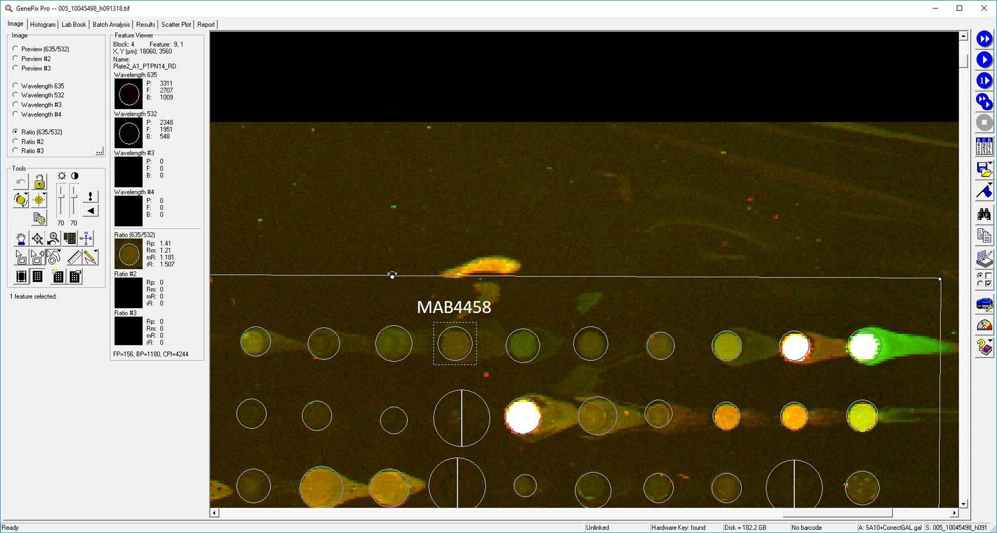
Application: MicroarraysSample Tested: EDTA PlasmaSpecies: HumanVerified Customer | Posted 03/11/2019
-
Application: MicroarraySample Tested: EDTA PlasmaSpecies: HumanVerified Customer | Posted 11/02/2018
There are no reviews that match your criteria.
Protocols
Find general support by application which include: protocols, troubleshooting, illustrated assays, videos and webinars.
- Antigen Retrieval Protocol (PIER)
- Antigen Retrieval for Frozen Sections Protocol
- Appropriate Fixation of IHC/ICC Samples
- Cellular Response to Hypoxia Protocols
- Chromogenic IHC Staining of Formalin-Fixed Paraffin-Embedded (FFPE) Tissue Protocol
- Chromogenic Immunohistochemistry Staining of Frozen Tissue
- ClariTSA™ Fluorophore Kits
- Detection & Visualization of Antibody Binding
- Fluorescent IHC Staining of Frozen Tissue Protocol
- Graphic Protocol for Heat-induced Epitope Retrieval
- Graphic Protocol for the Preparation and Fluorescent IHC Staining of Frozen Tissue Sections
- Graphic Protocol for the Preparation and Fluorescent IHC Staining of Paraffin-embedded Tissue Sections
- Graphic Protocol for the Preparation of Gelatin-coated Slides for Histological Tissue Sections
- IHC Sample Preparation (Frozen sections vs Paraffin)
- Immunofluorescent IHC Staining of Formalin-Fixed Paraffin-Embedded (FFPE) Tissue Protocol
- Immunohistochemistry (IHC) and Immunocytochemistry (ICC) Protocols
- Immunohistochemistry Frozen Troubleshooting
- Immunohistochemistry Paraffin Troubleshooting
- Preparing Samples for IHC/ICC Experiments
- Preventing Non-Specific Staining (Non-Specific Binding)
- Primary Antibody Selection & Optimization
- Protocol for Heat-Induced Epitope Retrieval (HIER)
- Protocol for Making a 4% Formaldehyde Solution in PBS
- Protocol for VisUCyte™ HRP Polymer Detection Reagent
- Protocol for the Preparation & Fixation of Cells on Coverslips
- Protocol for the Preparation and Chromogenic IHC Staining of Frozen Tissue Sections
- Protocol for the Preparation and Chromogenic IHC Staining of Frozen Tissue Sections - Graphic
- Protocol for the Preparation and Chromogenic IHC Staining of Paraffin-embedded Tissue Sections
- Protocol for the Preparation and Chromogenic IHC Staining of Paraffin-embedded Tissue Sections - Graphic
- Protocol for the Preparation and Fluorescent IHC Staining of Frozen Tissue Sections
- Protocol for the Preparation and Fluorescent IHC Staining of Paraffin-embedded Tissue Sections
- Protocol for the Preparation of Gelatin-coated Slides for Histological Tissue Sections
- R&D Systems Quality Control Western Blot Protocol
- TUNEL and Active Caspase-3 Detection by IHC/ICC Protocol
- The Importance of IHC/ICC Controls
- Troubleshooting Guide: Immunohistochemistry
- Troubleshooting Guide: Western Blot Figures
- Western Blot Conditions
- Western Blot Protocol
- Western Blot Protocol for Cell Lysates
- Western Blot Troubleshooting
- Western Blot Troubleshooting Guide
- View all Protocols, Troubleshooting, Illustrated assays and Webinars
Loading...