DYRK2 (Dual-specificity tyrosine [Y] phosphorylation regulated kinase 2) is a 58‑62 kDa member of the MNB/DYRK subfamily, CMGC Ser/Thr protein kinase family of enzymes. It is expressed in testis and shows dual substrate specificity; autophosphorylation on Tyr382 to self‑activate, and a Ser/Thr phosphorylation of target molecules. Substrates include NFATc, glycogen synthase, and p53. p53 phosphorylation on Ser46 initiates cell apoptosis. Human DYRK2 is 601 amino acids (aa) in length. It contains one kinase catalytic domain (aa 222‑535). One isoform variant shows an alternate start site at Met74. This shorter isoform was used for immunization. Thus, over aa 1‑107 (or aa 74‑180 of the long form), human DYRK2 shares 93% aa identity with mouse DYRK2.

Key Product Details
Species Reactivity
Human, Rat
Applications
Immunohistochemistry, Western Blot
Label
Unconjugated
Antibody Source
Monoclonal Mouse IgG2B Clone # 599542
Loading...
Product Specifications
Immunogen
E. coli-derived recombinant human DYRK2
aa 1-107
Accession # NP_003574
aa 1-107
Accession # NP_003574
Specificity
Detects human and rat DYRK2 in Western blots.
Clonality
Monoclonal
Host
Mouse
Isotype
IgG2B
Scientific Data Images for DYRK2 Antibody (599542)
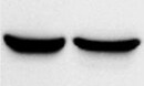
Detection of Human and Rat DYRK2 by Western Blot.
Western blot shows lysates of HepG2 human hepatocellular carcinoma cell line, CCD-1070Sk human foreskin fibroblast cell line, and L6 rat myoblast cell line. PVDF Membrane was probed with 1 µg/mL of Mouse Anti-Human/Rat DYRK2 Monoclonal Antibody (Catalog # MAB5408) followed by HRP-conjugated Anti-Mouse IgG Secondary Antibody (Catalog # HAF007). A specific band was detected for DYRK2 at approximately 60 kDa (as indicated). This experiment was conducted under reducing conditions and using Immunoblot Buffer Group 1.Applications for DYRK2 Antibody (599542)
Application
Recommended Usage
Immunohistochemistry
8-25 µg/mL
Sample: Immersion fixed paraffin-embedded testis
Sample: Immersion fixed paraffin-embedded testis
Western Blot
1 µg/mL
Sample: HepG2 human hepatocellular carcinoma cell line, CCD‑1070Sk human foreskin fibroblast cell line, and L6 rat myoblast cell line
Sample: HepG2 human hepatocellular carcinoma cell line, CCD‑1070Sk human foreskin fibroblast cell line, and L6 rat myoblast cell line
Reviewed Applications
Read 1 review rated 5 using MAB5408 in the following applications:
Formulation, Preparation, and Storage
Purification
Protein A or G purified from hybridoma culture supernatant
Reconstitution
Reconstitute at 0.5 mg/mL in sterile PBS. For liquid material, refer to CoA for concentration.
Loading...
Formulation
Lyophilized from a 0.2 μm filtered solution in PBS with Trehalose. *Small pack size (SP) is supplied either lyophilized or as a 0.2 µm filtered solution in PBS.
Shipping
Lyophilized product is shipped at ambient temperature. Liquid small pack size (-SP) is shipped with polar packs. Upon receipt, store immediately at the temperature recommended below.
Stability & Storage
Use a manual defrost freezer and avoid repeated freeze-thaw cycles.
- 12 months from date of receipt, -20 to -70 °C as supplied.
- 1 month, 2 to 8 °C under sterile conditions after reconstitution.
- 6 months, -20 to -70 °C under sterile conditions after reconstitution.
Calculators
Background: DYRK2
Long Name
Dual-specificity Tyrosine-[Y]-phosphorylation Regulated Kinase 2
Alternate Names
dual specificity tyrosine-phosphorylation-regulated kinase 2, dual-specificity tyrosine-(Y)-phosphorylation regulated kinase 2, EC 2.7.12, EC 2.7.12.1, FLJ21217, FLJ21365
Gene Symbol
DYRK2
UniProt
Additional DYRK2 Products
Product Documents for DYRK2 Antibody (599542)
Certificate of Analysis
To download a Certificate of Analysis, please enter a lot or batch number in the search box below.
Note: Certificate of Analysis not available for kit components.
Product Specific Notices for DYRK2 Antibody (599542)
For research use only
Related Research Areas
Customer Reviews for DYRK2 Antibody (599542) (1)
5 out of 5
1 Customer Rating
Have you used DYRK2 Antibody (599542)?
Submit a review and receive an Amazon gift card!
$25/€18/£15/$25CAN/¥2500 Yen for a review with an image
$10/€7/£6/$10CAN/¥1110 Yen for a review without an image
Submit a review
Customer Images
Showing
1
-
1 of
1 review
Showing All
Filter By:
-
Application: Western BlotSample Tested: HepG2 human hepatocellular carcinoma cell lineSpecies: HumanVerified Customer | Posted 07/07/2022

There are no reviews that match your criteria.
Protocols
Find general support by application which include: protocols, troubleshooting, illustrated assays, videos and webinars.
- Antigen Retrieval Protocol (PIER)
- Antigen Retrieval for Frozen Sections Protocol
- Appropriate Fixation of IHC/ICC Samples
- Cellular Response to Hypoxia Protocols
- Chromogenic IHC Staining of Formalin-Fixed Paraffin-Embedded (FFPE) Tissue Protocol
- Chromogenic Immunohistochemistry Staining of Frozen Tissue
- ClariTSA™ Fluorophore Kits
- Detection & Visualization of Antibody Binding
- Fluorescent IHC Staining of Frozen Tissue Protocol
- Graphic Protocol for Heat-induced Epitope Retrieval
- Graphic Protocol for the Preparation and Fluorescent IHC Staining of Frozen Tissue Sections
- Graphic Protocol for the Preparation and Fluorescent IHC Staining of Paraffin-embedded Tissue Sections
- Graphic Protocol for the Preparation of Gelatin-coated Slides for Histological Tissue Sections
- IHC Sample Preparation (Frozen sections vs Paraffin)
- Immunofluorescent IHC Staining of Formalin-Fixed Paraffin-Embedded (FFPE) Tissue Protocol
- Immunohistochemistry (IHC) and Immunocytochemistry (ICC) Protocols
- Immunohistochemistry Frozen Troubleshooting
- Immunohistochemistry Paraffin Troubleshooting
- Preparing Samples for IHC/ICC Experiments
- Preventing Non-Specific Staining (Non-Specific Binding)
- Primary Antibody Selection & Optimization
- Protocol for Heat-Induced Epitope Retrieval (HIER)
- Protocol for Making a 4% Formaldehyde Solution in PBS
- Protocol for VisUCyte™ HRP Polymer Detection Reagent
- Protocol for the Preparation & Fixation of Cells on Coverslips
- Protocol for the Preparation and Chromogenic IHC Staining of Frozen Tissue Sections
- Protocol for the Preparation and Chromogenic IHC Staining of Frozen Tissue Sections - Graphic
- Protocol for the Preparation and Chromogenic IHC Staining of Paraffin-embedded Tissue Sections
- Protocol for the Preparation and Chromogenic IHC Staining of Paraffin-embedded Tissue Sections - Graphic
- Protocol for the Preparation and Fluorescent IHC Staining of Frozen Tissue Sections
- Protocol for the Preparation and Fluorescent IHC Staining of Paraffin-embedded Tissue Sections
- Protocol for the Preparation of Gelatin-coated Slides for Histological Tissue Sections
- R&D Systems Quality Control Western Blot Protocol
- TUNEL and Active Caspase-3 Detection by IHC/ICC Protocol
- The Importance of IHC/ICC Controls
- Troubleshooting Guide: Immunohistochemistry
- Troubleshooting Guide: Western Blot Figures
- Western Blot Conditions
- Western Blot Protocol
- Western Blot Protocol for Cell Lysates
- Western Blot Troubleshooting
- Western Blot Troubleshooting Guide
- View all Protocols, Troubleshooting, Illustrated assays and Webinars
Loading...