Siglecs (1) (sialic acid binding Ig-like lectins) are I-type (Ig-type) lectins (2) belonging to the Ig superfamily. They are characterized by an N-terminal Ig-like V-type domain which mediates sialic acid binding (3), followed by varying numbers of Ig-like C2-type domains (1, 4). Eleven human Siglecs have been cloned and characterized (1, 4). They are sialoadhesin/CD169/Siglec-1, CD22/Siglec-2, CD33/Siglec-3, Myelin-Associated Glycoprotein (MAG/Siglec-4a) and the Siglec-5 to 11 (4, 5, 7). To date, no Siglec has been shown to recognized any cell surface ligand other than sialic acids, suggesting that interactions with glycans containing this carbohydrate are important in mediating the biological functions of Siglecs. Siglec-5 to 11 share a high degree of sequence similarity with CD33/Siglec-3 both in their extracellular and intracellular regions. They are collectively referred to as CD33-related Siglecs. One remarkable feature of the CD33-related Siglecs is their differential expression pattern within the hematopoietic system (4, 5). This fact, together with the presence of two conserved immunoreceptor tyrosine-based inhibition motifs (ITIMs) in their cytoplasma tails, suggests that CD33-related Siglecs are involved in the regulation of cellular activation within the immune system. Human Siglec-5 cDNA encodes a 551 amino acid (aa) polypeptide with a hydrophobic signal peptide, an N-terminal Ig-like V-type domain, three Ig-like C2-type domains, a transmembrane region and a cytoplasma tail (6). Siglec-5 exists as a disulfide-linked homodimer on the cell surface and is expressed on monocytes, neutrophils and B cells (4, 5, 6). It binds equally well to both alpha 2,3- and alpha 2,6-linked sialic acid (6).

Key Product Details
Species Reactivity
Validated:
Human
Cited:
Human, Bovine, Ovine
Applications
Validated:
Immunohistochemistry, Western Blot
Cited:
Immunohistochemistry, Western Blot, Neutralization, Immunocytochemistry
Label
Unconjugated
Antibody Source
Monoclonal Mouse IgG2B Clone # 194117
Loading...
Product Specifications
Immunogen
Mouse myeloma cell line NS0-derived recombinant human Siglec‑5/CD170
Lys18-Thr434
Accession # O15389
Lys18-Thr434
Accession # O15389
Specificity
Detects human Siglec‑5/CD170 in direct ELISAs and Western blots. In direct ELISAs and Western blots, no cross-reactivity with recombinant human Siglec-2, -3, -7, -9, or -14 is observed.
Clonality
Monoclonal
Host
Mouse
Isotype
IgG2B
Scientific Data Images for Human Siglec‑5/CD170 Antibody
Detection of Siglec‑5/CD170 in Human Spleen.
Siglec‑5/CD170 was detected in immersion fixed paraffin-embedded sections of human spleen using Mouse Anti-Human Siglec‑5/CD170 Monoclonal Antibody (Catalog # MAB1072) at 5 µg/ml for 1 hour at room temperature followed by incubation with the HRP-conjugated Anti-Mouse IgG Secondary Antibody (Catalog # HAF007) or the Anti-Mouse IgG VisUCyte™ HRP Polymer Antibody (Catalog # VC001). Before incubation with the primary antibody, tissue was subjected to heat-induced epitope retrieval using VisUCyte Antigen Retrieval Reagent-Basic (Catalog # VCTS021). Tissue was stained using DAB (brown) and counterstained with hematoxylin (blue). Specific staining was localized to the cell membrane. View our protocol for Chromogenic IHC Staining of Paraffin-embedded Tissue Sections.Applications for Human Siglec‑5/CD170 Antibody
Application
Recommended Usage
Immunohistochemistry
8-25 µg/mL
Sample: Immersion fixed paraffin-embedded sections of human spleen
Sample: Immersion fixed paraffin-embedded sections of human spleen
Western Blot
1 µg/mL
Sample: Recombinant Human Siglec‑5 Fc Chimera (Catalog # 1072-SL)
Sample: Recombinant Human Siglec‑5 Fc Chimera (Catalog # 1072-SL)
Reviewed Applications
Read 4 reviews rated 4 using MAB1072 in the following applications:
Formulation, Preparation, and Storage
Purification
Protein A or G purified from hybridoma culture supernatant
Reconstitution
Reconstitute at 0.5 mg/mL in sterile PBS. For liquid material, refer to CoA for concentration.
Loading...
Formulation
Lyophilized from a 0.2 μm filtered solution in PBS with Trehalose. See Certificate of Analysis for details.
*Small pack size (-SP) is supplied either lyophilized or as a 0.2 µm filtered solution in PBS.
*Small pack size (-SP) is supplied either lyophilized or as a 0.2 µm filtered solution in PBS.
Shipping
Lyophilized product is shipped at ambient temperature. Liquid small pack size (-SP) is shipped with polar packs. Upon receipt, store immediately at the temperature recommended below.
Stability & Storage
Use a manual defrost freezer and avoid repeated freeze-thaw cycles.
- 12 months from date of receipt, -20 to -70 °C as supplied.
- 1 month, 2 to 8 °C under sterile conditions after reconstitution.
- 6 months, -20 to -70 °C under sterile conditions after reconstitution.
Calculators
Background: Siglec-5/CD170
References
- Crocker, P.R. et al. (1998) Glycobiology 8:v.
- Powell, L.D. et al. (1995) J. Biol. Chem. 270:14243.
- May, A.R. et al. (1998) Mol. Cell 1998. 1:719.
- Crocker, P.R. and A. Varki (2001) Trends Immunol. 22:337.
- Crocker, P.R. et al. (2001) Immunology 103:137.
- Cornish, A.L. et al. (1998) Blood 92:2123.
- Angata, T. et al. (2002) J. Biol Chem. 277:24466.
Long Name
Sialic Acid Binding Ig-like Lectin 5
Alternate Names
CD170, CD33L2, OBBP2, Siglec5
Entrez Gene IDs
8778 (Human)
Gene Symbol
SIGLEC5
UniProt
Additional Siglec-5/CD170 Products
Product Documents for Human Siglec‑5/CD170 Antibody
Certificate of Analysis
To download a Certificate of Analysis, please enter a lot or batch number in the search box below.
Note: Certificate of Analysis not available for kit components.
Product Specific Notices for Human Siglec‑5/CD170 Antibody
For research use only
Citations for Human Siglec‑5/CD170 Antibody
 Powered by Bioz
Customer Reviews for Human Siglec‑5/CD170 Antibody (4)
4 out of 5
4 Customer Ratings
Have you used Human Siglec‑5/CD170 Antibody?
Submit a review and receive an Amazon gift card!
$25/€18/£15/$25CAN/¥2500 Yen for a review with an image
$10/€7/£6/$10CAN/¥1110 Yen for a review without an image
Submit a review
Customer Images

Showing
1
-
4 of
4 reviews
Showing All
Filter By:
-
Application: Western BlotSample Tested: PlasmaSpecies: HumanVerified Customer | Posted 10/14/2021

-
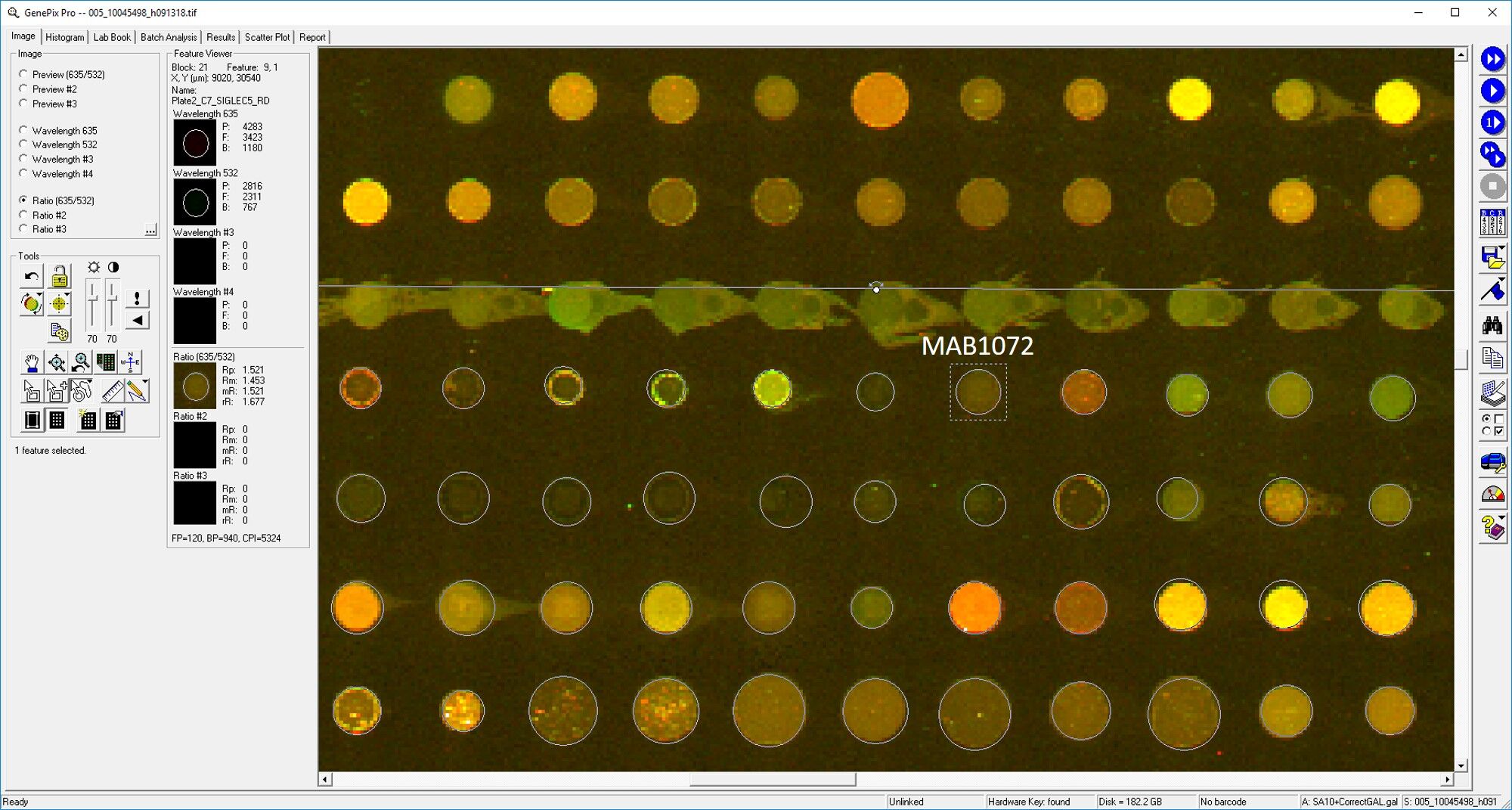
Application: MicroarraysSample Tested: EDTA PlasmaSpecies: HumanVerified Customer | Posted 03/11/2019
-
Application: MicroarraySample Tested: EDTA PlasmaSpecies: HumanVerified Customer | Posted 11/20/2018
-
Application: ELISASample Tested: PlasmaSpecies: HumanVerified Customer | Posted 11/10/2018
There are no reviews that match your criteria.
Protocols
Find general support by application which include: protocols, troubleshooting, illustrated assays, videos and webinars.
- Antigen Retrieval Protocol (PIER)
- Antigen Retrieval for Frozen Sections Protocol
- Appropriate Fixation of IHC/ICC Samples
- Cellular Response to Hypoxia Protocols
- Chromogenic IHC Staining of Formalin-Fixed Paraffin-Embedded (FFPE) Tissue Protocol
- Chromogenic Immunohistochemistry Staining of Frozen Tissue
- ClariTSA™ Fluorophore Kits
- Detection & Visualization of Antibody Binding
- Fluorescent IHC Staining of Frozen Tissue Protocol
- Graphic Protocol for Heat-induced Epitope Retrieval
- Graphic Protocol for the Preparation and Fluorescent IHC Staining of Frozen Tissue Sections
- Graphic Protocol for the Preparation and Fluorescent IHC Staining of Paraffin-embedded Tissue Sections
- Graphic Protocol for the Preparation of Gelatin-coated Slides for Histological Tissue Sections
- IHC Sample Preparation (Frozen sections vs Paraffin)
- Immunofluorescent IHC Staining of Formalin-Fixed Paraffin-Embedded (FFPE) Tissue Protocol
- Immunohistochemistry (IHC) and Immunocytochemistry (ICC) Protocols
- Immunohistochemistry Frozen Troubleshooting
- Immunohistochemistry Paraffin Troubleshooting
- Preparing Samples for IHC/ICC Experiments
- Preventing Non-Specific Staining (Non-Specific Binding)
- Primary Antibody Selection & Optimization
- Protocol for Heat-Induced Epitope Retrieval (HIER)
- Protocol for Making a 4% Formaldehyde Solution in PBS
- Protocol for VisUCyte™ HRP Polymer Detection Reagent
- Protocol for the Preparation & Fixation of Cells on Coverslips
- Protocol for the Preparation and Chromogenic IHC Staining of Frozen Tissue Sections
- Protocol for the Preparation and Chromogenic IHC Staining of Frozen Tissue Sections - Graphic
- Protocol for the Preparation and Chromogenic IHC Staining of Paraffin-embedded Tissue Sections
- Protocol for the Preparation and Chromogenic IHC Staining of Paraffin-embedded Tissue Sections - Graphic
- Protocol for the Preparation and Fluorescent IHC Staining of Frozen Tissue Sections
- Protocol for the Preparation and Fluorescent IHC Staining of Paraffin-embedded Tissue Sections
- Protocol for the Preparation of Gelatin-coated Slides for Histological Tissue Sections
- R&D Systems Quality Control Western Blot Protocol
- TUNEL and Active Caspase-3 Detection by IHC/ICC Protocol
- The Importance of IHC/ICC Controls
- Troubleshooting Guide: Immunohistochemistry
- Troubleshooting Guide: Western Blot Figures
- Western Blot Conditions
- Western Blot Protocol
- Western Blot Protocol for Cell Lysates
- Western Blot Troubleshooting
- Western Blot Troubleshooting Guide
- View all Protocols, Troubleshooting, Illustrated assays and Webinars
Loading...