VSIG1 (V-set and Ig domain-containing protein 1; also Glycoprotein A34) is a variably glycosylated 55‑70 kDa member of the JAM family of proteins. It has restricted expression, being limited to testicular germ cells plus pancreatic duct and gastric epithelium. VSIG1 is likely to serve as an adhesion molecule. Mature human VSIG1 is 366 amino acids (aa) in length. It is a type I transmembrane glycoprotein that contains a 211 aa extracellular domain (ECD). The ECD contains one V-type (aa 22‑132) and one C2-type Ig-like domain (aa 140‑227). Over aa 22‑234, human VSIG1 is 83% aa identical to both mouse and canine VSIG1. At least one potential splice variant exists in human. It shows an insertion of 36 aa after Ser72 and a deletion of aa 133‑387.

Key Product Details
Species Reactivity
Human
Applications
Immunohistochemistry, Western Blot, Simple Western
Label
Unconjugated
Antibody Source
Monoclonal Mouse IgG2B Clone # 771506
Loading...
Product Specifications
Immunogen
Mouse myeloma cell line NS0-derived recombinant human VSIG1
Val22-Gly234
Accession # Q86XK7
Val22-Gly234
Accession # Q86XK7
Specificity
Detects human VSIG1 in ELISAs. In direct ELISAs, no cross-reactivity with recombinant mouse VSIG1 or recombinant human VSIG2, 3, or 4 is observed.
Clonality
Monoclonal
Host
Mouse
Isotype
IgG2B
Scientific Data Images for Human VSIG1 Antibody (771506)
Detection of Human VSIG1 by Western Blot.
Western blot shows lysates of human stomach tissue. PVDF membrane was probed with 2 µg/mL of Mouse Anti-Human VSIG1 Monoclonal Antibody (Catalog # MAB4818) followed by HRP-conjugated Anti-Mouse IgG Secondary Antibody (Catalog # HAF007). A specific band was detected for VSIG1 at approximately 50 kDa (as indicated). This experiment was conducted under reducing conditions and using Immunoblot Buffer Group 1.
VSIG1 in Human Testis.
VSIG1 was detected in immersion fixed paraffin-embedded sections of human testis using Mouse Anti-Human VSIG1 Monoclonal Antibody (Catalog # MAB4818) at 15 µg/mL overnight at 4 °C. Before incubation with the primary antibody, tissue was subjected to heat-induced epitope retrieval using Antigen Retrieval Reagent-Basic (Catalog # CTS013). Tissue was stained using the Anti-Mouse HRP-DAB Cell & Tissue Staining Kit (brown; Catalog # CTS002) and counterstained with hematoxylin (blue). Specific staining was localized to plasma membranes in spermatocytes. View our protocol for Chromogenic IHC Staining of Paraffin-embedded Tissue Sections.
Detection of Human VSIG1 by Simple WesternTM.
Simple Western lane view shows lysates of human stomach tissue, loaded at 0.5 mg/mL. A specific band was detected for VSIG1 at approximately 48 kDa (as indicated) using 100 µg/mL of Mouse Anti-Human VSIG1 Monoclonal Antibody (Catalog # MAB4818). This experiment was conducted under reducing conditions and using the 12-230 kDa separation system. Non-specific interaction with the 230 kDa Simple Western standard may be seen with this antibody.Applications for Human VSIG1 Antibody (771506)
Application
Recommended Usage
Immunohistochemistry
8-25 µg/mL
Sample: Immersion fixed paraffin-embedded sections of human testis
Sample: Immersion fixed paraffin-embedded sections of human testis
Simple Western
100 µg/mL
Sample: Human stomach tissue
Sample: Human stomach tissue
Western Blot
2 µg/mL
Sample: Human stomach tissue
Sample: Human stomach tissue
Reviewed Applications
Read 1 review rated 5 using MAB4818 in the following applications:
Formulation, Preparation, and Storage
Purification
Protein A or G purified from hybridoma culture supernatant
Reconstitution
Sterile PBS to a final concentration of 0.5 mg/mL. For liquid material, refer to CoA for concentration.
Loading...
Formulation
Lyophilized from a 0.2 μm filtered solution in PBS with Trehalose. *Small pack size (SP) is supplied either lyophilized or as a 0.2 µm filtered solution in PBS.
Shipping
Lyophilized product is shipped at ambient temperature. Liquid small pack size (-SP) is shipped with polar packs. Upon receipt, store immediately at the temperature recommended below.
Stability & Storage
Use a manual defrost freezer and avoid repeated freeze-thaw cycles.
- 12 months from date of receipt, -20 to -70 °C as supplied.
- 1 month, 2 to 8 °C under sterile conditions after reconstitution.
- 6 months, -20 to -70 °C under sterile conditions after reconstitution.
Calculators
Background: VSIG1
Long Name
V-Set and Immunoglobulin Domain Containing 1
Alternate Names
GPA34
Gene Symbol
VSIG1
UniProt
Additional VSIG1 Products
Product Documents for Human VSIG1 Antibody (771506)
Certificate of Analysis
To download a Certificate of Analysis, please enter a lot or batch number in the search box below.
Note: Certificate of Analysis not available for kit components.
Product Specific Notices for Human VSIG1 Antibody (771506)
For research use only
Related Research Areas
Customer Reviews for Human VSIG1 Antibody (771506) (1)
5 out of 5
1 Customer Rating
Have you used Human VSIG1 Antibody (771506)?
Submit a review and receive an Amazon gift card!
$25/€18/£15/$25CAN/¥2500 Yen for a review with an image
$10/€7/£6/$10CAN/¥1110 Yen for a review without an image
Submit a review
Customer Images
Showing
1
-
1 of
1 review
Showing All
Filter By:
-
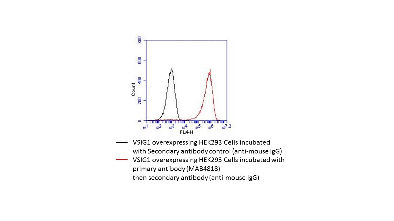
Application: Flow CytometrySample Tested: HEK293T VSIG1 overexpressingSpecies: HumanVerified Customer | Posted 06/01/2018This antibody reacts very well with VSIG1 overexpressing HEK293 cells in flow assay.

There are no reviews that match your criteria.
Protocols
Find general support by application which include: protocols, troubleshooting, illustrated assays, videos and webinars.
- Antigen Retrieval Protocol (PIER)
- Antigen Retrieval for Frozen Sections Protocol
- Appropriate Fixation of IHC/ICC Samples
- Cellular Response to Hypoxia Protocols
- Chromogenic IHC Staining of Formalin-Fixed Paraffin-Embedded (FFPE) Tissue Protocol
- Chromogenic Immunohistochemistry Staining of Frozen Tissue
- ClariTSA™ Fluorophore Kits
- Detection & Visualization of Antibody Binding
- Fluorescent IHC Staining of Frozen Tissue Protocol
- Graphic Protocol for Heat-induced Epitope Retrieval
- Graphic Protocol for the Preparation and Fluorescent IHC Staining of Frozen Tissue Sections
- Graphic Protocol for the Preparation and Fluorescent IHC Staining of Paraffin-embedded Tissue Sections
- Graphic Protocol for the Preparation of Gelatin-coated Slides for Histological Tissue Sections
- IHC Sample Preparation (Frozen sections vs Paraffin)
- Immunofluorescent IHC Staining of Formalin-Fixed Paraffin-Embedded (FFPE) Tissue Protocol
- Immunohistochemistry (IHC) and Immunocytochemistry (ICC) Protocols
- Immunohistochemistry Frozen Troubleshooting
- Immunohistochemistry Paraffin Troubleshooting
- Preparing Samples for IHC/ICC Experiments
- Preventing Non-Specific Staining (Non-Specific Binding)
- Primary Antibody Selection & Optimization
- Protocol for Heat-Induced Epitope Retrieval (HIER)
- Protocol for Making a 4% Formaldehyde Solution in PBS
- Protocol for VisUCyte™ HRP Polymer Detection Reagent
- Protocol for the Preparation & Fixation of Cells on Coverslips
- Protocol for the Preparation and Chromogenic IHC Staining of Frozen Tissue Sections
- Protocol for the Preparation and Chromogenic IHC Staining of Frozen Tissue Sections - Graphic
- Protocol for the Preparation and Chromogenic IHC Staining of Paraffin-embedded Tissue Sections
- Protocol for the Preparation and Chromogenic IHC Staining of Paraffin-embedded Tissue Sections - Graphic
- Protocol for the Preparation and Fluorescent IHC Staining of Frozen Tissue Sections
- Protocol for the Preparation and Fluorescent IHC Staining of Paraffin-embedded Tissue Sections
- Protocol for the Preparation of Gelatin-coated Slides for Histological Tissue Sections
- R&D Systems Quality Control Western Blot Protocol
- TUNEL and Active Caspase-3 Detection by IHC/ICC Protocol
- The Importance of IHC/ICC Controls
- Troubleshooting Guide: Immunohistochemistry
- Troubleshooting Guide: Western Blot Figures
- Western Blot Conditions
- Western Blot Protocol
- Western Blot Protocol for Cell Lysates
- Western Blot Troubleshooting
- Western Blot Troubleshooting Guide
- View all Protocols, Troubleshooting, Illustrated assays and Webinars
Loading...