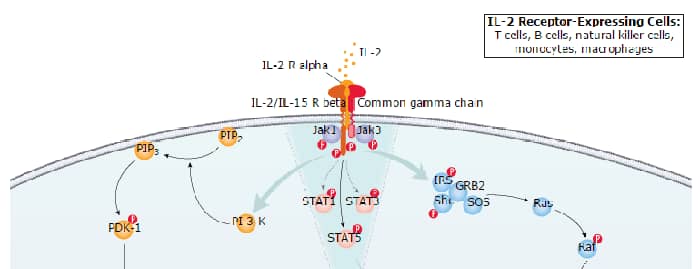
IL-2 receptor alpha (IL-2 R alpha ), also known as CD25, is a 55 kDa type I membrane glycoprotein that belongs to the family of cytokine receptors that utilize the common gamma chain subunit ( gamma c). IL-2 R alpha is primarily expressed on activated T cells and on regulatory T cells (Treg) (1-3). The mouse IL-2 R alpha cDNA encodes a 268 amino acid (aa) precursor that includes a 21 aa signal peptide, a 215 aa extracellular domain (ECD) with two Sushi domains, a 21 aa transmembrane segment, and an 11 aa cytoplasmic domain (4, 5). Within the ECD, mouse IL-2 R alpha shares 81% and 58% aa sequence identity with rat and human IL-2 R alpha, respectively. It shares approximately 15% aa sequence identity with IL-4, -7, -9, -15, and -21 receptor subunits that also complex with gamma c. IL-2 R beta (CD122) and gamma c (IL-2 R gamma /CD132) dimerize to form a constitutively expressed intermediate affinity IL-2 receptor (6, 7). By itself, IL-2 R alpha binds IL-2 with low affinity. It associates with IL-2 R beta and gamma c to generate a ternary high affinity IL-2 receptor complex (8). A soluble form of IL-2 R alpha can be generated by proteolytic cleavage of the cell surface receptor, rendering the T cell unresponsive to IL-2 (9, 10). Increased serum levels of soluble IL-2 R alpha are found in some cancers and immune disorders (11). IL-2 R alpha is required for activation induced cell death (AICD) of naive T cells, a mechanism responsible for deleting autoreactive T cell clones (12, 13). IL-2 R alpha is also required for the development of CD4+CD25+ Treg which suppress autoreactive CD4+ T cells, thereby contributing to peripheral T cell homeostasis (12-14).
Mouse CD25/IL-2R alpha Antibody
R&D Systems | Catalog # AF2438

Scientific images may be resized, cropped, or adjusted for brightness or contrast for presentation purposes. These updates are limited to presentation and do not affect the underlying scientific interpretation of the data. Where available, additional source data may be provided upon request.
Key Product Details
Species Reactivity
Validated:
Cited:
Applications
Validated:
Cited:
Label
Antibody Source
Product Specifications
Immunogen
Glu22-Lys236
Accession # Q544I2
Specificity
Clonality
Host
Isotype
Endotoxin Level
Scientific Data Images for Mouse CD25/IL-2R alpha Antibody
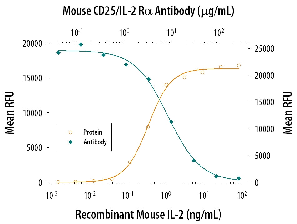
Cell Proliferation Induced by IL‑2 and Neutralization by Mouse CD25IL‑2 R alpha Antibody.
Recombinant Mouse IL-2 (402-ML) stimulates proliferation in the CTLL-2 mouse cytotoxic T cell line in a dose-dependent manner (orange line). Proliferation elicited by Recombinant Mouse IL-2 (2 ng/mL) is neutralized (green line) by increasing concentrations of Goat Anti-Mouse CD25/IL-2 Ra Antigen Affinity-purified Polyclonal Antibody (Catalog # AF2438). The ND50 is typically 5-20 µg/mL.
CD25/IL‑2 R alpha in Mouse Spleen.
CD25/IL-2 Ra was detected in immersion fixed frozen sections of mouse spleen using 5 µg/mL Goat Anti-Mouse CD25/IL-2 Ra Antigen Affinity-purified Polyclonal Antibody (Catalog # AF2438) overnight at 4 °C. Tissue was stained with the NorthernLights™ 557-conjugated Anti-Goat IgG Secondary Antibody (red; NL001) and counterstained (green). View our protocol for Fluorescent IHC Staining of Frozen Tissue Sections.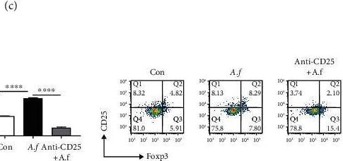
Detection of Mouse CD25/IL-2R alpha by Flow Cytometry
The persistent presence of fungi in lung injury induced by Aspergillus fumigatus is related to Tregs. (a) Increased CD4+CD25+Foxp3+ Treg proportion significant in PBMCs of patients infected with A. fumigatus (n = 7). (b) Foxp3 mRNA levels in the lungs were measured with q-PCR. Relative expression levels of the genes were expressed with the GAPDH housekeeping gene as an internal reference (n = 5/group). The expression of Foxp3 protein levels in the lungs was measured with Western blotting. Relative expression levels were expressed with the GAPDH as an internal reference (n = 3/group). (c) CD4+CD25+ T cells and CD4+CD25+Foxp3+ Tregs in the spleen were detected by flowcytometry, and FlowJo10 analyzed the proportions as prior described in Section 2. Increased CD4+CD25+ T cell and CD4+CD25+Foxp3+ Treg proportion significantly in the spleen of A. fumigatus infection mice (n = 5/group).Decreased susceptibility of mice to A. fumigatus infection after Treg depletion. (d) After pretreatment with CD25-neutralizing antibodies, the number of Treg cells in the spleen of mice with A. fumigatus infection group was significantly reduced, each group (n = 5/group). (e) Fungal load (colony-forming unit) after 1 day of infection (n = 4/group). Experiments were done at least three times. Data are presented as mean ± standard deviation. ∗Statistically significant difference (p < 0.05) against noninfected control. (f) Lungs from each experimental group were processed for histological examination after H&E staining. Lung injury scores were evaluated by the method described previously. ∗p < 0.05, ∗∗p < 0.01, ∗∗∗p < 0.001, and ∗∗∗∗p < 0.0001 by Student's unpaired two-tailed t test and the one-way ANOVA followed by the Tukey post hoc test. Error bars represent SEM. Image collected and cropped by CiteAb from the following open publication (https://pubmed.ncbi.nlm.nih.gov/34222495), licensed under a CC-BY license. Not internally tested by R&D Systems.
Mouse CD25 / IL-2 R alpha ELISA Standard Curve
Recombinant Mouse CD25/IL-2R alpha (Catalog # 2438-RM) was serially diluted and captured by Goat Anti-Mouse CD25/IL-2R alpha Antigen Affinity-purified Polyclonal Antibody (Catalog # AF2438) coated on a Clear Polystyrene Microplate (Catalog # DY990). Goat Anti-Mouse CD25/IL-2R alpha Antigen Affinity-purified Polyclonal Antibody (Catalog # AF2438) was biotinylated and incubated with the protein captured on the plate. Detection of the standard curve was achieved by incubating Streptavidin-HRP (Catalog # DY998)
Mouse CD25 / IL-2 R alpha ELISA Standard Curve
Recombinant Mouse CD25/IL-2R alpha (Catalog # 2438-RM) was serially diluted and captured by Goat Anti-Mouse CD25/IL-2R alpha Antigen Affinity-purified Polyclonal Antibody (Catalog # AF2438) coated on a Clear Polystyrene Microplate (Catalog # DY990). Goat Anti-Mouse CD25/IL-2R alpha Antigen Affinity-purified Polyclonal Antibody (Catalog # AF2438) was biotinylated and incubated with the protein captured on the plate. Detection of the standard curve was achieved by incubating Streptavidin-HRP (Catalog # DY998)Applications for Mouse CD25/IL-2R alpha Antibody
Immunohistochemistry
Sample: Immersion fixed frozen sections of mouse spleen
Western Blot
Sample: Recombinant Mouse CD25/IL‑2 R alpha (Catalog # 2438-RM)
Neutralization
Mouse CD25/IL-2 R alpha Sandwich Immunoassay
Formulation, Preparation, and Storage
Purification
Reconstitution
Reconstitute at 0.2 mg/mL in sterile PBS. For liquid material, refer to CoA for concentration.
Formulation
Shipping
Stability & Storage
- 12 months from date of receipt, -20 to -70 °C as supplied.
- 1 month, 2 to 8 °C under sterile conditions after reconstitution.
- 6 months, -20 to -70 °C under sterile conditions after reconstitution.
Calculators
Background: CD25/IL-2R alpha
References
- Minami, Y. et al. (1993) Annu. Rev. Immunol. 11:245.
- Kovanen, P.E. and W.J. Leonard (2004) Immunol. Rev. 202:67.
- Bluestone, J.A. and Q. Tang (2005) Curr. Opin. Immunol. 17:638.
- Miller, J. et al. (1985) J. Immunol. 134:4212.
- Shimuzu, A. et al. (1985) Nucleic Acids Res. 13:1505.
- Hatakeyama, M. et al. (1989) Science 244:551.
- Takeshita, T. et al. (1992) Science 257:379.
- Wang, X. et al. (2005) Science 310:1159.
- Wagner, D.K. et al. (1986) J. Immunol. 137:592.
- Schulz, O. et al. (1998) J. Exp. Med. 187:271.
- Witkowska, A.M. (2005) Mediat. Inflamm. 2005:121.
- Willerford, D.M. et al. (1995) Immunity 3:521.
- Van Parijs, L. et al. (1997) J. Immunol. 158:3738.
- Almeida, A.R.M. et al. (2002) J. Immunol. 169:4850.
Long Name
Alternate Names
Entrez Gene IDs
Gene Symbol
UniProt
Additional CD25/IL-2R alpha Products
Product Documents for Mouse CD25/IL-2R alpha Antibody
Certificate of Analysis
To download a Certificate of Analysis, please enter a lot or batch number in the search box below.
Note: Certificate of Analysis not available for kit components.
Product Specific Notices for Mouse CD25/IL-2R alpha Antibody
For research use only
Related Research Areas
Citations for Mouse CD25/IL-2R alpha Antibody
 Powered by Bioz
Customer Reviews for Mouse CD25/IL-2R alpha Antibody
There are currently no reviews for this product. Be the first to review Mouse CD25/IL-2R alpha Antibody and earn rewards!
Have you used Mouse CD25/IL-2R alpha Antibody?
Submit a review and receive an Amazon gift card!
$25/€18/£15/$25CAN/¥2500 for a review with an image
$10/€7/£6/$10CAN/¥1110 for a review without an image
Submit a review
Protocols
Find general support by application which include: protocols, troubleshooting, illustrated assays, videos and webinars.
- Antigen Retrieval Protocol (PIER)
- Antigen Retrieval for Frozen Sections Protocol
- Appropriate Fixation of IHC/ICC Samples
- Cellular Response to Hypoxia Protocols
- Chromogenic IHC Staining of Formalin-Fixed Paraffin-Embedded (FFPE) Tissue Protocol
- Chromogenic Immunohistochemistry Staining of Frozen Tissue
- ClariTSA™ Fluorophore Kits
- Detection & Visualization of Antibody Binding
- Fluorescent IHC Staining of Frozen Tissue Protocol
- Graphic Protocol for Heat-induced Epitope Retrieval
- Graphic Protocol for the Preparation and Fluorescent IHC Staining of Frozen Tissue Sections
- Graphic Protocol for the Preparation and Fluorescent IHC Staining of Paraffin-embedded Tissue Sections
- Graphic Protocol for the Preparation of Gelatin-coated Slides for Histological Tissue Sections
- IHC Sample Preparation (Frozen sections vs Paraffin)
- Immunofluorescent IHC Staining of Formalin-Fixed Paraffin-Embedded (FFPE) Tissue Protocol
- Immunohistochemistry (IHC) and Immunocytochemistry (ICC) Protocols
- Immunohistochemistry Frozen Troubleshooting
- Immunohistochemistry Paraffin Troubleshooting
- Preparing Samples for IHC/ICC Experiments
- Preventing Non-Specific Staining (Non-Specific Binding)
- Primary Antibody Selection & Optimization
- Protocol for Heat-Induced Epitope Retrieval (HIER)
- Protocol for Making a 4% Formaldehyde Solution in PBS
- Protocol for VisUCyte™ HRP Polymer Detection Reagent
- Protocol for the Preparation & Fixation of Cells on Coverslips
- Protocol for the Preparation and Chromogenic IHC Staining of Frozen Tissue Sections
- Protocol for the Preparation and Chromogenic IHC Staining of Frozen Tissue Sections - Graphic
- Protocol for the Preparation and Chromogenic IHC Staining of Paraffin-embedded Tissue Sections
- Protocol for the Preparation and Chromogenic IHC Staining of Paraffin-embedded Tissue Sections - Graphic
- Protocol for the Preparation and Fluorescent IHC Staining of Frozen Tissue Sections
- Protocol for the Preparation and Fluorescent IHC Staining of Paraffin-embedded Tissue Sections
- Protocol for the Preparation of Gelatin-coated Slides for Histological Tissue Sections
- R&D Systems Quality Control Western Blot Protocol
- TUNEL and Active Caspase-3 Detection by IHC/ICC Protocol
- The Importance of IHC/ICC Controls
- Troubleshooting Guide: Immunohistochemistry
- Troubleshooting Guide: Western Blot Figures
- Western Blot Conditions
- Western Blot Protocol
- Western Blot Protocol for Cell Lysates
- Western Blot Troubleshooting
- Western Blot Troubleshooting Guide
- View all Protocols, Troubleshooting, Illustrated assays and Webinars
Associated Pathways