Mouse CXCL10/IP-10/CRG-2 Antibody
R&D Systems | Catalog # AF-466-NA

Key Product Details
Validated by
Knockout/Knockdown
Species Reactivity
Validated:
Mouse
Cited:
Human, Mouse, Rat
Applications
Validated:
Western Blot, Neutralization
Cited:
Immunohistochemistry, Immunohistochemistry-Paraffin, Immunohistochemistry-Frozen, Western Blot, Neutralization, Flow Cytometry, Immunocytochemistry, Functional Assay, Tissue Culture
Label
Unconjugated
Antibody Source
Polyclonal Goat IgG
Loading...
Product Specifications
Immunogen
E. coli-derived recombinant mouse CXCL10/IP-10/CRG-2
Ile22-Pro98
Accession # P17515
Ile22-Pro98
Accession # P17515
Specificity
Detects mouse CXCL10/IP-10/CRG-2 in direct ELISAs and Western blots. In direct ELISAs, less than 10% cross-reactivity with recombinant human CXCL10 is observed.
Clonality
Polyclonal
Host
Goat
Isotype
IgG
Endotoxin Level
<0.10 EU per 1 μg of the antibody by the LAL method.
Scientific Data Images for Mouse CXCL10/IP-10/CRG-2 Antibody
Detection of Mouse CXCL10/IP‑10/CRG‑2 by Western Blot.
Western blot shows lysates of RAW 264.7 mouse monocyte/macrophage cell line. PVDF membrane was probed with 1 µg/mL of Goat Anti-Mouse CXCL10/IP-10/CRG-2 Antigen Affinity-purified Polyclonal Antibody (Catalog # AF-466-NA) followed by HRP-conjugated Anti-Goat IgG Secondary Antibody (HAF017). A specific band was detected for CXCL10/IP-10/CRG-2 at approximately 15 kDa (as indicated). This experiment was conducted under reducing conditions and using Immunoblot Buffer Group 1.
Chemotaxis Induced by CXCL10/CRG‑2 and Neutral-ization by Mouse CXCL10/ CRG‑2 Antibody.
Recombinant Mouse CXCL10/ CRG-2 (466-CR) chemoattracts the BaF3 mouse pro-B cell line transfected with human CXCR3 in a dose-dependent manner (orange line). The amount of cells that migrated through to the lower chemotaxis chamber was measured by Resazurin (AR002). Chemotaxis elicited by Recombinant Mouse CXCL10/ CRG-2 (0.5 µg/mL) is neutral-ized (green line) by increasing concentrations of Goat Anti-Mouse CXCL10/CRG-2 Antigen Affinity-purified Polyclonal Antibody (Catalog # AF-466-NA). The ND50 is typically 2.00‑25.0 µg/mL.
Detection of Mouse CXCL10/IP-10/CRG-2 by Western Blot
Conditional deletion of astroglial CXCL10. (A) Location of loxP sites in the CXCL10 locus in CXCL10fl/fl mice. (B) Day 17 post-MOG peptide injection (dpi) representative images of immunostaining for CXCL10-Red, GFAP-Green, and DAPI-Blue. (C) Quantification of images for CXCL10 integrated density at 17 dpi (n = 4–7 mice/group). (D and E) qRT/PCR for CXCL10 and CXCL9 in spinal cord of astroglial CXCL10 knockout and littermate control mice at 17 dpi (n = 6 mice/group). (F) Quantification of CXCL10 protein levels in the spinal cord of astroglial CXCL10 knockout and control mice at 14 dpi, by western blot analysis (n = 4 mice/group). (G) Data were normalized to GAPDH, P = 0.046. Vertical bars = SEMs. Image collected and cropped by CiteAb from the following publication (https://pubmed.ncbi.nlm.nih.gov/24924222), licensed under a CC-BY license. Not internally tested by R&D Systems.
Detection of Mouse CXCL10/IP-10/CRG-2 by Immunocytochemistry/Immunofluorescence
Conditional deletion of astroglial CXCL10. (A) Location of loxP sites in the CXCL10 locus in CXCL10fl/fl mice. (B) Day 17 post-MOG peptide injection (dpi) representative images of immunostaining for CXCL10-Red, GFAP-Green, and DAPI-Blue. (C) Quantification of images for CXCL10 integrated density at 17 dpi (n = 4–7 mice/group). (D and E) qRT/PCR for CXCL10 and CXCL9 in spinal cord of astroglial CXCL10 knockout and littermate control mice at 17 dpi (n = 6 mice/group). (F) Quantification of CXCL10 protein levels in the spinal cord of astroglial CXCL10 knockout and control mice at 14 dpi, by western blot analysis (n = 4 mice/group). (G) Data were normalized to GAPDH, P = 0.046. Vertical bars = SEMs. Image collected and cropped by CiteAb from the following publication (https://pubmed.ncbi.nlm.nih.gov/24924222), licensed under a CC-BY license. Not internally tested by R&D Systems.
Detection of Mouse CXCL10/IP-10/CRG-2 by Immunohistochemistry
Correlations between neutrophil/mast cell numbers and CXCL10 expression in the fracture hematoma during the early inflammatory phase. Correlation analysis by simple linear regression was performed between the parameters neutrophil numbers, mast cell numbers and local CXCL10 protein expression in all samples. (A) Neutrophil number/CXCL10 correlation. (B) Mast cell number/CXCL10 correlation. (C) Mast cell number/neutrophil number correlation. (D) Immunofluoresence double staining for mast cells (Avidin staining, red) and CXCL10 (green). DNA was counterstained with Hoechst (blue). Scale bar = 50 µm. E) Black box marked the area which is shown in (E). (F) Single fluorescent channels for Avidin (red) and CXCL10 (green). Scale bar = 50 µm. Image collected and cropped by CiteAb from the following open publication (https://pubmed.ncbi.nlm.nih.gov/36761764), licensed under a CC-BY license. Not internally tested by R&D Systems.
Mouse CXCL10 / IP-10 / CRG-2 ELISA Standard Curve
Recombinant Mouse CXCL10/IP‑10/CRG‑2 (Catalog # 466-CR) was serially diluted and captured by Rat Anti-Mouse CXCL10/IP‑10/CRG‑2 Monoclonal Antibody (Catalog # MAB466) coated on a Clear Polystyrene Microplate (Catalog # DY990). Goat Anti-Mouse CXCL10/IP‑10/CRG‑2 Antigen Affinity-purified Polyclonal Antibody (Catalog # AF-466-NA) was biotinylated and incubated with the protein captured on the plate. Detection of the standard curve was achieved by incubating Streptavidin-HRP (Catalog # DY998)Applications for Mouse CXCL10/IP-10/CRG-2 Antibody
Application
Recommended Usage
Western Blot
1 µg/mL
Sample: RAW 264.7 mouse monocyte/macrophage cell line
Sample: RAW 264.7 mouse monocyte/macrophage cell line
Neutralization
Measured by its ability to neutralize CXCL10/IP‑10/CRG‑2-induced chemotaxis in the BaF3 mouse pro‑B cell line transfected with human CXCR3. The Neutralization Dose (ND50) is typically 2.00-25.0 µg/mL in the presence of 0.5 µg/mL Recombinant Mouse CXCL10/IP‑10/CRG‑2.
Reviewed Applications
Read 3 reviews rated 2.7 using AF-466-NA in the following applications:
Formulation, Preparation, and Storage
Purification
Antigen Affinity-purified
Reconstitution
Reconstitute at 0.2 mg/mL in sterile PBS. For liquid material, refer to CoA for concentration.
Loading...
Formulation
Lyophilized from a 0.2 μm filtered solution in PBS with Trehalose. See Certificate of Analysis for details.
*Small pack size (-SP) is supplied either lyophilized or as a 0.2 µm filtered solution in PBS.
*Small pack size (-SP) is supplied either lyophilized or as a 0.2 µm filtered solution in PBS.
Shipping
Lyophilized product is shipped at ambient temperature. Liquid small pack size (-SP) is shipped with polar packs. Upon receipt, store immediately at the temperature recommended below.
Stability & Storage
Use a manual defrost freezer and avoid repeated freeze-thaw cycles.
- 12 months from date of receipt, -20 to -70 °C as supplied.
- 1 month, 2 to 8 °C under sterile conditions after reconstitution.
- 6 months, -20 to -70 °C under sterile conditions after reconstitution.
Calculators
Background: CXCL10/IP-10/CRG-2
References
- Loetscher, M. et al. (1996) J. Exp. Med. 184:963.
- Vanguri, P. (1996) J. Neuroimmunol. 56:35.
- Sgadari, C. et al. (1996) Blood 87:3877.
Alternate Names
CRG-2, CRG2, IP-10
Gene Symbol
CXCL10
UniProt
Additional CXCL10/IP-10/CRG-2 Products
Product Documents for Mouse CXCL10/IP-10/CRG-2 Antibody
Certificate of Analysis
To download a Certificate of Analysis, please enter a lot or batch number in the search box below.
Note: Certificate of Analysis not available for kit components.
Product Specific Notices for Mouse CXCL10/IP-10/CRG-2 Antibody
For research use only
Citations for Mouse CXCL10/IP-10/CRG-2 Antibody
 Powered by Bioz
Customer Reviews for Mouse CXCL10/IP-10/CRG-2 Antibody (3)
2.7 out of 5
3 Customer Ratings
Have you used Mouse CXCL10/IP-10/CRG-2 Antibody?
Submit a review and receive an Amazon gift card!
$25/€18/£15/$25CAN/¥2500 Yen for a review with an image
$10/€7/£6/$10CAN/¥1110 Yen for a review without an image
Submit a review
Customer Images
Showing
1
-
3 of
3 reviews
Showing All
Filter By:
-
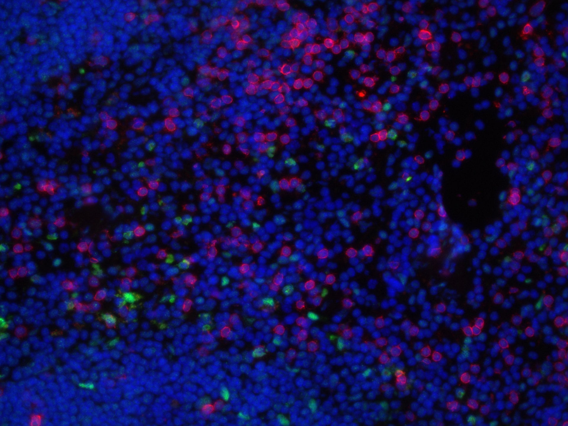
Application: Immunohistochemistry-FrozenSample Tested: Mouse SpleenSpecies: MouseVerified Customer | Posted 12/05/2022Murine spleen with CD3 (red), CXCL10 (green) and DAPI (blue)

-
Application: ELISASample Tested: Cell Culture SamplesSpecies: MouseVerified Customer | Posted 12/16/2021Antibody initially had high non-specific background (picture) and then antibody function decreased over a course of time when stored at 4C long term.
-
Application: Immunocytochemistry/ImmunofluorescenceSample Tested: Spleen tissueSpecies: MouseVerified Customer | Posted 05/09/2019
There are no reviews that match your criteria.
Protocols
Find general support by application which include: protocols, troubleshooting, illustrated assays, videos and webinars.
- Cellular Response to Hypoxia Protocols
- R&D Systems Quality Control Western Blot Protocol
- Troubleshooting Guide: Western Blot Figures
- Western Blot Conditions
- Western Blot Protocol
- Western Blot Protocol for Cell Lysates
- Western Blot Troubleshooting
- Western Blot Troubleshooting Guide
- View all Protocols, Troubleshooting, Illustrated assays and Webinars
Loading...
Associated Pathways
