Neprilysin (NEP, neutral endopeptidase 24.11, EC 3.4.24.11) is a zinc metallopeptidase expressed at the cell surface of a variety of cells. The enzyme functions both as an endopeptidase with a thermolysin-like specificity and as a dipeptidylcarboxypeptidase. NEP has been shown to be involved in the degradation of enkephalins in the mammalian brain and the inactivation of circulating atrial natriuretic peptide (1, 2). NEP has also been identified as the common acute lymphoblastic leukemia antigen (CALLA), and to be expressed on the surface of lymphocytes in some disease states (3). These and other observations have resulted in considerable clinical interest in NEP as a potential target for analgesics and antihypertensive drugs. NEP is also a major degrading enzyme of amyloid beta peptide (A beta ) in the brain, indicating that down-regulation of NEP activity, which could be caused by aging, can contribute to the development of Alzheimer’s disease by promoting A beta accumulation (4).

Key Product Details
Species Reactivity
Validated:
Mouse
Cited:
Mouse, Rat, Transgenic Mouse, Xenograft
Applications
Validated:
Immunohistochemistry, Western Blot, ELISA Capture (Matched Antibody Pair), Immunoprecipitation
Cited:
Immunohistochemistry, Immunohistochemistry-Paraffin, Immunohistochemistry-Frozen, Western Blot, ELISA Capture
Label
Unconjugated
Antibody Source
Polyclonal Goat IgG
Loading...
Product Specifications
Immunogen
S. frugiperda insect ovarian cell line Sf 21-derived recombinant mouse Neprilysin/CD10
Tyr52-Trp750
Accession # AAA37386
Tyr52-Trp750
Accession # AAA37386
Specificity
Detects mouse Neprilysin/CD10 in ELISAs and Western blots. In sandwich immunoassays, less than 35% cross-reactivity with recombinant human (rh) Neprilysin is observed and less than 0.2% cross-reactivity with rhNeprilysin-2 is observed.
Clonality
Polyclonal
Host
Goat
Isotype
IgG
Scientific Data Images for Mouse Neprilysin/CD10 Antibody
Neprilysin/CD10 in Mouse Brain.
Neprilysin/CD10 was detected in perfusion fixed frozen sections of mouse brain (glial cell in hippocampus) using 15 µg/mL Goat Anti-Mouse Neprilysin/CD10 Antigen Affinity-purified Polyclonal Antibody (Catalog # AF1126) overnight at 4 °C. Tissue was stained (red) and counterstained (green). View our protocol for Fluorescent IHC Staining of Frozen Tissue Sections.
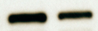
Detection of Mouse Neprilysin/CD10 by Western Blot
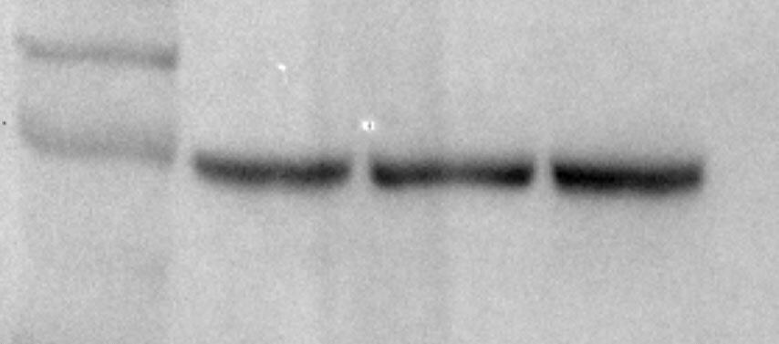
Astrocyte‐specific Stat3 deletion increases microglial A beta internalization and degradation, and reduces apoE expression, dystrophic neurites, and detrimental cytokinesAInternalization of A beta (stained with IC16 antibody or methoxy‐XO4) was assessed using an engulfment assay, in which glial and A beta structures were surface‐rendered and A beta volumes co‐localized with glial volumes were quantified. Scale bars, 10 μm.B, CMicroglia (left Y axes) from APP/PS1 mice internalized significantly more A beta positive for IC16 or methoxy‐XO4 when Stat3 was deleted in astrocytes (*P < 0.05, Mann–Whitney test), whereas no changes were seen in astrocytes (right axes; APP/PS1‐Stat3WT, n = 8 (four females and four males) mice; APP/PS1‐Stat3KO, n = 11 (five females and six males) mice; age, 11 months; Mann–Whitney test).D–H(D–F) Western blot quantification of protein levels of the A beta ‐degrading enzymes neprilysin/CD10 and CD36, as well as the A beta ‐binding apolipoprotein E (apoE), revealed a significantly increased expression of neprilysin and CD36 and a decreased expression of apoE (APP/PS1‐Stat3WT, n = 9 (five females and four males) mice; APP/PS1‐Stat3KO, n = 9 (five females and four males) mice; age, 11 months; *P < 0.05, Mann–Whitney test for all comparisons). (G) In contrast, TREM2 expression remained unchanged (APP/PS1‐Stat3WT, n = 8 (four females and four males) mice; APP/PS1‐Stat3KO, n = 7 (four females and three males) mice; age, 11 months; Mann–Whitney test). (H) Western blots for proteins analyzed in (D‐G).Data information: Data are represented as mean ± SEM.Source data are available online for this figure. Image collected and cropped by CiteAb from the following open publication (https://pubmed.ncbi.nlm.nih.gov/30617153), licensed under a CC-BY license. Not internally tested by R&D Systems.
Detection of Mouse Neprilysin/CD10 by Western Blot
Astrocyte‐specific Stat3 deletion increases microglial A beta internalization and degradation, and reduces apoE expression, dystrophic neurites, and detrimental cytokinesAInternalization of A beta (stained with IC16 antibody or methoxy‐XO4) was assessed using an engulfment assay, in which glial and A beta structures were surface‐rendered and A beta volumes co‐localized with glial volumes were quantified. Scale bars, 10 μm.B, CMicroglia (left Y axes) from APP/PS1 mice internalized significantly more A beta positive for IC16 or methoxy‐XO4 when Stat3 was deleted in astrocytes (*P < 0.05, Mann–Whitney test), whereas no changes were seen in astrocytes (right axes; APP/PS1‐Stat3WT, n = 8 (four females and four males) mice; APP/PS1‐Stat3KO, n = 11 (five females and six males) mice; age, 11 months; Mann–Whitney test).D–H(D–F) Western blot quantification of protein levels of the A beta ‐degrading enzymes neprilysin/CD10 and CD36, as well as the A beta ‐binding apolipoprotein E (apoE), revealed a significantly increased expression of neprilysin and CD36 and a decreased expression of apoE (APP/PS1‐Stat3WT, n = 9 (five females and four males) mice; APP/PS1‐Stat3KO, n = 9 (five females and four males) mice; age, 11 months; *P < 0.05, Mann–Whitney test for all comparisons). (G) In contrast, TREM2 expression remained unchanged (APP/PS1‐Stat3WT, n = 8 (four females and four males) mice; APP/PS1‐Stat3KO, n = 7 (four females and three males) mice; age, 11 months; Mann–Whitney test). (H) Western blots for proteins analyzed in (D‐G).Data information: Data are represented as mean ± SEM.Source data are available online for this figure. Image collected and cropped by CiteAb from the following open publication (https://pubmed.ncbi.nlm.nih.gov/30617153), licensed under a CC-BY license. Not internally tested by R&D Systems.
Immersion fixed paraffin-embedded sections of mouse kidney
Neprilysin/CD10 was detected in immersion fixed paraffin-embedded sections of mouse kidney using Goat Anti-Mouse Neprilysin/CD10 Antigen Affinity-purified Polyclonal Antibody (Catalog # AF1126) at 1 µg/ml overnight at 4 °C. Before incubation with the primary antibody, tissue was subjected to heat-induced epitope retrieval using VisUCyte Antigen Retrieval Reagent-Basic (Catalog # VCTS021). Tissue was stained using the HRP-conjugated Anti-Goat IgG Secondary Antibody (Catalog # HAF017) and counterstained with hematoxylin (blue). Specific staining was localized to the membrane. View our protocol for Chromogenic IHC Staining of Paraffin-embedded Tissue Sections.
Immersion fixed paraffin-embedded sections of mouse spinal cord
Neprilysin/CD10 was detected in immersion fixed paraffin-embedded sections of mouse spinal cord using Goat Anti-Mouse Neprilysin/CD10 Antigen Affinity-purified Polyclonal Antibody (Catalog # AF1126) at 1 µg/ml overnight at 4 °C. Before incubation with the primary antibody, tissue was subjected to heat-induced epitope retrieval using VisUCyte Antigen Retrieval Reagent-Basic (Catalog # VCTS021). Tissue was stained using the HRP-conjugated Anti-Goat IgG Secondary Antibody (Catalog # HAF017) and counterstained with hematoxylin (blue). Specific staining was localized to the membrane. View our protocol for Chromogenic IHC Staining of Paraffin-embedded Tissue Sections.
Mouse Neprilysin / CD10 ELISA Standard Curve
Mouse Neprilysin/CD10 was serially diluted and captured by Goat Anti-Mouse Neprilysin/CD10 Antigen Affinity-purified Polyclonal Antibody (Catalog # AF1126) coated on a Clear Polystyrene Microplate (Catalog # DY990). Goat Anti-Mouse Neprilysin/CD10 Antigen Affinity-purified Polyclonal Antibody (Catalog # AF1126) was biotinylated and incubated with the protein captured on the plate. Detection of the standard curve was achieved by incubating Streptavidin-HRP (Catalog # DY998)
Mouse Neprilysin / CD10 ELISA Standard Curve
Rat C-Reactive Protein/CRP was serially diluted and captured by Goat Anti-Mouse Neprilysin/CD10 Antigen Affinity-purified Polyclonal Antibody (Catalog # AF1126) coated on a Clear Polystyrene Microplate (Catalog # DY990). Goat Anti-Mouse Neprilysin/CD10 Antigen Affinity-purified Polyclonal Antibody (Catalog # AF1126) was biotinylated and incubated with the protein captured on the plate. Detection of the standard curve was achieved by incubating Streptavidin-HRP (Catalog # DY998)Applications for Mouse Neprilysin/CD10 Antibody
Application
Recommended Usage
Immunohistochemistry
0.25-25 µg/mL
Sample: Perfusion fixed frozen sections of mouse brain (glial cell in hippocampus), Immersion fixed paraffin-embedded sections of mouse spinal cord, and of mouse kidney
Sample: Perfusion fixed frozen sections of mouse brain (glial cell in hippocampus), Immersion fixed paraffin-embedded sections of mouse spinal cord, and of mouse kidney
Immunoprecipitation
25 µg/mL
Sample: Conditioned cell culture medium spiked with Recombinant Mouse Neprilysin/CD10 (Catalog # 1126-ZN), see our available Western blot detection antibodies
Sample: Conditioned cell culture medium spiked with Recombinant Mouse Neprilysin/CD10 (Catalog # 1126-ZN), see our available Western blot detection antibodies
Western Blot
0.1 µg/mL
Sample: Recombinant Mouse Neprilysin/CD10 (Catalog # 1126-ZN)
Sample: Recombinant Mouse Neprilysin/CD10 (Catalog # 1126-ZN)
Mouse Neprilysin/CD10 Sandwich Immunoassay
Please Note: Optimal dilutions of this antibody should be experimentally determined.
Reviewed Applications
Read 2 reviews rated 5 using AF1126 in the following applications:
Formulation, Preparation, and Storage
Purification
Antigen Affinity-purified
Reconstitution
Reconstitute at 0.2 mg/mL in sterile PBS. For liquid material, refer to CoA for concentration.
Loading...
Formulation
Lyophilized from a 0.2 μm filtered solution in PBS with Trehalose. See Certificate of Analysis for details.
*Small pack size (-SP) is supplied either lyophilized or as a 0.2 µm filtered solution in PBS.
*Small pack size (-SP) is supplied either lyophilized or as a 0.2 µm filtered solution in PBS.
Shipping
Lyophilized product is shipped at ambient temperature. Liquid small pack size (-SP) is shipped with polar packs. Upon receipt, store immediately at the temperature recommended below.
Stability & Storage
Use a manual defrost freezer and avoid repeated freeze-thaw cycles.
- 12 months from date of receipt, -20 to -70 °C as supplied.
- 1 month, 2 to 8 °C under sterile conditions after reconstitution.
- 6 months, -20 to -70 °C under sterile conditions after reconstitution.
Calculators
Background: Neprilysin/CD10
References
- Malfroy, B. et al. (1978) Nature 276:523.
- Kenny, A.J. and S.L. Stephenson (1988) FEBS Lett. 232:1.
- LeTarte, M. et al. (1988) J. Exp. Med. 168:1247.
- Itwata, N. et al. (2001) Science 292:1550.
Alternate Names
CALLA, CD10, Enkephalinase, Leu-19, MME, Neutral Endopeptidase 24.11, NKH1
Gene Symbol
MME
UniProt
Additional Neprilysin/CD10 Products
Product Documents for Mouse Neprilysin/CD10 Antibody
Certificate of Analysis
To download a Certificate of Analysis, please enter a lot or batch number in the search box below.
Note: Certificate of Analysis not available for kit components.
Product Specific Notices for Mouse Neprilysin/CD10 Antibody
For research use only
Citations for Mouse Neprilysin/CD10 Antibody
 Powered by Bioz
Customer Reviews for Mouse Neprilysin/CD10 Antibody (2)
5 out of 5
2 Customer Ratings
Have you used Mouse Neprilysin/CD10 Antibody?
Submit a review and receive an Amazon gift card!
$25/€18/£15/$25CAN/¥2500 Yen for a review with an image
$10/€7/£6/$10CAN/¥1110 Yen for a review without an image
Submit a review
Customer Images
Showing
1
-
2 of
2 reviews
Showing All
Filter By:
-
Application: Western BlotSample Tested: Kidney tissueSpecies: RatVerified Customer | Posted 08/26/2016

-
Application: Western BlotSample Tested: Kidney tissueSpecies: MouseVerified Customer | Posted 04/26/2016
There are no reviews that match your criteria.
Protocols
Find general support by application which include: protocols, troubleshooting, illustrated assays, videos and webinars.
- Antigen Retrieval Protocol (PIER)
- Antigen Retrieval for Frozen Sections Protocol
- Appropriate Fixation of IHC/ICC Samples
- Cellular Response to Hypoxia Protocols
- Chromogenic IHC Staining of Formalin-Fixed Paraffin-Embedded (FFPE) Tissue Protocol
- Chromogenic Immunohistochemistry Staining of Frozen Tissue
- ClariTSA™ Fluorophore Kits
- Detection & Visualization of Antibody Binding
- Fluorescent IHC Staining of Frozen Tissue Protocol
- Graphic Protocol for Heat-induced Epitope Retrieval
- Graphic Protocol for the Preparation and Fluorescent IHC Staining of Frozen Tissue Sections
- Graphic Protocol for the Preparation and Fluorescent IHC Staining of Paraffin-embedded Tissue Sections
- Graphic Protocol for the Preparation of Gelatin-coated Slides for Histological Tissue Sections
- IHC Sample Preparation (Frozen sections vs Paraffin)
- Immunofluorescent IHC Staining of Formalin-Fixed Paraffin-Embedded (FFPE) Tissue Protocol
- Immunohistochemistry (IHC) and Immunocytochemistry (ICC) Protocols
- Immunohistochemistry Frozen Troubleshooting
- Immunohistochemistry Paraffin Troubleshooting
- Immunoprecipitation Protocol
- Preparing Samples for IHC/ICC Experiments
- Preventing Non-Specific Staining (Non-Specific Binding)
- Primary Antibody Selection & Optimization
- Protocol for Heat-Induced Epitope Retrieval (HIER)
- Protocol for Making a 4% Formaldehyde Solution in PBS
- Protocol for VisUCyte™ HRP Polymer Detection Reagent
- Protocol for the Preparation & Fixation of Cells on Coverslips
- Protocol for the Preparation and Chromogenic IHC Staining of Frozen Tissue Sections
- Protocol for the Preparation and Chromogenic IHC Staining of Frozen Tissue Sections - Graphic
- Protocol for the Preparation and Chromogenic IHC Staining of Paraffin-embedded Tissue Sections
- Protocol for the Preparation and Chromogenic IHC Staining of Paraffin-embedded Tissue Sections - Graphic
- Protocol for the Preparation and Fluorescent IHC Staining of Frozen Tissue Sections
- Protocol for the Preparation and Fluorescent IHC Staining of Paraffin-embedded Tissue Sections
- Protocol for the Preparation of Gelatin-coated Slides for Histological Tissue Sections
- R&D Systems Quality Control Western Blot Protocol
- TUNEL and Active Caspase-3 Detection by IHC/ICC Protocol
- The Importance of IHC/ICC Controls
- Troubleshooting Guide: Immunohistochemistry
- Troubleshooting Guide: Western Blot Figures
- Western Blot Conditions
- Western Blot Protocol
- Western Blot Protocol for Cell Lysates
- Western Blot Troubleshooting
- Western Blot Troubleshooting Guide
- View all Protocols, Troubleshooting, Illustrated assays and Webinars


