Mouse B7 homolog 1(B7-H1), also called programmed death ligand 1 (PD-L1) and programmed cell death 1 ligand 1 (PDCD1L1), is a member of the B7 family of proteins that provide signals for regulating T-cell activation and tolerance (1‑4). Other family members include B7-1, B7-2, B7-H2, B7-H3 and PD-L2. B7 proteins are immunoglobulin (Ig) superfamily members with extracellular Ig-V-like and Ig-C-like domains and a short cytoplasmic region. Among the family members, they share from 20‑40% amino acid (aa) sequence identity. The cloned mouse B7-H1/PD-L1 cDNA encodes a 290 aa type I membrane precursor protein with a putative 18 aa signal peptide, a 220 aa extracellular region containing one V-like and one C-like Ig domain, a 22 aa transmembrane region, and a 30 aa cytoplasmic domain. Mouse and human B7-H1/PD-L1 share approximately 70% aa sequence identity. B7-H1/PD-L1 is one of two ligands for programmed death-1 (PD-1), a member of the CD28 family of immunoreceptors. The other identified ligand is PD-L2. Mouse B7-H1/PD-L1 and PD-L2 share approximately 34% aa sequence identity and have similar functions. B7-H1/PD-L1 is constitutively expressed in various lymphoid and non-lymphoid organs including placenta, heart, pancreas, lung, liver, and endothelium (1‑4). The expression of B7-H1/PD-L1 is detected on B cells, T cells, monocytes, dendritic cells and thymic epithelial cells. IFN-gamma treatment induces B7‑H1/PD‑L1 expression in monocytes, dendritic cells, and endothelial cells. B7-H1/PD-L1 expression is also upregulated in a variety of tumor cell lines. On previously activated T cells, B7-H1/PD-L1 interaction with PD-1 inhibits TCR-mediated proliferation and cytokine production, suggesting an inhibitory role in regulating immune responses. In contrast, a costimulatory function for the PD-1 ligands on resting T cells has also been reported (1‑4).

Key Product Details
Validated by
Biological Validation
Species Reactivity
Validated:
Mouse
Cited:
Human, Mouse, Transgenic Mouse
Applications
Validated:
Immunohistochemistry, Western Blot, Flow Cytometry, Dual RNAscope ISH-IHC Compatible, CyTOF-ready
Cited:
Immunohistochemistry, Immunohistochemistry-Paraffin, Immunohistochemistry-Frozen, Western Blot, ELISA, Flow Cytometry, Immunocytochemistry
Label
Unconjugated
Antibody Source
Polyclonal Goat IgG
Loading...
Product Specifications
Immunogen
Mouse myeloma cell line NS0-derived recombinant mouse PD-L1/B7-H1
Phe19-Thr238
Accession # Q9EP73
Phe19-Thr238
Accession # Q9EP73
Specificity
Detects mouse PD-L1/B7-H1 in direct ELISAs and Western blots.
Clonality
Polyclonal
Host
Goat
Isotype
IgG
Scientific Data Images for Mouse PD-L1/B7-H1 Antibody
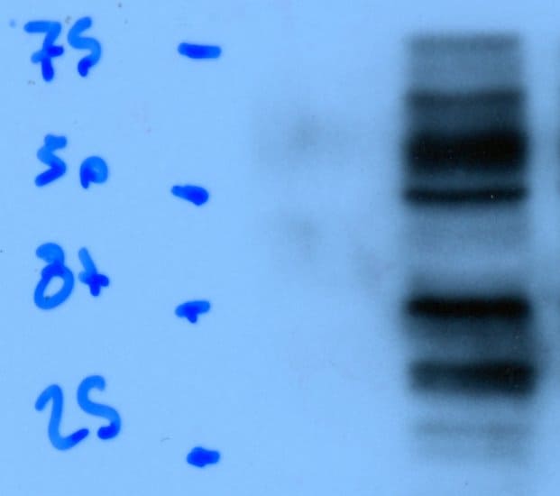
Detection of Mouse PD-L1/B7-H1 by Western Blot.
Western blot shows lysates of RAW 264.7 mouse monocyte/macrophage cell line untreated (-) or treated (+) with 10 µg/mL LPS for 4 hours. PVDF membrane was probed with 0.5 µg/mL of Goat Anti-Mouse PD-L1/B7-H1 Antigen Affinity-purified Polyclonal Antibody (Catalog # AF1019) followed by HRP-conjugated Anti-Goat IgG Secondary Antibody (Catalog # HAF017). A specific band was detected for PD-L1/B7-H1 at approximately 50-55 kDa (as indicated). This experiment was conducted under reducing conditions and using Immunoblot Buffer Group 1.
Detection of PD-L1/B7-H1 in RAW 264.7 Mouse Cell Line by Flow Cytometry.
RAW 264.7 mouse monocyte/macrophage cell line either treated with LPS overnight (filled histogram) or untreated (open histogram) was stained with Goat Anti-Mouse PD-L1/B7-H1 Antigen Affinity-purified Polyclonal Antibody (Catalog # AF1019), followed by Allophycocyanin-conjugated Anti-Goat IgG Secondary Antibody (Catalog # F0108). View our protocol for Staining Membrane-associated Proteins.
Detection of PD-L1/B7-H1 in HEK293 Human Cell Line Transfected with Mouse PD-L1/B7-H1 and eGFP by Flow Cytometry.
HEK293 human embryonic kidney cell line transfected with either (A) mouse PD-L1/B7-H1 or (B) irrelevant transfectants and eGFP was stained with Goat Anti-Mouse PD-L1/B7-H1 Antigen Affinity-purified Polyclonal Antibody (Catalog # AF1019) followed by Allophycocyanin-conjugated Anti-Goat IgG Secondary Antibody (Catalog # F0108). Quadrant markers were set based on control antibody staining (Catalog # AB-108-C). View our protocol for Staining Membrane-associated Proteins.
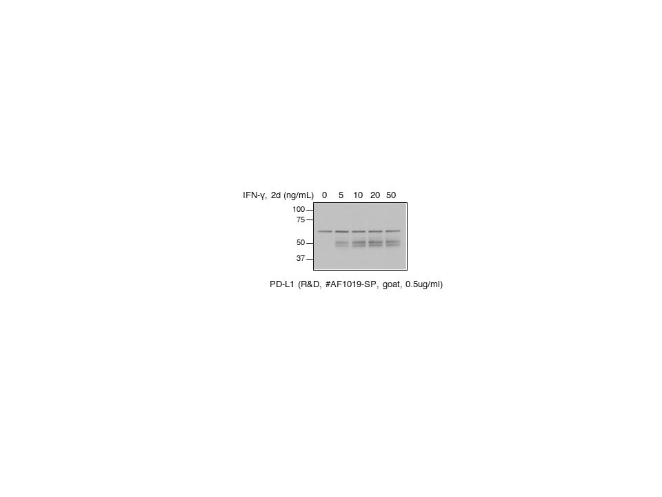
Detection of Mouse PD-L1 by Western Blot
Pyruvate kinase isoform M2 (PKM2) is required for LPS-induced expression of PD-L1. RAW cells (0.5 × 10⁶ cells/ml) (A), bone marrow-derived macrophages (BMDMs) (B), or bone marrow-derived dendritic cells (C) were treated with TEPP-46 at 50 µM for 1 h prior to stimulation with LPS (100 ng/ml, 24 h), lysed and assayed for expression of PD-L1 by flow cytometry [(A–C) top panels] and pdl1 mRNA by RT-PCR [(A,B) bottom panels]. Statistical analysis was performed on cells from three separate experiments. Error bars represent mean ± SEM. BMDM cells were transfected with scrambled control (SC) or anti-PKM2 siRNA (D). 24 h post-transfection, cells were stimulated with LPS (100 ng/ml) for 24 h after which expression of PKM2 (top panel) and PDL1 (middle panel) was measured by western blot. Image collected and cropped by CiteAb from the following publication (https://pubmed.ncbi.nlm.nih.gov/29081778), licensed under a CC-BY license. Not internally tested by R&D Systems.
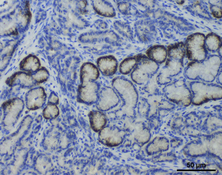
Detection of PD-L1/B7-H1 in Mouse Intestine.
Formalin-fixed paraffin-embedded tissue sections of mouse intestines were probed for PD-L1 mRNA (ACD RNAScope Probe, catalog #420508; Fast Red chromogen, ACD catalog # 322750). Adjacent tissue section was processed for immunohistochemistry using goat anti-mouse PD-L1 polyclonal antibody (R&D Systems catalog # AF1019) at 3ug/mL with one-hour incubation at room temperature followed by incubation with anti-goat IgG VisUCyte HRP Polymer Antibody (Catalog # VC004) and DAB chromogen (yellow-brown). Tissue was counterstained with hematoxylin (blue). Specific staining was localized to lamina propria of villi.
Detection of Mouse Mouse PD-L1/B7-H1 Antibody by Immunohistochemistry
Protumor roles for TAMs and granulocytic myeloid-derived suppressor cells are obviated in neoantigen+ PDA.(A) Immunofluorescence staining of CB+ (nAg+) or CB– (nAg–) orthotopic tumors on day 21. Arrows show CD8+ T cell contact with a macrophage. Scale bar: 50 μm. Original magnification of insets, 2.25×. The number of CD8+ T cell and TAM (F4/80+) contacts per field of view. The box plots depict the minimum and maximum values (whiskers), the upper and lower quartiles, and the median. The length of the box represents the interquartile range. Image collected and cropped by CiteAb from the following publication (https://pubmed.ncbi.nlm.nih.gov/35393950), licensed under a CC-BY license. Not internally tested by R&D Systems.
Detection of Mouse Mouse PD-L1/B7-H1 Antibody by Immunohistochemistry
Representative images (×20 magnification) of H&E and PD-L1 IHC-stained sections (A-C): (A) Nude mouse lymph node showing normal histology (H&E) and diffuse staining for PD-L1 (mouse) with vessels and cells within the paracortical regions displaying increased staining intensity; (B) Nude mouse spleen showing normal histology (H&E) and intense membranous staining for PD-L1 (mouse) with the most intense staining at the periphery of the white pulp; (C) MDA-MB231 tumor showing a viable region (H&E necrotic regions were observed in all sections) with membranous and cytoplasmic staining for PD-L1 (human) on the tumor cells; (D) IHC quantitative analysis (staining intensity score, H-score) of PD-L1 expression levels in lymph nodes, spleen, and MDA-MB231 tumors from mouse xenografts; each bar represents the mean H-score ± standard deviation (SD; n = 3, spleen and lymph nodes; n= 6, tumors). Image collected and cropped by CiteAb from the following publication (https://pubmed.ncbi.nlm.nih.gov/31044647), licensed under a CC-BY license. Not internally tested by R&D Systems.
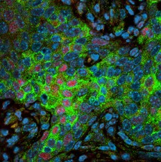
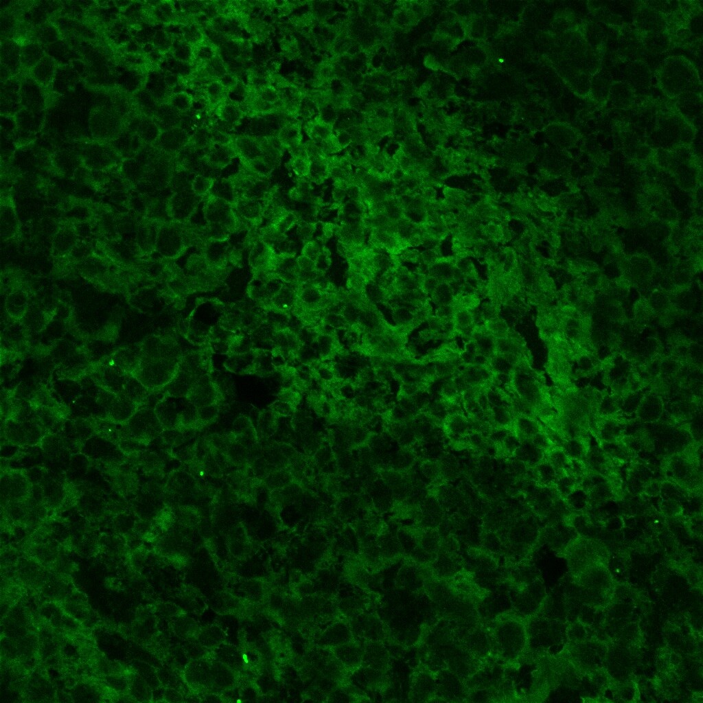
Detection of Human PD-L1 by Immunocytochemistry/ Immunofluorescence
Human pancreas tissue from type 1 diabetic subjects express PD-L1. Human pancreas sections from type 1 diabetic (T1D), autoantibody positive (AA+), type 2 diabetic (T2D), and non-diabetic controls (NDB) were obtained from the Network for Pancreatic Organ Donation (nPOD) and stained for T cell markers (CD4, CD8), insulin, and PD-L1. Shown are representative islets from each group with 7–15 unique islets analyzed from three independent experiments with one patient per group. Scale bar corresponds to 20 µm. Image collected and cropped by CiteAb from the following open publication (https://pubmed.ncbi.nlm.nih.gov/29844327), licensed under a CC-BY license. Not internally tested by R&D Systems.Applications for Mouse PD-L1/B7-H1 Antibody
Application
Recommended Usage
CyTOF-ready
Ready to be labeled using established conjugation methods. No BSA or other carrier proteins that could interfere with conjugation.
Dual RNAscope ISH-IHC Compatible
5-15 µg/mL
Sample: Immersion fixed paraffin-embedded sections of mouse intestine
Sample: Immersion fixed paraffin-embedded sections of mouse intestine
Flow Cytometry
0.25 µg/106 cells
Sample: RAW 264.7 mouse monocyte/macrophage cell line treated with LPS and HEK293 human embryonic kidney cell line transfected with mouse B7-H1/PD-L1
Sample: RAW 264.7 mouse monocyte/macrophage cell line treated with LPS and HEK293 human embryonic kidney cell line transfected with mouse B7-H1/PD-L1
Immunohistochemistry
5-15 µg/mL
Sample: Perfusion fixed frozen sections of mouse small intestine (Peyer's patch) and thymus
Sample: Perfusion fixed frozen sections of mouse small intestine (Peyer's patch) and thymus
Western Blot
0.5 µg/mL
Sample: RAW 264.7 mouse monocyte/macrophage cell line treated with LPS
Sample: RAW 264.7 mouse monocyte/macrophage cell line treated with LPS
Reviewed Applications
Read 13 reviews rated 4.5 using AF1019 in the following applications:
Flow Cytometry Panel Builder
Bio-Techne Knows Flow Cytometry
Save time and reduce costly mistakes by quickly finding compatible reagents using the Panel Builder Tool.
Advanced Features
- Spectra Viewer - Custom analysis of spectra from multiple fluorochromes
- Spillover Popups - Visualize the spectra of individual fluorochromes
- Antigen Density Selector - Match fluorochrome brightness with antigen density

Formulation, Preparation, and Storage
Purification
Antigen Affinity-purified
Reconstitution
Reconstitute at 0.2 mg/mL in sterile PBS. For liquid material, refer to CoA for concentration.
Loading...
Formulation
Lyophilized from a 0.2 μm filtered solution in PBS with Trehalose. See Certificate of Analysis for details.
*Small pack size (-SP) is supplied either lyophilized or as a 0.2 µm filtered solution in PBS.
*Small pack size (-SP) is supplied either lyophilized or as a 0.2 µm filtered solution in PBS.
Shipping
Lyophilized product is shipped at ambient temperature. Liquid small pack size (-SP) is shipped with polar packs. Upon receipt, store immediately at the temperature recommended below.
Stability & Storage
Use a manual defrost freezer and avoid repeated freeze-thaw cycles.
- 12 months from date of receipt, -20 to -70 °C as supplied.
- 1 month, 2 to 8 °C under sterile conditions after reconstitution.
- 6 months, -20 to -70 °C under sterile conditions after reconstitution.
Calculators
Background: PD-L1/B7-H1
References
- Tamura, H. et al. (2001) Blood 97:1809.
- Freeman, G. et al. (2000) J. Exp. Med. 192:1027.
- Sharpe, A.H. and G. J. Freeman (2002) Nat. Rev. Immunol. 2:116.
- Coyle, A. and J. Gutierrez-Ramos (2001) Nat. Immunol. 2:203.
Long Name
Programmed Death Ligand 1
Alternate Names
B7-H1, B7H1, CD274, PDCD1L1, PDCD1LG1, PDL1
Entrez Gene IDs
Gene Symbol
CD274
UniProt
Additional PD-L1/B7-H1 Products
Product Documents for Mouse PD-L1/B7-H1 Antibody
Certificate of Analysis
To download a Certificate of Analysis, please enter a lot or batch number in the search box below.
Note: Certificate of Analysis not available for kit components.
Product Specific Notices for Mouse PD-L1/B7-H1 Antibody
For research use only
Citations for Mouse PD-L1/B7-H1 Antibody
 Powered by Bioz
Customer Reviews for Mouse PD-L1/B7-H1 Antibody (13)
4.5 out of 5
13 Customer Ratings
Have you used Mouse PD-L1/B7-H1 Antibody?
Submit a review and receive an Amazon gift card!
$25/€18/£15/$25CAN/¥2500 Yen for a review with an image
$10/€7/£6/$10CAN/¥1110 Yen for a review without an image
Submit a review
Customer Images


Showing
1
-
5 of
13 reviews
Showing All
Filter By:
-
Application: ImmunohistochemistrySample Tested: Breast cancer tissueSpecies: MouseVerified Customer | Posted 07/31/2020

-
Application: Western BlotSample Tested: BT549 and input sample type hereSpecies: HumanVerified Customer | Posted 04/04/2020
-
Application: Immunohistochemistry-ParaffinSample Tested: Breast cancer tissueSpecies: MouseVerified Customer | Posted 04/03/2020
-
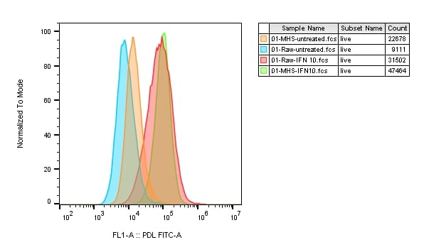
Application: Flow CytometrySample Tested: RAW 264.7 mouse monocyte/macrophage cell line and MH-S mouse macrophage cell lineSpecies: MouseVerified Customer | Posted 01/21/2020MH-S and RAW264.7 cells were treated with 10ug/ml concentration of IFN alpha. Cells were analyzed by flow cytometer (surface staining) for PD-L1 expression using a labelled secondary antibody. Protocol was followed as given with the article. Worked exactly as expected.
-
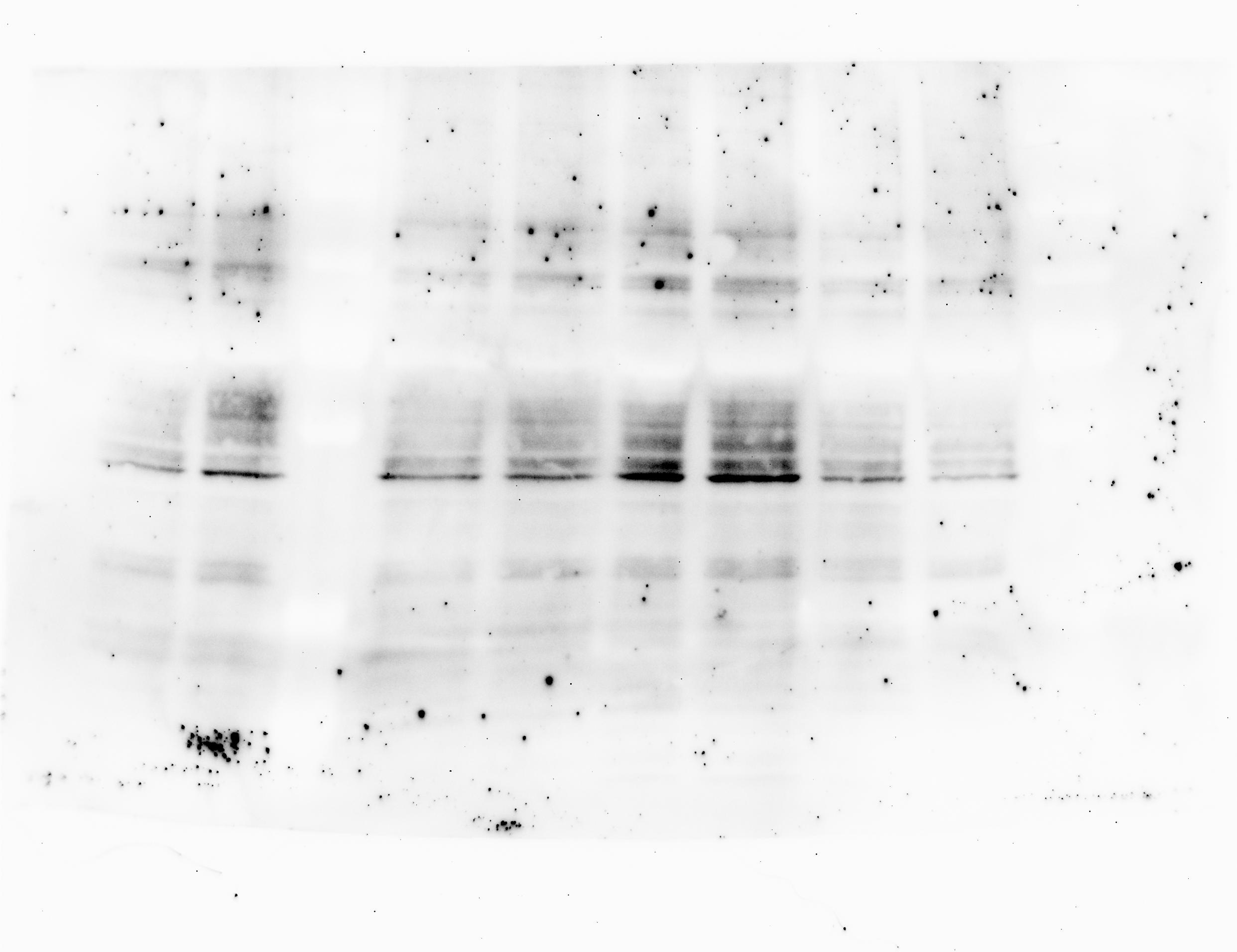
Application: Western BlotSample Tested: Cell LysatesSpecies: MouseVerified Customer | Posted 12/31/2019MH-S mice macrophage cell lysates. Dark bands in the middle are PD-L1
-
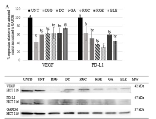
Application: Western BlotSample Tested: HCT-116 human colorectal carcinoma cell lineSpecies: HumanVerified Customer | Posted 03/20/2019
-
Application: Western BlotSample Tested: mouse beast cancer cell line Eo771Species: MouseVerified Customer | Posted 02/28/2019
-
Application: Western BlotSample Tested: EMT6 cells and 4T1 mouse breast cancer cell lineSpecies: MouseVerified Customer | Posted 10/28/2018
-
Application: ImmunohistochemistrySample Tested: frozen sectionSpecies: MouseVerified Customer | Posted 03/22/2018
-
Application: Western BlotSample Tested: cultured mouse mammary gland tumor and mouse mammary gland tumor cellsSpecies: MouseVerified Customer | Posted 11/26/2017
Bio-Techne ResponseTechnical Service will be following up with the customer.
-
Application: ImmunohistochemistrySample Tested: Pancreas tissueSpecies: MouseVerified Customer | Posted 10/17/2017
-
Application: Western BlotSample Tested: Mouse lymphocytesSpecies: MouseVerified Customer | Posted 10/26/2015Buffer: tbst<br />Dilution: 1/1000
-
Application: Western BlotSample Tested: See PMID 23593203Species: MouseVerified Customer | Posted 01/05/2015
There are no reviews that match your criteria.
Protocols
Find general support by application which include: protocols, troubleshooting, illustrated assays, videos and webinars.
- 7-Amino Actinomycin D (7-AAD) Cell Viability Flow Cytometry Protocol
- Antigen Retrieval Protocol (PIER)
- Antigen Retrieval for Frozen Sections Protocol
- Appropriate Fixation of IHC/ICC Samples
- Cellular Response to Hypoxia Protocols
- Chromogenic IHC Staining of Formalin-Fixed Paraffin-Embedded (FFPE) Tissue Protocol
- Chromogenic Immunohistochemistry Staining of Frozen Tissue
- ClariTSA™ Fluorophore Kits
- Detection & Visualization of Antibody Binding
- Extracellular Membrane Flow Cytometry Protocol
- Flow Cytometry Protocol for Cell Surface Markers
- Flow Cytometry Protocol for Staining Membrane Associated Proteins
- Flow Cytometry Staining Protocols
- Flow Cytometry Troubleshooting Guide
- Fluorescent IHC Staining of Frozen Tissue Protocol
- Graphic Protocol for Heat-induced Epitope Retrieval
- Graphic Protocol for the Preparation and Fluorescent IHC Staining of Frozen Tissue Sections
- Graphic Protocol for the Preparation and Fluorescent IHC Staining of Paraffin-embedded Tissue Sections
- Graphic Protocol for the Preparation of Gelatin-coated Slides for Histological Tissue Sections
- IHC Sample Preparation (Frozen sections vs Paraffin)
- ISH-IHC Protocol for Chromogenic Detection on Formalin Fixed Paraffin Embedded (FFPE) Tissue
- Immunofluorescent IHC Staining of Formalin-Fixed Paraffin-Embedded (FFPE) Tissue Protocol
- Immunohistochemistry (IHC) and Immunocytochemistry (ICC) Protocols
- Immunohistochemistry Frozen Troubleshooting
- Immunohistochemistry Paraffin Troubleshooting
- Intracellular Flow Cytometry Protocol Using Alcohol (Methanol)
- Intracellular Flow Cytometry Protocol Using Detergents
- Intracellular Nuclear Staining Flow Cytometry Protocol Using Detergents
- Intracellular Staining Flow Cytometry Protocol Using Alcohol Permeabilization
- Intracellular Staining Flow Cytometry Protocol Using Detergents to Permeabilize Cells
- Preparing Samples for IHC/ICC Experiments
- Preventing Non-Specific Staining (Non-Specific Binding)
- Primary Antibody Selection & Optimization
- Propidium Iodide Cell Viability Flow Cytometry Protocol
- Protocol for Heat-Induced Epitope Retrieval (HIER)
- Protocol for Liperfluo
- Protocol for Making a 4% Formaldehyde Solution in PBS
- Protocol for VisUCyte™ HRP Polymer Detection Reagent
- Protocol for the Characterization of Human Th22 Cells
- Protocol for the Characterization of Human Th9 Cells
- Protocol for the Preparation & Fixation of Cells on Coverslips
- Protocol for the Preparation and Chromogenic IHC Staining of Frozen Tissue Sections
- Protocol for the Preparation and Chromogenic IHC Staining of Frozen Tissue Sections - Graphic
- Protocol for the Preparation and Chromogenic IHC Staining of Paraffin-embedded Tissue Sections
- Protocol for the Preparation and Chromogenic IHC Staining of Paraffin-embedded Tissue Sections - Graphic
- Protocol for the Preparation and Fluorescent IHC Staining of Frozen Tissue Sections
- Protocol for the Preparation and Fluorescent IHC Staining of Paraffin-embedded Tissue Sections
- Protocol for the Preparation of Gelatin-coated Slides for Histological Tissue Sections
- Protocol: Annexin V and PI Staining by Flow Cytometry
- Protocol: Annexin V and PI Staining for Apoptosis by Flow Cytometry
- R&D Systems Quality Control Western Blot Protocol
- TUNEL and Active Caspase-3 Detection by IHC/ICC Protocol
- The Importance of IHC/ICC Controls
- Troubleshooting Guide: Fluorokine Flow Cytometry Kits
- Troubleshooting Guide: Immunohistochemistry
- Troubleshooting Guide: Western Blot Figures
- Western Blot Conditions
- Western Blot Protocol
- Western Blot Protocol for Cell Lysates
- Western Blot Troubleshooting
- Western Blot Troubleshooting Guide
- View all Protocols, Troubleshooting, Illustrated assays and Webinars
Loading...