TRANCE (receptor activator of NF-kappa B ligand [RANK L], also called TNF-related activation-induced cytokines, osteoprotegerin ligand [OPGL], and osteoclast differentiation factor [ODF]), is a member of the tumor necrosis factor (TNF) family. In the TNF superfamily nomenclature, TRANCE is referred to as TNFSF11. TRANCE was originally identified as an immediate early gene upregulated by T cell receptor stimulation. The murine TRANCE cDNA encodes a type II transmembrane protein of 316 amino acids with a predicted cytoplasmic domain of 48 amino acids and an extracellular domain of 247 amino acids. The extracellular domain contains two potential N-linked glycosylation sites. Mouse and human TRANCE share 85% amino acid identity. TRANCE is primarily expressed in T cells and T cell rich organs, such as thymus and lymph nodes. The multi-functions of TRANCE include induction of activation of the c-jun N-terminal kinase, enhancement of T cell growth and dendritic cell function, induction of osteoclastogenesis, and lymph node organogenesis. RANK is the cell surface signaling receptor of TRANCE. RANK has been shown to undergo receptor clustering during signal transduction. Osteoprotegerin, a soluble member of the TNF receptor family which binds TRANCE, is a naturally occurring decoy receptor that counterbalances the effects of TRANCE.
Mouse TRANCE/TNFSF11/RANK L Antibody
R&D Systems | Catalog # AF462

Key Product Details
Validated by
Biological Validation
Species Reactivity
Validated:
Mouse
Cited:
Human, Mouse, Transgenic Mouse
Applications
Validated:
Immunohistochemistry, Western Blot, ELISA Capture (Matched Antibody Pair), Neutralization
Cited:
Immunohistochemistry, Immunohistochemistry-Paraffin, Western Blot, Neutralization, Immunocytochemistry, ELISA Development, Proximity Ligation Assay
Label
Unconjugated
Antibody Source
Polyclonal Goat IgG
Loading...
Product Specifications
Immunogen
Mouse myeloma cell line NS0-derived recombinant mouse TRANCE/TNFSF11/RANK L
Arg72-Asp316
Accession # O35235
Arg72-Asp316
Accession # O35235
Specificity
Detects mouse TRANCE/TNFSF11/RANK L indirect ELISAs and Western blots. In direct ELISA's, less than 5% cross-reactivity with recombinant mouse (rm)CD27, rmCD40, and rmFas is observed and less than 1% cross-reactivity with rmCD30, recombinant human (rh)DR6, rmGITR, rmTNF RI, and rmTNF RII is observed.
Clonality
Polyclonal
Host
Goat
Isotype
IgG
Endotoxin Level
<0.10 EU per 1 μg of the antibody by the LAL method.
Scientific Data Images for Mouse TRANCE/TNFSF11/RANK L Antibody
Osteoclast-like Cell Formation Induced by TRANCE/ TNFSF11/RANK L and Neutralization by Mouse TRANCE/TNFSF11/RANK L Antibody.
In the presence of Recombinant Mouse M-CSF (20 ng/mL, 416-ML), Recombinant Mouse TRANCE/TNFSF11/RANK L (462-TR) induces osteoclast-like cell formation in RAW 264.7 cells in a dose-dependent manner (orange line), as measured by TRAP (tartrate-resistant acid phosphatase) solution assay. Under these conditions, osteoclast-like cell formation elicited by Recombinant Mouse TRANCE/TNFSF11/ RANK L (30 ng/mL) is neutralized (green line) by increasing concentrations of Goat Anti-Mouse TRANCE/TNFSF11/RANK L Antigen Affinity-purified Polyclonal Antibody (Catalog # AF462). The ND50 is typicallyDetection of Mouse TRANCE/TNFSF11/RANK L by Immunohistochemistry-Paraffin
MMP2, MMP9, and RANKL expression in AAAs.A. Immunohistochemical staining for indicated proteins of serial sections of aortas one week after CaCl2 treatment. Elastic van Gieson staining is also shown. SM alpha -actin and F4/80 were stained to locate SMCs and macrophages, respectively. Shown are representative images of 4 or more samples in each group. Scale bars, 50 µm. B. Relative positive staining area of MMP2, MMP9, and RANKL in sections from control diet and EPA diet groups. n = 4–5. *P<0.05. Image collected and cropped by CiteAb from the following open publication (https://dx.plos.org/10.1371/journal.pone.0096286), licensed under a CC-BY license. Not internally tested by R&D Systems.Detection of TRANCE/TNFSF11/RANK L by Western Blot
Tnfaip3 I325N mice exhibit impaired mammary development, elevated non-canonical RankL signalling and dampened non-canonical NF-kappa B signalling. (A) Mammary gland whole mounts of the 3rd and 4th mammary gland from 14-week virgin Tnfaip3I325N mice of indicated genotype. * denotes empty fat pad space. LN = lymph node. (B) Representative H&E of mammary gland from does 8.5 days post-partum, and (C) pup weight of resulting pairs, normalised to litter size. (D, E) Quantified positive immunohistochemistry staining for (D) progesterone receptor (PR) or (E) estrogen receptor (ER) (n = 9 Tnfaip3+/+ and 9 Tnfaip3I325N/I325N). (F) 4th Mammary gland from 10-week-old mice were collected and lysed for immunoblot assessment of paracrine hormones OPG and RANKL with Beta-actin used as the loading control.* denotes non-specific band and arrow head denotes specific band. Molecular weight in Kilodaltons is shown to the right of each blot. (G–I) 4th Mammary gland and its lymph node (J–L) from 10-week-old mice were collected for immunoblot (G, J) and densitometry analysis (H, I, K, L) for non-canonical NF-kappa B components NIK, p100/p52 and RelB. Immunoblots are representative of 2 independent experiments with n=6 (H, I) or n = 3 (K, L) biological replicates for both WT and HOM donors. Densitometry values were normalised to the average WT value of each blot to allow cumulative quantification. Error bars represent s.e.m and Student’s T test used for significance analysis * = P < 0.05. Image collected and cropped by CiteAb from the following open publication (https://pubmed.ncbi.nlm.nih.gov/35464428), licensed under a CC-BY license. Not internally tested by R&D Systems.Immersion fixed paraffin-embedded sections of mouse spleen
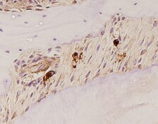
TRANCE/TNFSF11/RANK L was detected in immersion fixed paraffin-embedded sections of mouse spleen using Goat Anti-Mouse TRANCE/TNFSF11/RANK L Antigen Affinity-purified Polyclonal Antibody (Catalog # AF462) at 0.25 µg/ml overnight at 4 °C. Before incubation with the primary antibody, tissue was subjected to heat-induced epitope retrieval using VisUCyte Antigen Retrieval Reagent-Basic (VCTS021). Tissue was stained using the HRP-conjugated Anti-Goat IgG Secondary Antibody (HAF109) and counterstained with hematoxylin (blue). Specific staining was localized to the membrane. View our protocol for Chromogenic IHC Staining of Paraffin-embedded Tissue Sections.Applications for Mouse TRANCE/TNFSF11/RANK L Antibody
Application
Recommended Usage
Immunohistochemistry
0.25-25 µg/mL
Sample: Immersion fixed paraffin-embedded sections of mouse spleen
Sample: Immersion fixed paraffin-embedded sections of mouse spleen
Western Blot
0.1 µg/mL
Sample: Recombinant Mouse TRANCE/TNFSF11/RANK L (Catalog # 462-TR)
Sample: Recombinant Mouse TRANCE/TNFSF11/RANK L (Catalog # 462-TR)
Neutralization
Measured by its ability to neutralize TRANCE/TNFSF11/RANK L-induced osteoclast-like cell formation in RAW 264.7 cells. Nakagawa, N. et al. (1998) Biochim. Biophys. Res. Commun. 253:395. The Neutralization Dose (ND50) is typically <0.500 µg/mL in the presence of 30 ng/mL Recombinant Mouse TRANCE/TNFSF11/RANK L.
Mouse TRANCE/TNFSF11/RANK L Sandwich Immunoassay
Please Note: Optimal dilutions of this antibody should be experimentally determined.
Reviewed Applications
Read 2 reviews rated 5 using AF462 in the following applications:
Formulation, Preparation, and Storage
Purification
Antigen Affinity-purified
Reconstitution
Reconstitute at 0.2 mg/mL in sterile PBS. For liquid material, refer to CoA for concentration.
Loading...
Formulation
Lyophilized from a 0.2 μm filtered solution in PBS with Trehalose. See Certificate of Analysis for details.
*Small pack size (-SP) is supplied either lyophilized or as a 0.2 µm filtered solution in PBS.
*Small pack size (-SP) is supplied either lyophilized or as a 0.2 µm filtered solution in PBS.
Shipping
Lyophilized product is shipped at ambient temperature. Liquid small pack size (-SP) is shipped with polar packs. Upon receipt, store immediately at the temperature recommended below.
Stability & Storage
Use a manual defrost freezer and avoid repeated freeze-thaw cycles.
- 12 months from date of receipt, -20 to -70 °C as supplied.
- 1 month, 2 to 8 °C under sterile conditions after reconstitution.
- 6 months, -20 to -70 °C under sterile conditions after reconstitution.
Calculators
Background: TRANCE/TNFSF11/RANK L
References
- Wong, B.R. et al. (1997) J. Biol. Chem. 272:25190.
- Anderson, D.M. et al. (1997) Nature 390:175.
- Nakagawa, N. et al. (1998) Biochem. Biophys. Res. Commun. 245:382.
- Kong, Y-Y. et al. (1999) Nature 397:315.
Long Name
TNF-related Activation-induced Cytokine
Alternate Names
CD254, ODF, OPGL, RANK L, RANKL, TNFSF11
Gene Symbol
TNFSF11
UniProt
Additional TRANCE/TNFSF11/RANK L Products
Product Documents for Mouse TRANCE/TNFSF11/RANK L Antibody
Certificate of Analysis
To download a Certificate of Analysis, please enter a lot or batch number in the search box below.
Note: Certificate of Analysis not available for kit components.
Product Specific Notices for Mouse TRANCE/TNFSF11/RANK L Antibody
For research use only
Related Research Areas
Citations for Mouse TRANCE/TNFSF11/RANK L Antibody
 Powered by Bioz
Customer Reviews for Mouse TRANCE/TNFSF11/RANK L Antibody (2)
5 out of 5
2 Customer Ratings
Have you used Mouse TRANCE/TNFSF11/RANK L Antibody?
Submit a review and receive an Amazon gift card!
$25/€18/£15/$25CAN/¥2500 Yen for a review with an image
$10/€7/£6/$10CAN/¥1110 Yen for a review without an image
Submit a review
Customer Images
Showing
1
-
2 of
2 reviews
Showing All
Filter By:
-
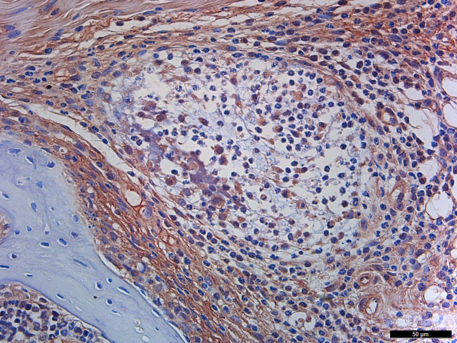
Application: ImmunohistochemistrySample Tested: Cartilage tissueSpecies: MouseVerified Customer | Posted 03/13/2023doi.org/10.3390/ijms24021137

-
Application: Immunocytochemistry/ImmunofluorescenceSample Tested: BoneSpecies: MouseVerified Customer | Posted 07/06/2021
There are no reviews that match your criteria.
Protocols
Find general support by application which include: protocols, troubleshooting, illustrated assays, videos and webinars.
- Antigen Retrieval Protocol (PIER)
- Antigen Retrieval for Frozen Sections Protocol
- Appropriate Fixation of IHC/ICC Samples
- Cellular Response to Hypoxia Protocols
- Chromogenic IHC Staining of Formalin-Fixed Paraffin-Embedded (FFPE) Tissue Protocol
- Chromogenic Immunohistochemistry Staining of Frozen Tissue
- ClariTSA™ Fluorophore Kits
- Detection & Visualization of Antibody Binding
- Fluorescent IHC Staining of Frozen Tissue Protocol
- Graphic Protocol for Heat-induced Epitope Retrieval
- Graphic Protocol for the Preparation and Fluorescent IHC Staining of Frozen Tissue Sections
- Graphic Protocol for the Preparation and Fluorescent IHC Staining of Paraffin-embedded Tissue Sections
- Graphic Protocol for the Preparation of Gelatin-coated Slides for Histological Tissue Sections
- IHC Sample Preparation (Frozen sections vs Paraffin)
- Immunofluorescent IHC Staining of Formalin-Fixed Paraffin-Embedded (FFPE) Tissue Protocol
- Immunohistochemistry (IHC) and Immunocytochemistry (ICC) Protocols
- Immunohistochemistry Frozen Troubleshooting
- Immunohistochemistry Paraffin Troubleshooting
- Preparing Samples for IHC/ICC Experiments
- Preventing Non-Specific Staining (Non-Specific Binding)
- Primary Antibody Selection & Optimization
- Protocol for Heat-Induced Epitope Retrieval (HIER)
- Protocol for Making a 4% Formaldehyde Solution in PBS
- Protocol for VisUCyte™ HRP Polymer Detection Reagent
- Protocol for the Preparation & Fixation of Cells on Coverslips
- Protocol for the Preparation and Chromogenic IHC Staining of Frozen Tissue Sections
- Protocol for the Preparation and Chromogenic IHC Staining of Frozen Tissue Sections - Graphic
- Protocol for the Preparation and Chromogenic IHC Staining of Paraffin-embedded Tissue Sections
- Protocol for the Preparation and Chromogenic IHC Staining of Paraffin-embedded Tissue Sections - Graphic
- Protocol for the Preparation and Fluorescent IHC Staining of Frozen Tissue Sections
- Protocol for the Preparation and Fluorescent IHC Staining of Paraffin-embedded Tissue Sections
- Protocol for the Preparation of Gelatin-coated Slides for Histological Tissue Sections
- R&D Systems Quality Control Western Blot Protocol
- TUNEL and Active Caspase-3 Detection by IHC/ICC Protocol
- The Importance of IHC/ICC Controls
- Troubleshooting Guide: Immunohistochemistry
- Troubleshooting Guide: Western Blot Figures
- Western Blot Conditions
- Western Blot Protocol
- Western Blot Protocol for Cell Lysates
- Western Blot Troubleshooting
- Western Blot Troubleshooting Guide
- View all Protocols, Troubleshooting, Illustrated assays and Webinars
Loading...
Associated Pathways
