3-Nitrotyrosine is formed when tyrosine is reacted with peroxynitrite. Since peroxynitrite is formed from nitric oxide and superoxide anion, nitrotyrosine adducts on proteins have been used as markers of oxidative cellular damage and macrophage activation. Elevated nitrotyrosine immunoreactivity has been found in inflammation, osteoarthritis, neurodegenerative diseases, and ischemic damage to the heart and brain.
Nitrotyrosine Antibody (306507)
R&D Systems | Catalog # MAB3248

Key Product Details
Validated by
Biological Validation
Species Reactivity
Validated:
Multi-Species
Cited:
Human, Mouse, Rat
Applications
Validated:
Western Blot, Immunocytochemistry
Cited:
Immunohistochemistry, Immunohistochemistry-Frozen, Western Blot, Immunocytochemistry, ELISA Capture
Label
Unconjugated
Antibody Source
Monoclonal Mouse IgG3 Clone # 306507
Loading...
Product Specifications
Immunogen
Nitrotyrosine-modified KLH
Specificity
Detects Nitrotyrosine adducts on proteins in Western blots. It does not cross-react with phosphotyrosine or 4‑hydroxynonenal adducts. Unfixed cells, tissues, and proteins can be treated with Peroxynitrite (Catalog # AR006) for use as positive controls with this antibody.
Clonality
Monoclonal
Host
Mouse
Isotype
IgG3
Scientific Data Images for Nitrotyrosine Antibody (306507)
Detection of Nitrotyrosine by Western Blot.
Western blot shows lysates of NIH-3T3 mouse embryonic fibroblast cell line untreated (-) or treated (+) with 3 mM peroxynitrite, 3 mM inactivated peroxynitrite, or 100 µM peroxyvanadate for 1 hour and recombinantE. coliDNAK treated with 1 mM 4-hydroxynonenal or 1 mM peroxyynitrite for 1 hour. PVDF membrane was probed with 1 µg/mL of Mouse Anti-Nitrotyrosine Monoclonal Antibody (Catalog # MAB3248), followed by HRP-conjugated Anti-Mouse IgG Secondary Antibody (HAF007). The lysates were also probed with Phospho-Tyrosine Monoclonal Antibody (MAB1676). This experiment was conducted under reducing conditions and using Western Blot Buffer Group 1.
Nitrotyrosine in Human PBMCs.
Nitrotyrosine was detected in immersion fixed human peripheral blood mononuclear cells (PBMCs) using 25 µg/mL Mouse Anti-Nitrotyrosine Monoclonal Antibody (Catalog # MAB3248) for 3 hours at room temperature. Cells were stained with the NorthernLights™ 557-conjugated Anti-Mouse IgG Secondary Antibody (red; NL007) and counterstained (green). View our protocol for Fluorescent ICC Staining of Non-adherent Cells.
Detection of Nitrotyrosine in A549 human lung carcinoma cells
Nitrotyrosine was detected in immersion fixed A549 human lung carcinoma cells using Mouse Anti-Nitrotyrosine Monoclonal Antibody (Catalog # MAB3248) at 8 µg/mL for 3 hours at room temperature. Cells were stained using the NorthernLights™ 557-conjugated Anti-Mouse IgG Secondary Antibody (red; Catalog # NL007) and counterstained with DAPI (blue). Specific staining was localized to Cytoplasmic. View our protocol for Fluorescent ICC Staining of Cells on Coverslips.
Detection of Nitrotyrosine in HepG2 cells
Nitrotyrosine was detected in immersion fixed HepG2 human hepatocellular carcinoma cells using Mouse Anti-Nitrotyrosine Monoclonal Antibody (Catalog # MAB3248) at 8 µg/mL for 3 hours at room temperature. Cells were stained using the NorthernLights™ 557-conjugated Anti-Mouse IgG Secondary Antibody (red; Catalog # NL007) and counterstained with DAPI (blue). Specific staining was localized to Cytoplasmic. View our protocol for Fluorescent ICC Staining of Cells on Coverslips.Applications for Nitrotyrosine Antibody (306507)
Application
Recommended Usage
Immunocytochemistry
8-25 µg/mL
Sample: Immersion fixed human peripheral blood mononuclear cells (PBMCs), A549 human lung carcinoma cells (Positive)immersion fixed HepG2 human hepatocellular carcinoma cells (Positive)
Sample: Immersion fixed human peripheral blood mononuclear cells (PBMCs), A549 human lung carcinoma cells (Positive)immersion fixed HepG2 human hepatocellular carcinoma cells (Positive)
Western Blot
1 µg/mL
Sample: Peroxynitrite or peroxyvanadate-treated NIH‑3T3 mouse embryonic fibroblast cell line
Sample: Peroxynitrite or peroxyvanadate-treated NIH‑3T3 mouse embryonic fibroblast cell line
Reviewed Applications
Read 1 review rated 4 using MAB3248 in the following applications:
Formulation, Preparation, and Storage
Purification
Protein A or G purified from hybridoma culture supernatant
Reconstitution
Reconstitute at 0.5 mg/mL in sterile PBS. For liquid material, refer to CoA for concentration.
Loading...
Formulation
Lyophilized from a 0.2 μm filtered solution in TBS with Trehalose. See Certificate of Analysis for details.
*Small pack size (-SP) is supplied either lyophilized or as a 0.2 µm filtered solution in PBS.
*Small pack size (-SP) is supplied either lyophilized or as a 0.2 µm filtered solution in PBS.
Shipping
Lyophilized product is shipped at ambient temperature. Liquid small pack size (-SP) is shipped with polar packs. Upon receipt, store immediately at the temperature recommended below.
Stability & Storage
Use a manual defrost freezer and avoid repeated freeze-thaw cycles.
- 12 months from date of receipt, -20 to -70 °C as supplied.
- 1 month, 2 to 8 °C under sterile conditions after reconstitution.
- 6 months, -20 to -70 °C under sterile conditions after reconstitution.
Calculators
Background: Nitrotyrosine
Alternate Names
Nitrotyrosine
Additional Nitrotyrosine Products
Product Documents for Nitrotyrosine Antibody (306507)
Certificate of Analysis
To download a Certificate of Analysis, please enter a lot or batch number in the search box below.
Note: Certificate of Analysis not available for kit components.
Product Specific Notices for Nitrotyrosine Antibody (306507)
For research use only
Related Research Areas
Citations for Nitrotyrosine Antibody (306507)
 Powered by Bioz
Customer Reviews for Nitrotyrosine Antibody (306507) (1)
4 out of 5
1 Customer Rating
Have you used Nitrotyrosine Antibody (306507)?
Submit a review and receive an Amazon gift card!
$25/€18/£15/$25CAN/¥2500 Yen for a review with an image
$10/€7/£6/$10CAN/¥1110 Yen for a review without an image
Submit a review
Customer Images
Showing
1
-
1 of
1 review
Showing All
Filter By:
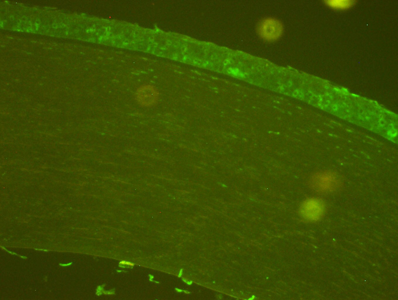
-
Application: Immunohistochemistry-ParaffinSample Tested: human corneaSpecies: HumanVerified Customer | Posted 10/11/2017Corneal section stained with anti-nitrotyrosine antibody (1:100) followed by alexa fluor 488.

There are no reviews that match your criteria.
Protocols
Find general support by application which include: protocols, troubleshooting, illustrated assays, videos and webinars.
- Appropriate Fixation of IHC/ICC Samples
- Cellular Response to Hypoxia Protocols
- ClariTSA™ Fluorophore Kits
- Detection & Visualization of Antibody Binding
- ICC Cell Smear Protocol for Suspension Cells
- ICC Immunocytochemistry Protocol Videos
- ICC for Adherent Cells
- Immunocytochemistry (ICC) Protocol
- Immunocytochemistry Troubleshooting
- Immunofluorescence of Organoids Embedded in Cultrex Basement Membrane Extract
- Immunohistochemistry (IHC) and Immunocytochemistry (ICC) Protocols
- Preparing Samples for IHC/ICC Experiments
- Preventing Non-Specific Staining (Non-Specific Binding)
- Primary Antibody Selection & Optimization
- Protocol for VisUCyte™ HRP Polymer Detection Reagent
- Protocol for the Fluorescent ICC Staining of Cell Smears - Graphic
- Protocol for the Fluorescent ICC Staining of Cultured Cells on Coverslips - Graphic
- Protocol for the Preparation and Fluorescent ICC Staining of Cells on Coverslips
- Protocol for the Preparation and Fluorescent ICC Staining of Non-adherent Cells
- Protocol for the Preparation and Fluorescent ICC Staining of Stem Cells on Coverslips
- Protocol for the Preparation of a Cell Smear for Non-adherent Cell ICC - Graphic
- R&D Systems Quality Control Western Blot Protocol
- TUNEL and Active Caspase-3 Detection by IHC/ICC Protocol
- The Importance of IHC/ICC Controls
- Troubleshooting Guide: Western Blot Figures
- Western Blot Conditions
- Western Blot Protocol
- Western Blot Protocol for Cell Lysates
- Western Blot Troubleshooting
- Western Blot Troubleshooting Guide
- View all Protocols, Troubleshooting, Illustrated assays and Webinars
Loading...