Phospho-Tyrosine Antibody (179003)
R&D Systems | Catalog # MAB1676

Key Product Details
Validated by
Biological Validation
Species Reactivity
Validated:
Multi-Species
Cited:
Human, Transgenic Mouse
Applications
Validated:
Western Blot, Immunocytochemistry, Simple Western, Immunoprecipitation
Cited:
Immunohistochemistry, Western Blot
Label
Unconjugated
Antibody Source
Monoclonal Mouse IgG1 Clone # 179003
Loading...
Product Specifications
Immunogen
KLH-coupled phospho-tyrosine synthetic peptide
Specificity
Detects proteins containing phosphorylated tyrosine residues. ELISA and Western blot analyses using pervanadate-treated cell lysates indicate that clone 179003 binds phospho-tyrosine in a broad manner largely independent of the surrounding amino acid sequence.No cross‑reactivity with proteins or peptides containing phosphorylated serine or threonine residues is observed.
Clonality
Monoclonal
Host
Mouse
Isotype
IgG1
Scientific Data Images for Phospho-Tyrosine Antibody (179003)
Detection of Phospho-Tyrosine by Western Blot.
Western blot shows lysates of A431 human epithelial carcinoma cell line untreated (-) or treated (+) with 50 µM pervanadate (PV) for 15 minutes. PVDF membrane was probed with 1 µg/mL of Mouse Anti-Phospho-Tyrosine Monoclonal Antibody (Catalog # MAB1676), followed by HRP-conjugated Anti-Mouse IgG Secondary Antibody (HAF007). Tyrosine-phosphorylated proteins were detected (as indicated). This experiment was conducted under reducing conditions and using Immunoblot Buffer Group 10.
Detection of Human Phospho-Tyrosine by Simple WesternTM.
Simple Western lane view shows lysates of HUVEC human umbilical vein endothelial cells untreated (-) or treated (+) with 50 µM Pervanadate (PV) for 15 minutes, loaded at 0.2 mg/mL. Tyrosine-phosphorylated proteins were detected (as indicated) using 50 µg/mL of Mouse Anti-Phospho-Tyrosine Monoclonal Antibody (Catalog # MAB1676). This experiment was conducted under reducing conditions and using the 12-230 kDa separation system.
Phospho-Tyrosine in A431 Human Cell Line.
Phospho-Tyrosine was detected in immersion fixed A431 human epithelial carcinoma cell line treated with 50 ng/mL Recombinant Human EGF (left panel; 236-EG) or untreated (right panel) using Mouse Anti-Phospho-Tyrosine Monoclonal Antibody (Catalog # MAB1676) at 10 µg/mL for 3 hours at room temperature. Cells were stained using the NorthernLights™ 557-conjugated Anti-Mouse IgG Secondary Antibody (red; NL007) and counterstained with DAPI (blue). Specific staining was localized to the cytoplasm and cell surface. View our protocol for Fluorescent ICC Staining of Cells on Coverslips.
Detection of Tyrosine in A431 Human Cell Line.
Tyrosine was detected in Immersion fixed A431 human epithelial carcinoma cell line treated with 100μM Pervanadate (PV) for 10 minutes (left panel) or untreated (right panel) using Mouse Anti-Phospho-Tyrosine Monoclonal Antibody (Catalog # MAB1676) at 10 µg/ml for 3 hours at room temperature. Cells were stained using the NorthernLights™ 557-conjugated Anti-Mouse IgG Secondary Antibody (red; Catalog # NL007) and counterstained with DAPI (blue). Specific staining was localized to the cytoplasm of treated cells. View our protocol for Fluorescent ICC Staining of Cells on Coverslips.Applications for Phospho-Tyrosine Antibody (179003)
Application
Recommended Usage
Immunocytochemistry
8-25 µg/mL
Sample: Immersion fixed A431 human epithelial carcinoma cell line treated with recombinant human EGF (Catalog # 236-EG) or Pervanadate
Sample: Immersion fixed A431 human epithelial carcinoma cell line treated with recombinant human EGF (Catalog # 236-EG) or Pervanadate
Immunoprecipitation
5 µg/500 µg cell lysate
Sample: A431 human epithelial carcinoma cell line, see our available Western blot detection antibodies
Sample: A431 human epithelial carcinoma cell line, see our available Western blot detection antibodies
Simple Western
50 µg/mL
Sample: HUVEC human umbilical vein endothelial cells treated with Pervanadate (PV)
Sample: HUVEC human umbilical vein endothelial cells treated with Pervanadate (PV)
Western Blot
1 µg/mL
Sample: Pervanadate-treated A431 human epithelial carcinoma cell line
Sample: Pervanadate-treated A431 human epithelial carcinoma cell line
Reviewed Applications
Read 2 reviews rated 4 using MAB1676 in the following applications:
Formulation, Preparation, and Storage
Purification
Protein A or G purified from hybridoma culture supernatant
Reconstitution
Reconstitute at 0.5 mg/mL in sterile PBS. For liquid material, refer to CoA for concentration.
Loading...
Formulation
Lyophilized from a 0.2 μm filtered solution in PBS with Trehalose. See Certificate of Analysis for details.
*Small pack size (-SP) is supplied either lyophilized or as a 0.2 µm filtered solution in PBS.
*Small pack size (-SP) is supplied either lyophilized or as a 0.2 µm filtered solution in PBS.
Shipping
Lyophilized product is shipped at ambient temperature. Liquid small pack size (-SP) is shipped with polar packs. Upon receipt, store immediately at the temperature recommended below.
Stability & Storage
Use a manual defrost freezer and avoid repeated freeze-thaw cycles.
- 12 months from date of receipt, -20 to -70 °C as supplied.
- 1 month, 2 to 8 °C under sterile conditions after reconstitution.
- 6 months, -20 to -70 °C under sterile conditions after reconstitution.
Calculators
Background: Phospho-Tyrosine
Alternate Names
PhosphoTyrosine
Additional Phospho-Tyrosine Products
Product Documents for Phospho-Tyrosine Antibody (179003)
Certificate of Analysis
To download a Certificate of Analysis, please enter a lot or batch number in the search box below.
Note: Certificate of Analysis not available for kit components.
Product Specific Notices for Phospho-Tyrosine Antibody (179003)
For research use only
Related Research Areas
Citations for Phospho-Tyrosine Antibody (179003)
 Powered by Bioz
Customer Reviews for Phospho-Tyrosine Antibody (179003) (2)
4 out of 5
2 Customer Ratings
Have you used Phospho-Tyrosine Antibody (179003)?
Submit a review and receive an Amazon gift card!
$25/€18/£15/$25CAN/¥2500 Yen for a review with an image
$10/€7/£6/$10CAN/¥1110 Yen for a review without an image
Submit a review
Customer Images
Showing
1
-
2 of
2 reviews
Showing All
Filter By:
-
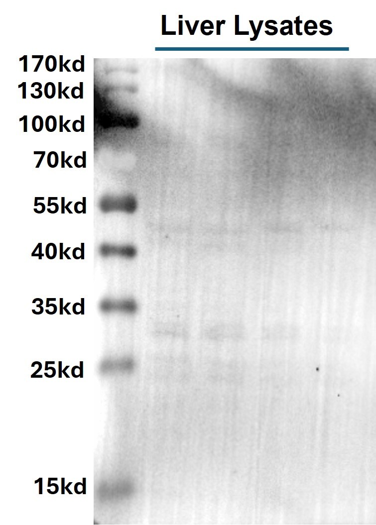
Application: Western BlotSample Tested: Liver tissueSpecies: MouseVerified Customer | Posted 12/10/2024WB_Phospho tyrosine - Liver tissueLiver tissue lysates were immunoblotted with the Phospho-Tyrosine Antibody at concentration of 1 ug/ml. Multiple bands of phosphorylated proteins were observed.

-
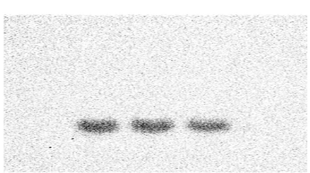
Application: ImmunoprecipitationSample Tested: 293T human embryonic kidney cell lineSpecies: HumanVerified Customer | Posted 06/28/2017Immunoprecipitation with antibody for target protein and Western Blotting detection of phospholation.
There are no reviews that match your criteria.
Protocols
Find general support by application which include: protocols, troubleshooting, illustrated assays, videos and webinars.
- Appropriate Fixation of IHC/ICC Samples
- Cellular Response to Hypoxia Protocols
- ClariTSA™ Fluorophore Kits
- Detection & Visualization of Antibody Binding
- ICC Cell Smear Protocol for Suspension Cells
- ICC Immunocytochemistry Protocol Videos
- ICC for Adherent Cells
- Immunocytochemistry (ICC) Protocol
- Immunocytochemistry Troubleshooting
- Immunofluorescence of Organoids Embedded in Cultrex Basement Membrane Extract
- Immunohistochemistry (IHC) and Immunocytochemistry (ICC) Protocols
- Immunoprecipitation Protocol
- Preparing Samples for IHC/ICC Experiments
- Preventing Non-Specific Staining (Non-Specific Binding)
- Primary Antibody Selection & Optimization
- Protocol for VisUCyte™ HRP Polymer Detection Reagent
- Protocol for the Fluorescent ICC Staining of Cell Smears - Graphic
- Protocol for the Fluorescent ICC Staining of Cultured Cells on Coverslips - Graphic
- Protocol for the Preparation and Fluorescent ICC Staining of Cells on Coverslips
- Protocol for the Preparation and Fluorescent ICC Staining of Non-adherent Cells
- Protocol for the Preparation and Fluorescent ICC Staining of Stem Cells on Coverslips
- Protocol for the Preparation of a Cell Smear for Non-adherent Cell ICC - Graphic
- R&D Systems Quality Control Western Blot Protocol
- TUNEL and Active Caspase-3 Detection by IHC/ICC Protocol
- The Importance of IHC/ICC Controls
- Troubleshooting Guide: Western Blot Figures
- Western Blot Conditions
- Western Blot Protocol
- Western Blot Protocol for Cell Lysates
- Western Blot Troubleshooting
- Western Blot Troubleshooting Guide
- View all Protocols, Troubleshooting, Illustrated assays and Webinars
Loading...