Human Annexin V Antibody (38701)
R&D Systems | Catalog # MAB3991

Key Product Details
Validated by
Knockout/Knockdown
Species Reactivity
Validated:
Human
Cited:
Human
Applications
Validated:
Knockout Validated, Immunohistochemistry, Western Blot, Simple Western
Cited:
Western Blot
Label
Unconjugated
Antibody Source
Monoclonal Mouse IgG2A Clone # 38701
Loading...
Product Specifications
Immunogen
E. coli-derived recombinant human Annexin V
Met1-Asp320
Accession # P08758
Met1-Asp320
Accession # P08758
Specificity
Detects human Annexin V in direct ELISAs.
Clonality
Monoclonal
Host
Mouse
Isotype
IgG2A
Scientific Data Images for Human Annexin V Antibody (38701)
Detection of Human Annexin V by Western Blot.
Western blot shows lysates of HepG2 human hepatocellular carcinoma cell line. PVDF membrane was probed with 0.25 µg/mL of Mouse Anti-Human Annexin V Monoclonal Antibody (Catalog # MAB3991) followed by HRP-conjugated Anti-Mouse IgG Secondary Antibody (Catalog # HAF018). A specific band was detected for Annexin V at approximately 32 kDa (as indicated). This experiment was conducted under reducing conditions and using Immunoblot Buffer Group 1.
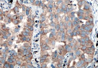
Annexin V in Human Placenta.
Annexin V was detected in immersion fixed paraffin-embedded sections of human placenta using Mouse Anti-Human Annexin V Monoclonal Antibody (Catalog # MAB3991) at 5 µg/mL for 1 hour at room temperature followed by incubation with the Anti-Mouse IgG VisUCyte™ HRP Polymer Antibody (Catalog # VC001). Tissue was stained using DAB (brown) and counterstained with hematoxylin (blue). Specific staining was localized to syncytiotrophoblasts. View our protocol for IHC Staining with VisUCyte HRP Polymer Detection Reagents.
Detection of Human Annexin V by Simple WesternTM.
Simple Western lane view shows lysates of human lung tissue, loaded at 0.2 mg/mL. A specific band was detected for Annexin V at approximately 39 kDa (as indicated) using 2.5 µg/mL of Mouse Anti-Human Annexin V Monoclonal Antibody (Catalog # MAB3991). This experiment was conducted under reducing conditions and using the 12-230 kDa separation system.
Detection of Human Annexin V by Simple WesternTM.
Simple Western lane view shows lysates of A549 human lung carcinoma cell line exosomes, HT‑29 human colon adenocarcinoma cell line exosomes, and COLO 205 human colorectal adenocarcinoma cell line whole cell lysate (WCL), loaded at 0.2 mg/mL. A specific band was detected for Annexin V at approximately 38 kDa (as indicated) using 50 µg/mL of Mouse Anti-Human Annexin V Monoclonal Antibody (Catalog # MAB3991). This experiment was conducted under reducing conditions and using the 12-230 kDa separation system.
Western Blot Shows Human Annexin V Specificity by Using Knockout Cell Line.
Western blot shows lysates of HEK293T human embryonic kidney parental cell line and Annexin V knockout HEK293T cell line (KO). PVDF membrane was probed with 0.25 µg/mL of Mouse Anti-Human Annexin V Monoclonal Antibody (Catalog # MAB3991) followed by HRP-conjugated Anti-Mouse IgG Secondary Antibody (Catalog # HAF018). A specific band was detected for Annexin V at approximately 32 kDa (as indicated) in the parental HEK293T cell line, but is not detectable in knockout HEK293T cell line. GAPDH (Catalog # MAB5718) is shown as a loading control. This experiment was conducted under reducing conditions and using Immunoblot Buffer Group 1.
Detection of Annexin V by Western Blot
Extracellular vesicle secretion induced by Sorafenib and their miRNA content. (A) Particle size and concentration analysis of Large, Small, and Very Small EVs obtained at 6 and 24 h. (B) Expression of EV markers and cellular contaminants in Large, Small, and Very Small EVs and cell lysates obtained at 24 h after Sorafenib treatment. Total lane protein content was used as the loading control. (C) Representative images of cryo-EM of Small and Very Small EVs obtained at 6 and 24 h from the control and Sorafenib-treated cells. (D) Assessment of EV size (nm) in the cryo-EM images. (E) Number of EVs quantified in the cryo-EM images. (F) miRNA expression in the three fractions of EVs at 6 h. (G) miRNA expression in the three fractions of EVs at 24 h. Fold-change values were calculated between Sorafenib and the control treated samples. Results are expressed as the mean ± SEM of six independent experiments. Ns, non-significant; * p ≤0.05, ** p ≤ 0.01, *** p ≤ 0.001, and **** p ≤ 0.0001 between the miRNA expression in the control and Sorafenib derived EVs. Multiple comparison test statistics are expressed with lower case a–i. Image collected and cropped by CiteAb from the following open publication (https://pubmed.ncbi.nlm.nih.gov/36078082), licensed under a CC-BY license. Not internally tested by R&D Systems.
Detection of Annexin V by Western Blot
Extracellular vesicle secretion induced by Sorafenib and their miRNA content. (A) Particle size and concentration analysis of Large, Small, and Very Small EVs obtained at 6 and 24 h. (B) Expression of EV markers and cellular contaminants in Large, Small, and Very Small EVs and cell lysates obtained at 24 h after Sorafenib treatment. Total lane protein content was used as the loading control. (C) Representative images of cryo-EM of Small and Very Small EVs obtained at 6 and 24 h from the control and Sorafenib-treated cells. (D) Assessment of EV size (nm) in the cryo-EM images. (E) Number of EVs quantified in the cryo-EM images. (F) miRNA expression in the three fractions of EVs at 6 h. (G) miRNA expression in the three fractions of EVs at 24 h. Fold-change values were calculated between Sorafenib and the control treated samples. Results are expressed as the mean ± SEM of six independent experiments. Ns, non-significant; * p ≤0.05, ** p ≤ 0.01, *** p ≤ 0.001, and **** p ≤ 0.0001 between the miRNA expression in the control and Sorafenib derived EVs. Multiple comparison test statistics are expressed with lower case a–i. Image collected and cropped by CiteAb from the following open publication (https://pubmed.ncbi.nlm.nih.gov/36078082), licensed under a CC-BY license. Not internally tested by R&D Systems.Applications for Human Annexin V Antibody (38701)
Application
Recommended Usage
Immunohistochemistry
5-25 µg/mL
Sample: Immersion fixed paraffin-embedded sections of human placenta
Sample: Immersion fixed paraffin-embedded sections of human placenta
Knockout Validated
Annexin V
is specifically detected in HEK293T human embryonic kidney parental cell line but is not detectable in
Annexin V knockout HEK293T cell line.
Simple Western
2.5-50 µg/mL
Sample: Human lung tissue, A549 human lung carcinoma cell line exosomes, HT‑29 human colon adenocarcinoma cell line exosomes, and COLO 205 human colorectal adenocarcinoma cell line whole cell lysate (WCL)
Sample: Human lung tissue, A549 human lung carcinoma cell line exosomes, HT‑29 human colon adenocarcinoma cell line exosomes, and COLO 205 human colorectal adenocarcinoma cell line whole cell lysate (WCL)
Western Blot
0.25 µg/mL
Sample: HepG2 human hepatocellular carcinoma cell line
Sample: HepG2 human hepatocellular carcinoma cell line
Reviewed Applications
Read 1 review rated 5 using MAB3991 in the following applications:
Formulation, Preparation, and Storage
Purification
Protein A or G purified from hybridoma culture supernatant
Reconstitution
Reconstitute at 0.5 mg/mL in sterile PBS. For liquid material, refer to CoA for concentration.
Loading...
Formulation
Lyophilized from a 0.2 μm filtered solution in PBS with Trehalose. *Small pack size (SP) is supplied either lyophilized or as a 0.2 µm filtered solution in PBS.
Shipping
Lyophilized product is shipped at ambient temperature. Liquid small pack size (-SP) is shipped with polar packs. Upon receipt, store immediately at the temperature recommended below.
Stability & Storage
Use a manual defrost freezer and avoid repeated freeze-thaw cycles.
- 12 months from date of receipt, -20 to -70 °C as supplied.
- 1 month, 2 to 8 °C under sterile conditions after reconstitution.
- 6 months, -20 to -70 °C under sterile conditions after reconstitution.
Calculators
Background: Annexin V
Additional Annexin V Products
Product Documents for Human Annexin V Antibody (38701)
Certificate of Analysis
To download a Certificate of Analysis, please enter a lot or batch number in the search box below.
Note: Certificate of Analysis not available for kit components.
Product Specific Notices for Human Annexin V Antibody (38701)
For research use only
Related Research Areas
Citations for Human Annexin V Antibody (38701)
 Powered by Bioz
Customer Reviews for Human Annexin V Antibody (38701) (1)
5 out of 5
1 Customer Rating
Have you used Human Annexin V Antibody (38701)?
Submit a review and receive an Amazon gift card!
$25/€18/£15/$25CAN/¥2500 Yen for a review with an image
$10/€7/£6/$10CAN/¥1110 Yen for a review without an image
Submit a review
Customer Images
Showing
1
-
1 of
1 review
Showing All
Filter By:
-
Application: ImmunohistochemistrySample Tested: Placental tissueSpecies: HumanVerified Customer | Posted 07/13/2022

There are no reviews that match your criteria.
Protocols
Find general support by application which include: protocols, troubleshooting, illustrated assays, videos and webinars.
- Antigen Retrieval Protocol (PIER)
- Antigen Retrieval for Frozen Sections Protocol
- Appropriate Fixation of IHC/ICC Samples
- Cellular Response to Hypoxia Protocols
- Chromogenic IHC Staining of Formalin-Fixed Paraffin-Embedded (FFPE) Tissue Protocol
- Chromogenic Immunohistochemistry Staining of Frozen Tissue
- ClariTSA™ Fluorophore Kits
- Detection & Visualization of Antibody Binding
- Fluorescent IHC Staining of Frozen Tissue Protocol
- Graphic Protocol for Heat-induced Epitope Retrieval
- Graphic Protocol for the Preparation and Fluorescent IHC Staining of Frozen Tissue Sections
- Graphic Protocol for the Preparation and Fluorescent IHC Staining of Paraffin-embedded Tissue Sections
- Graphic Protocol for the Preparation of Gelatin-coated Slides for Histological Tissue Sections
- IHC Sample Preparation (Frozen sections vs Paraffin)
- Immunofluorescent IHC Staining of Formalin-Fixed Paraffin-Embedded (FFPE) Tissue Protocol
- Immunohistochemistry (IHC) and Immunocytochemistry (ICC) Protocols
- Immunohistochemistry Frozen Troubleshooting
- Immunohistochemistry Paraffin Troubleshooting
- Preparing Samples for IHC/ICC Experiments
- Preventing Non-Specific Staining (Non-Specific Binding)
- Primary Antibody Selection & Optimization
- Protocol for Heat-Induced Epitope Retrieval (HIER)
- Protocol for Making a 4% Formaldehyde Solution in PBS
- Protocol for VisUCyte™ HRP Polymer Detection Reagent
- Protocol for the Preparation & Fixation of Cells on Coverslips
- Protocol for the Preparation and Chromogenic IHC Staining of Frozen Tissue Sections
- Protocol for the Preparation and Chromogenic IHC Staining of Frozen Tissue Sections - Graphic
- Protocol for the Preparation and Chromogenic IHC Staining of Paraffin-embedded Tissue Sections
- Protocol for the Preparation and Chromogenic IHC Staining of Paraffin-embedded Tissue Sections - Graphic
- Protocol for the Preparation and Fluorescent IHC Staining of Frozen Tissue Sections
- Protocol for the Preparation and Fluorescent IHC Staining of Paraffin-embedded Tissue Sections
- Protocol for the Preparation of Gelatin-coated Slides for Histological Tissue Sections
- R&D Systems Quality Control Western Blot Protocol
- TUNEL and Active Caspase-3 Detection by IHC/ICC Protocol
- The Importance of IHC/ICC Controls
- Troubleshooting Guide: Immunohistochemistry
- Troubleshooting Guide: Western Blot Figures
- Western Blot Conditions
- Western Blot Protocol
- Western Blot Protocol for Cell Lysates
- Western Blot Troubleshooting
- Western Blot Troubleshooting Guide
- View all Protocols, Troubleshooting, Illustrated assays and Webinars
Loading...