Human BRCA1 C-Terminus Antibody (440621)
R&D Systems | Catalog # MAB22101

Key Product Details
Species Reactivity
Validated:
Human
Cited:
Human, Mouse
Applications
Validated:
Immunohistochemistry, Western Blot
Cited:
Western Blot, Flow Cytometry, Immunoprecipitation
Label
Unconjugated
Antibody Source
Monoclonal Mouse IgG2B Clone # 440621
Loading...
Product Specifications
Immunogen
E. coli-derived recombinant human BRCA1 C-Terminus
Arg1634-Tyr1863
Accession # P38398
Arg1634-Tyr1863
Accession # P38398
Specificity
Detects human BRCA1 C-Terminus.
Clonality
Monoclonal
Host
Mouse
Isotype
IgG2B
Scientific Data Images for Human BRCA1 C-Terminus Antibody (440621)
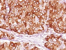
BRCA1 in Human Breast Cancer Tissue.
BRCA1 was detected in immersion fixed paraffin-embedded sections of human breast cancer tissue using Human BRCA1 Monoclonal Antibody (Catalog # MAB22101) at 5 µg/mL overnight at 4 °C. Before incubation with the primary antibody tissue was subjected to heat-induced epitope retrieval using Antigen Retrieval Reagent-Basic (CTS013). Tissue was stained using the Anti-Mouse HRP-DAB Cell & Tissue Staining Kit (brown; CTS002) and counterstained with hematoxylin (blue). View our protocol for Chromogenic IHC Staining of Paraffin-embedded Tissue Sections.
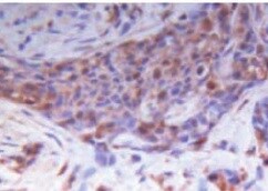
BRCA1 in Human Breast.
BRCA1 was detected in immersion fixed paraffin-embedded sections of human breast array using Human BRCA1 Monoclonal Antibody (Catalog # MAB22101) at 15 µg/mL overnight at 4 °C. Tissue was stained using the Anti-Mouse HRP-DAB Cell & Tissue Staining Kit (brown; CTS002) and counterstained with hematoxylin (blue). Lower panel shows a lack of labeling if primary antibodies are omitted and tissue is stained only with secondary antibody followed by incubation with detection reagents. View our protocol for Chromogenic IHC Staining of Paraffin-embedded Tissue Sections.
Detection of Human BRCA1 by Western Blot.
Western blot shows lysates of U2OS human osteosarcoma cell line and HeLa human cervical epithelial carcinoma cell line. PVDF membrane was probed with 1 µg/mL of Human BRCA1 Monoclonal Antibody (Catalog # MAB22101) followed by HRP-conjugated Anti-Mouse IgG Secondary Antibody (Catalog # HAF007). A specific band was detected for BRCA1 at approximately 240 kDa (as indicated). This experiment was conducted under reducing conditions and using Immunoblot Buffer Group 1.
Detection of BRCA1 C-Terminus by Western Blot
Microtubule-dependent nuclear deformations and DSBs mobility. A Immunoblot for BRCA1, CHK2 and phospho-CHK2 (P-CHK2) in p53-deleted (sg-P53) Brca1F/F Mouse Embryonic Fibroblasts (MEFs) 72 h after transduction with Hit&Run Cre and/or 6 h treatment with PARPi (0.5 μM). Actin is shown as loading control. b Examples of 10 min traces of mCherry-BP1-2 foci in Brca1F/F MEFs 72 h after Brca1 deletion, 6 h after PARPi addition in the absence or presence of taxol (1 h) or nocodazole (2 h). Scale bars: 10 μm. c MSD of mCherry-BP1-2 foci in the indicated MEFs as described in (b), with SD. Total foci analyzed: 1618 for DMSO, 1056 for taxol, and 1085 for nocodazole from 35, 30, 28 nuclei, respectively from n = 3 independent experiment. d Representative image of Brca1F/F MEFs without any treatment or 72 h after Cre-mediated deletion of Brca1, PARPi treatment for 6 h treatment and/or incubation with taxol for 1 h. Highlighted boxes indicate nuclear deformations as vertical invaginations (blue), light (green) and deep (yellow) lateral invaginations, and longitudinal invaginations (magenta). In black and gray boxes, the control nuclear edges and nuclear interior with no invaginations, respectively. Scale bars: 2 μm. Magnification ×4.25. (e) Quantification of nuclear deformations as in (d) from 40, 20 and 22 cells for each condition derived from one representative experiment. Statistical analysis by Kruskal–Wallis test for multiple comparisons. (*) P < 0.05 (p = 0.0381); P ≧ 0.05 are not significant. Source data are provided as a Source Data file. See also Supplementary Fig. 1. Image collected and cropped by CiteAb from the following open publication (https://pubmed.ncbi.nlm.nih.gov/40527886), licensed under a CC-BY license. Not internally tested by R&D Systems.Applications for Human BRCA1 C-Terminus Antibody (440621)
Application
Recommended Usage
Immunohistochemistry
8-25 µg/mL
Sample: Immersion fixed paraffin-embedded sections of human breast and human breast cancer tissue
Sample: Immersion fixed paraffin-embedded sections of human breast and human breast cancer tissue
Western Blot
1 µg/mL
Sample: U2OS human osteosarcoma cell line and HeLa human cervical epithelial carcinoma cell line
Sample: U2OS human osteosarcoma cell line and HeLa human cervical epithelial carcinoma cell line
Reviewed Applications
Read 2 reviews rated 5 using MAB22101 in the following applications:
Formulation, Preparation, and Storage
Purification
Protein A or G purified from hybridoma culture supernatant
Reconstitution
Reconstitute at 0.5 mg/mL in sterile PBS. For liquid material, refer to CoA for concentration.
Loading...
Formulation
Lyophilized from a 0.2 μm filtered solution in PBS with Trehalose. See Certificate of Analysis for details.
*Small pack size (-SP) is supplied either lyophilized or as a 0.2 µm filtered solution in PBS.
*Small pack size (-SP) is supplied either lyophilized or as a 0.2 µm filtered solution in PBS.
Shipping
Lyophilized product is shipped at ambient temperature. Liquid small pack size (-SP) is shipped with polar packs. Upon receipt, store immediately at the temperature recommended below.
Stability & Storage
Use a manual defrost freezer and avoid repeated freeze-thaw cycles.
- 12 months from date of receipt, -20 to -70 °C as supplied.
- 1 month, 2 to 8 °C under sterile conditions after reconstitution.
- 6 months, -20 to -70 °C under sterile conditions after reconstitution.
Calculators
Background: BRCA1
Long Name
Breast Cancer 1
Alternate Names
BRCAI, breast and ovarian cancer susceptibility protein 1, breast and ovarian cancer sususceptibility protein, breast cancer 1, early onset, breast cancer type 1 susceptibility protein, EC 6.3.2, EC 6.3.2.-, IRIS, PNCA4, PSCP, subunit 1
Gene Symbol
BRCA1
UniProt
Additional BRCA1 Products
Product Documents for Human BRCA1 C-Terminus Antibody (440621)
Certificate of Analysis
To download a Certificate of Analysis, please enter a lot or batch number in the search box below.
Note: Certificate of Analysis not available for kit components.
Product Specific Notices for Human BRCA1 C-Terminus Antibody (440621)
For research use only
Related Research Areas
Citations for Human BRCA1 C-Terminus Antibody (440621)
 Powered by Bioz
Customer Reviews for Human BRCA1 C-Terminus Antibody (440621) (2)
5 out of 5
2 Customer Ratings
Have you used Human BRCA1 C-Terminus Antibody (440621)?
Submit a review and receive an Amazon gift card!
$25/€18/£15/$25CAN/¥2500 Yen for a review with an image
$10/€7/£6/$10CAN/¥1110 Yen for a review without an image
Submit a review
Customer Images
Showing
1
-
2 of
2 reviews
Showing All
Filter By:
-
Application: ImmunohistochemistrySample Tested: Breast cancer tissueSpecies: HumanVerified Customer | Posted 04/20/2022

-
Application: ImmunohistochemistrySample Tested: Breast cancer tissueSpecies: HumanVerified Customer | Posted 11/09/2021
There are no reviews that match your criteria.
Protocols
Find general support by application which include: protocols, troubleshooting, illustrated assays, videos and webinars.
- Antigen Retrieval Protocol (PIER)
- Antigen Retrieval for Frozen Sections Protocol
- Appropriate Fixation of IHC/ICC Samples
- Cellular Response to Hypoxia Protocols
- Chromogenic IHC Staining of Formalin-Fixed Paraffin-Embedded (FFPE) Tissue Protocol
- Chromogenic Immunohistochemistry Staining of Frozen Tissue
- ClariTSA™ Fluorophore Kits
- Detection & Visualization of Antibody Binding
- Fluorescent IHC Staining of Frozen Tissue Protocol
- Graphic Protocol for Heat-induced Epitope Retrieval
- Graphic Protocol for the Preparation and Fluorescent IHC Staining of Frozen Tissue Sections
- Graphic Protocol for the Preparation and Fluorescent IHC Staining of Paraffin-embedded Tissue Sections
- Graphic Protocol for the Preparation of Gelatin-coated Slides for Histological Tissue Sections
- IHC Sample Preparation (Frozen sections vs Paraffin)
- Immunofluorescent IHC Staining of Formalin-Fixed Paraffin-Embedded (FFPE) Tissue Protocol
- Immunohistochemistry (IHC) and Immunocytochemistry (ICC) Protocols
- Immunohistochemistry Frozen Troubleshooting
- Immunohistochemistry Paraffin Troubleshooting
- Preparing Samples for IHC/ICC Experiments
- Preventing Non-Specific Staining (Non-Specific Binding)
- Primary Antibody Selection & Optimization
- Protocol for Heat-Induced Epitope Retrieval (HIER)
- Protocol for Making a 4% Formaldehyde Solution in PBS
- Protocol for VisUCyte™ HRP Polymer Detection Reagent
- Protocol for the Preparation & Fixation of Cells on Coverslips
- Protocol for the Preparation and Chromogenic IHC Staining of Frozen Tissue Sections
- Protocol for the Preparation and Chromogenic IHC Staining of Frozen Tissue Sections - Graphic
- Protocol for the Preparation and Chromogenic IHC Staining of Paraffin-embedded Tissue Sections
- Protocol for the Preparation and Chromogenic IHC Staining of Paraffin-embedded Tissue Sections - Graphic
- Protocol for the Preparation and Fluorescent IHC Staining of Frozen Tissue Sections
- Protocol for the Preparation and Fluorescent IHC Staining of Paraffin-embedded Tissue Sections
- Protocol for the Preparation of Gelatin-coated Slides for Histological Tissue Sections
- R&D Systems Quality Control Western Blot Protocol
- TUNEL and Active Caspase-3 Detection by IHC/ICC Protocol
- The Importance of IHC/ICC Controls
- Troubleshooting Guide: Immunohistochemistry
- Troubleshooting Guide: Western Blot Figures
- Western Blot Conditions
- Western Blot Protocol
- Western Blot Protocol for Cell Lysates
- Western Blot Troubleshooting
- Western Blot Troubleshooting Guide
- View all Protocols, Troubleshooting, Illustrated assays and Webinars
Loading...