Key Product Details

Validated by

Knockout/Knockdown, Biological Validation

Species Reactivity

Validated:

Human

Cited:

Human, Mouse, Rat, Avian - Chicken, Gerbil, Xenograft

Applications

Validated:

Knockout Validated, Immunohistochemistry, Western Blot, Immunocytochemistry, Simple Western, Immunoprecipitation

Cited:

Immunohistochemistry, Immunohistochemistry-Paraffin, Immunohistochemistry-Frozen, Western Blot, Immunocytochemistry

Label

Unconjugated

Antibody Source

Polyclonal Goat IgG
Loading...

Product Specifications

Immunogen

Mouse myeloma cell line NS0-derived recombinant human Cathepsin B
Arg18-Ile339
Accession # P07858

Specificity

Detects human Cathepsin B in direct ELISAs and Western blots. In direct ELISAs, approximately 35% cross-reactivity with recombinant mouse (rm) Cathepsin B is observed and less than 5% cross-reactivity with recombinant human (rh) Cathepsin C, rmCathepsin H, and rhCathepsin L is observed.

Clonality

Polyclonal

Host

Goat

Isotype

IgG

Scientific Data Images for Human Cathepsin B Antibody

Detection of Human Cathepsin B antibody by Western Blot.

Detection of Human Cathepsin B by Western Blot.

Western blot shows lysates of HepG2 human hepatocellular carcinoma cell line. PVDF membrane was probed with 0.25 µg/mL of Goat Anti-Human Cathepsin B Antigen Affinity-purified Polyclonal Antibody (Catalog # AF953) followed by HRP-conjugated Anti-Goat IgG Secondary Antibody (Catalog # HAF019). A specific band was detected for Cathepsin B at approximately 25-30 kDa (as indicated). This experiment was conducted under reducing conditions and using Immunoblot Buffer Group 1.

Cathepsin B antibody in Human Brain by Immunohistochemistry (IHC-P).

Cathepsin B in Human Brain.

Cathepsin B was detected in immersion fixed paraffin-embedded sections of human brain (cortex) using Goat Anti-Human Cathepsin B Antigen Affinity-purified Polyclonal Antibody (Catalog # AF953) at 10 µg/mL overnight at 4 °C. Tissue was stained using the Anti-Goat HRP-DAB Cell & Tissue Staining Kit (brown; CTS008) and counterstained with hematoxylin (blue). Lower panel shows a lack of labeling if primary antibodies are omitted and tissue is stained only with secondary antibody followed by incubation with detection reagents. View our protocol for Chromogenic IHC Staining of Paraffin-embedded Tissue Sections.

Detection of Human Cathepsin B antibody by Simple WesternTM.

Detection of Human Cathepsin B by Simple WesternTM.

Simple Western lane view shows lysates of HepG2 human hepatocellular carcinoma cell line, loaded at 0.2 mg/mL. A specific band was detected for Cathepsin B at approximately 34 kDa (as indicated) using 2.5 µg/mL of Goat Anti-Human Cathepsin B Antigen Affinity-purified Polyclonal Antibody (Catalog # AF953) followed by 1:50 dilution of HRP-conjugated Anti-Goat IgG Secondary Antibody (HAF109). This experiment was conducted under reducing conditions and using the 12-230 kDa separation system.

Western Blot Shows Human Cathepsin B Antibody Specificity by Using Knockout Cell Line.

Western Blot Shows Human Cathepsin B Specificity by Using Knockout Cell Line.

Western blot shows lysates of HeLa human cervical epithelial carcinoma parental cell line and Cathepsin B knockout HeLa cell line (KO). PVDF membrane was probed with 0.25 µg/mL of Goat Anti-Human Cathepsin B Antigen Affinity-purified Polyclonal Antibody (Catalog # AF953) followed by HRP-conjugated Anti-Goat IgG Secondary Antibody (HAF017). Specific bands were detected for Cathepsin B at approximately 25-45 kDa (as indicated) in the parental HeLa cell line, but is not detectable in knockout HeLa cell line. GAPDH (Catalog # AF5718) is shown as a loading control. This experiment was conducted under reducing conditions and using Immunoblot Buffer Group 1. New adjunct appears with knockout cell line.

Detection of Cathepsin B in U‑87 (Positive) & Daudi (Negative).

Cathepsin B was detected in immersion fixed U‑87 MG human glioblastoma/astrocytoma cell line (Positive) & absent in Daudi human Burkitt's lymphoma cell line (Negative) using Goat Anti-Human Cathepsin B Antigen Affinity-purified Polyclonal Antibody (Catalog # AF953) at 5 µg/mL for 3 hours at room temperature. Cells were stained using the NorthernLights™ 557-conjugated Anti-Goat IgG Secondary Antibody (red; Catalog # NL001) and counterstained with DAPI (blue). Specific staining was localized to cell surface. View our protocol for Fluorescent ICC Staining of Cells on Coverslips.

Detection of Human Cathepsin B by Western Blot

Detection of Human Cathepsin B by Western Blot

Legumain, cathepsin B and L expressions in cytosolic and membrane fractions of myotubes.Myotubes were treated with (+) or without (−) 30 µM simvastatin for 48 h before subcellular fractionation was performed. One representative immunoblot of legumain, cathepsin B and L in the cytosolic and membrane fractions is shown. All lanes were loaded with equal amount of total proteins and probed with antibodies as indicated. LAMP-2 and alpha -tubulin are shown as cell compartment controls (n = 3). Image collected and cropped by CiteAb from the following open publication (https://pubmed.ncbi.nlm.nih.gov/24416446), licensed under a CC-BY license. Not internally tested by R&D Systems.
Detection of Human Cathepsin B by Western Blot

Detection of Human Cathepsin B by Western Blot

CTSS inhibition does not impair lysosomal activities.(a) OEC-M1 cells were pretreated with vesicle or 100 nM BAF for 1 h and then incubated with 20 μM 6r for 1 h further. Lysosomal proteolytic activities were determined using BODIPY–BSA and quantified through flow cytometry. (b) After 48 h of siRNA knockdown of CTSS and CTSB, the relative expression of CTSS and CTSB were determined through Western blotting (right panel). Lysosomal proteolytic activities were determined using BODIPY–BSA and quantified through flow cytometry (right panel). Data represent the mean ± SD of three independent experiments. Differences were found to be statistically significant at ***P < 0.001. Image collected and cropped by CiteAb from the following open publication (https://www.nature.com/articles/srep29256), licensed under a CC-BY license. Not internally tested by R&D Systems.

Detection of Cathepsin B by Immunoprecipitation.

HAP1 cell line lysates were prepared and immunoprecipitation was performed using 2.0 µg of Goat Anti-Human Cathepsin B Antigen Affinity-purified Polyclonal Antibody (Catalog # AF953) pre-coupled to Dynabeads Protein A. Immunoprecipitated Cathepsin B was detected in Western Blot with a rabbit anti-Cathepsin B antibody used at 1/500. The Ponceau stained transfer of the blot is shown. SM=4% starting material; UB=4% unbound fraction; IP=immunoprecipitate; HC=antibody heavy chain. Image, protocol and testing courtesy of YCharOS Inc. (ycharos.com).

Applications for Human Cathepsin B Antibody

Application
Recommended Usage

Immunocytochemistry

5-25 µg/mL
Sample: Immersion fixed U‑87 MG human glioblastoma/astrocytoma cell line (Positive) & Daudi human Burkitt's lymphoma cell line (Negative)

Immunohistochemistry

5-15 µg/mL
Sample: Immersion fixed paraffin-embedded sections of human normal brain (cortex) and bladder cancer tissue

Immunoprecipitation

25 µg/mL
Sample: Conditioned cell culture medium spiked with Recombinant Human Cathepsin B (Catalog # 953-CY), see our available Western blot detection antibodies
HAP1 cell line.

Knockout Validated

Cathepsin B is specifically detected in HeLa human cervical epithelial carcinoma parental cell line but is not detectable in Cathepsin B knockout HeLa cell line.

Simple Western

2.5 µg/mL
Sample: HepG2 human hepatocellular carcinoma cell line

Western Blot

0.25 µg/mL
Sample: HepG2 human hepatocellular carcinoma cell line

Reviewed Applications

Read 3 reviews rated 5 using AF953 in the following applications:

Formulation, Preparation, and Storage

Purification

Antigen Affinity-purified

Reconstitution

Reconstitute at 0.2 mg/mL in sterile PBS. For liquid material, refer to CoA for concentration.


Loading...

Formulation

Lyophilized from a 0.2 μm filtered solution in PBS with Trehalose. See Certificate of Analysis for details.
*Small pack size (-SP) is supplied either lyophilized or as a 0.2 µm filtered solution in PBS.

Shipping

Lyophilized product is shipped at ambient temperature. Liquid small pack size (-SP) is shipped with polar packs. Upon receipt, store immediately at the temperature recommended below.

Stability & Storage

Use a manual defrost freezer and avoid repeated freeze-thaw cycles.
  • 12 months from date of receipt, -20 to -70 °C as supplied.
  • 1 month, 2 to 8 °C under sterile conditions after reconstitution.
  • 6 months, -20 to -70 °C under sterile conditions after reconstitution.

Calculators

The reconstitution calculator allows you to quickly calculate the volume of a reagent to reconstitute your vial. Simply enter the mass of reagent and the target concentration and the calculator will determine the rest.

=
÷

Background: Cathepsin B

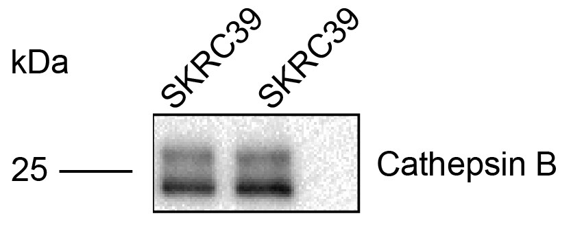
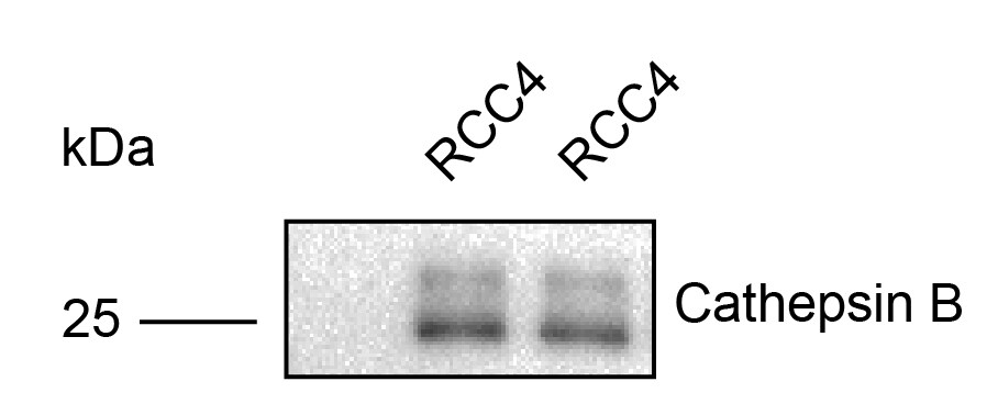
Cathepsin B is the first described member of the family of lysosomal cysteine proteases (1). Cathepsin B possesses both endopeptidase and exopeptidase activities, in the latter case acting as a peptidyl-dipeptidase. It is known to process a number of proteins, including pro and active caspases, prorenin, and secretory leucoprotease inhibitor (SLPI) (2-4). Therefore, Cathepsin B may play a role in activation and inactivation of caspases, activation of renin and inactivation of SLPI, the key steps in apoptosis, angiotensin production, and progression of emphysema, respectively. Because of its increased levels and redistribution of the enzyme in human and animal tumors, Cathepsin B may also have role in invasion and metastasis (5).

In addition to lysosome, Cathepsin B can be secreted or associated with plasma membrane, cytoplasm, and nucleus. It is synthesized as a preproenzyme. Following removal of the signal peptide, the inactive proenzyme undergoes further modifications including removal of the pro region to result in the active enzyme (1).

References

  1. Mort, J.S. (2004) in Handbook of Proteolytic Enzymes. Barrett, A.J. et al. (eds): Academic Press, San Diego, p. 1079.
  2. Vancompernolle, K. et al. (1998) FEBS Lett. 438:150.
  3. Jutras, I. and T.L. Reudelhuber (1999) FEBS Lett. 443:48.
  4. Taggart, C.C. et al. (2001) J. Biol. Chem. 276:33345.
  5. Bergquin, I.M. and B.F. Sloane (1996) Adv. Exp. Med. Biol. 389:281.

Alternate Names

CTSB

Entrez Gene IDs

1508 (Human); 13030 (Mouse)

Gene Symbol

CTSB

UniProt

Additional Cathepsin B Products

Product Documents for Human Cathepsin B Antibody

Certificate of Analysis

To download a Certificate of Analysis, please enter a lot or batch number in the search box below.

Note: Certificate of Analysis not available for kit components.

Product Specific Notices for Human Cathepsin B Antibody

For research use only

Citations for Human Cathepsin B Antibody

Customer Reviews for Human Cathepsin B Antibody (3)

5 out of 5
3 Customer Ratings
5 Stars
100%
4 Stars
0%
3 Stars
0%
2 Stars
0%
1 Stars
0%

Have you used Human Cathepsin B Antibody?

Submit a review and receive an Amazon gift card!

$25/€18/£15/$25CAN/¥2500 Yen for a review with an image

$10/€7/£6/$10CAN/¥1110 Yen for a review without an image

Submit a review
Amazon Gift Card

Customer Images


Showing  1 - 3 of 3 reviews Showing All
Filter By:
  • Human Cathepsin B Antibody
    Name: Anonymous
    Application: Western Blot
    Sample Tested: renal carcinoma
    Species: Human
    Verified Customer | Posted 08/06/2021
    1:500 dilution is good.
    Human Cathepsin B Antibody AF953
  • Human Cathepsin B Antibody
    Name: Anonymous
    Application: It worked very good in human kidney cell lines
    Sample Tested: Renal cancer tissue and renal cancer cell
    Species: Human
    Verified Customer | Posted 08/06/2021
    It's better to use 12% SDS-PAGE
    Human Cathepsin B Antibody AF953
  • Human Cathepsin B Antibody
    Name: Anonymous
    Application: Western Blot
    Sample Tested: ARP-1 human myeloma cell line
    Species: Human
    Verified Customer | Posted 11/12/2020
    Human Cathepsin B Antibody AF953

There are no reviews that match your criteria.

Showing  1 - 3 of 3 reviews Showing All

Protocols

Find general support by application which include: protocols, troubleshooting, illustrated assays, videos and webinars.

FAQs

No product specific FAQs exist for this product.

View all FAQs for Antibodies