CCL19, also known as MIP-3 beta and ELC (EBI1-Ligand Chemokine), is a 77 amino acid (aa) beta chemokine that is distantly related to other beta chemokines (20-30% aa sequence identity). The gene for MIP-3 beta has been mapped to chromosome 9p13 rather than chromosome 17 where the genes for many human beta chemokines are clustered. MIP-3 beta is constitutively expressed in various lymphoid tissues (including thymus, lymph nodes, appendix and spleen). The expression of MIP-3 beta is
down‑regulated by the anti-inflammatory cytokine IL-10. Recombinant MIP-3 beta is chemotactic for cultured human lymphocytes. MIP-3 beta is a ligand for CCR7 (previously referred to as the Epstein-Barr virus-induced gene 1 (EBI1) orphan receptor), a chemokine receptor that is expressed in various lymphoid tissues and activated B and T lymphocytes. CCR7 is strongly up-regulated in B cells infected with Epstein-Barr virus and T cells infected with herpesvirus 6 or 7.
Human CCL19/MIP-3 beta Antibody (54909)
R&D Systems | Catalog # MAB361

Key Product Details
Species Reactivity
Validated:
Human
Cited:
Human, Mouse
Applications
Validated:
Immunohistochemistry, Western Blot, Intracellular Staining by Flow Cytometry, Immunocytochemistry
Cited:
Immunohistochemistry, Immunohistochemistry-Paraffin, Immunohistochemistry-Frozen, Western Blot, Flow Cytometry, Immunocytochemistry, ELISA Development
Label
Unconjugated
Antibody Source
Monoclonal Mouse IgG2B Clone # 54909
Loading...
Product Specifications
Immunogen
E. coli-derived recombinant human CCL19/MIP-3 beta
Gly22-Ser98
Accession # Q99731.1
Gly22-Ser98
Accession # Q99731.1
Specificity
Detects human CCL19/MIP-3 beta in direct ELISAs and Western blots. In direct ELISAs, no cross-reactivity with recombinant
human CCL1, 2, 3, 4, 5, 7, 8, 11, 13, 14, 15, 16, 17, 18, 20, 21, 22, 23, 24,
25, recombinant mouse CCL2, 3, 4, 5, 6, 7, 9, 11, 12, 21, 22, or 25 is observed.
Clonality
Monoclonal
Host
Mouse
Isotype
IgG2B
Scientific Data Images for Human CCL19/MIP-3 beta Antibody (54909)
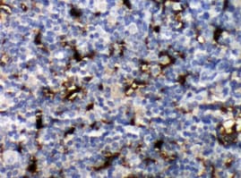
CCL19/MIP‑3 beta in Human Tonsil.
CCL19/MIP-3 beta was detected in immersion fixed paraffin-embedded sections of human tonsil using 25 µg/mL Mouse Anti-Human CCL19/ MIP-3 beta Monoclonal Antibody (Catalog # MAB361) overnight at 4 °C. Tissue was stained with the Anti-Mouse HRP-DAB Cell & Tissue Staining Kit (brown; Catalog # CTS002) and counter-stained with hematoxylin (blue). View our protocol for Chromogenic IHC Staining of Paraffin-embedded Tissue Sections.
CCL19/MIP‑3 beta in Human PBMCs.
CCL19/MIP-3 beta was detected in immersion fixed human peripheral blood mononuclear cells (PBMCs) using 10 µg/mL Mouse Anti-Human CCL19/MIP-3 beta Monoclonal Anti-body (Catalog # MAB361) for 3 hours at room temperature. Cells were stained with the NorthernLights™ 557-conjugated Anti-Mouse IgG Secondary Antibody (red; Catalog # NL007) and counter-stained with DAPI (blue). View our protocol for Fluorescent ICC Staining of Non-adherent Cells.
Detection of CCL19/MIP‑3 beta in Human Dendritic Cells by Flow Cytometry.
Human monocyte-derived dendritic cells were stained with Mouse Anti-Human CCL19/MIP-3 beta Monoclonal Antibody (Catalog # MAB361, filled histogram) or isotype control antibody (Catalog # MAB0041, open histogram), followed by Phycoerythrin-conjugated Anti-Mouse IgG Secondary Antibody (Catalog # F0102B). To facilitate intracellular staining, cells were fixed with Flow Cytometry Fixation Buffer (Catalog # FC004) and permeabilized with Flow Cytometry Permeabilization/Wash Buffer I (Catalog # FC005).Applications for Human CCL19/MIP-3 beta Antibody (54909)
Application
Recommended Usage
Immunocytochemistry
8-25 µg/mL
Sample: Immersion fixed human peripheral blood mononuclear cells (PBMCs)
Sample: Immersion fixed human peripheral blood mononuclear cells (PBMCs)
Immunohistochemistry
8-25 µg/mL
Sample: Immersion fixed paraffin-embedded sections of human tonsil
Sample: Immersion fixed paraffin-embedded sections of human tonsil
Intracellular Staining by Flow Cytometry
0.25 µg/106 cells
Sample: Human monocyte-derived dendritic cells
Sample: Human monocyte-derived dendritic cells
Western Blot
1 µg/mL
Sample: Recombinant Human CCL19/MIP‑3 beta (Catalog # 361-MI)
Sample: Recombinant Human CCL19/MIP‑3 beta (Catalog # 361-MI)
Reviewed Applications
Read 1 review rated 5 using MAB361 in the following applications:
Flow Cytometry Panel Builder
Bio-Techne Knows Flow Cytometry
Save time and reduce costly mistakes by quickly finding compatible reagents using the Panel Builder Tool.
Advanced Features
- Spectra Viewer - Custom analysis of spectra from multiple fluorochromes
- Spillover Popups - Visualize the spectra of individual fluorochromes
- Antigen Density Selector - Match fluorochrome brightness with antigen density

Formulation, Preparation, and Storage
Purification
Protein A or G purified from ascites
Reconstitution
For liquid material, refer to CoA for concentration.
Formulation
Supplied as a 0.2 μm filtered solution in Tris and Acetic Acid. *Small pack size (SP) is supplied either lyophilized or as a 0.2 µm filtered solution in PBS.
Shipping
Lyophilized product is shipped at ambient temperature. Liquid small pack size (-SP) is shipped with polar packs. Upon receipt, store immediately at the temperature recommended below.
Stability & Storage
Use a manual defrost freezer and avoid repeated freeze-thaw cycles.
- 12 months from date of receipt, -20 to -70 °C, as supplied.
- 1 month, 2 to 8 °C under sterile conditions after opening.
- 6 months, -20 to -70 °C under sterile conditions after opening.
Calculators
Background: CCL19/MIP-3 beta
References
- Yoshida, R. et al. (1997) J. Biol. Chem. 272:13803.
Alternate Names
ELC, MIP-3 beta, MIP3 beta
Gene Symbol
CCL19
UniProt
Additional CCL19/MIP-3 beta Products
Product Documents for Human CCL19/MIP-3 beta Antibody (54909)
Certificate of Analysis
To download a Certificate of Analysis, please enter a lot or batch number in the search box below.
Note: Certificate of Analysis not available for kit components.
Product Specific Notices for Human CCL19/MIP-3 beta Antibody (54909)
For research use only
Citations for Human CCL19/MIP-3 beta Antibody (54909)
 Powered by Bioz
Customer Reviews for Human CCL19/MIP-3 beta Antibody (54909) (1)
5 out of 5
1 Customer Rating
Have you used Human CCL19/MIP-3 beta Antibody (54909)?
Submit a review and receive an Amazon gift card!
$25/€18/£15/$25CAN/¥2500 Yen for a review with an image
$10/€7/£6/$10CAN/¥1110 Yen for a review without an image
Submit a review
Customer Images
Showing
1
-
1 of
1 review
Showing All
Filter By:
-
Application: ImmunohistochemistrySample Tested: Tonsil tissueSpecies: HumanVerified Customer | Posted 03/13/2022

There are no reviews that match your criteria.
Protocols
Find general support by application which include: protocols, troubleshooting, illustrated assays, videos and webinars.
- 7-Amino Actinomycin D (7-AAD) Cell Viability Flow Cytometry Protocol
- Antigen Retrieval Protocol (PIER)
- Antigen Retrieval for Frozen Sections Protocol
- Appropriate Fixation of IHC/ICC Samples
- Cellular Response to Hypoxia Protocols
- Chromogenic IHC Staining of Formalin-Fixed Paraffin-Embedded (FFPE) Tissue Protocol
- Chromogenic Immunohistochemistry Staining of Frozen Tissue
- ClariTSA™ Fluorophore Kits
- Detection & Visualization of Antibody Binding
- Extracellular Membrane Flow Cytometry Protocol
- Flow Cytometry Protocol for Cell Surface Markers
- Flow Cytometry Protocol for Staining Membrane Associated Proteins
- Flow Cytometry Staining Protocols
- Flow Cytometry Troubleshooting Guide
- Fluorescent IHC Staining of Frozen Tissue Protocol
- Graphic Protocol for Heat-induced Epitope Retrieval
- Graphic Protocol for the Preparation and Fluorescent IHC Staining of Frozen Tissue Sections
- Graphic Protocol for the Preparation and Fluorescent IHC Staining of Paraffin-embedded Tissue Sections
- Graphic Protocol for the Preparation of Gelatin-coated Slides for Histological Tissue Sections
- ICC Cell Smear Protocol for Suspension Cells
- ICC Immunocytochemistry Protocol Videos
- ICC for Adherent Cells
- IHC Sample Preparation (Frozen sections vs Paraffin)
- Immunocytochemistry (ICC) Protocol
- Immunocytochemistry Troubleshooting
- Immunofluorescence of Organoids Embedded in Cultrex Basement Membrane Extract
- Immunofluorescent IHC Staining of Formalin-Fixed Paraffin-Embedded (FFPE) Tissue Protocol
- Immunohistochemistry (IHC) and Immunocytochemistry (ICC) Protocols
- Immunohistochemistry Frozen Troubleshooting
- Immunohistochemistry Paraffin Troubleshooting
- Intracellular Flow Cytometry Protocol Using Alcohol (Methanol)
- Intracellular Flow Cytometry Protocol Using Detergents
- Intracellular Nuclear Staining Flow Cytometry Protocol Using Detergents
- Intracellular Staining Flow Cytometry Protocol Using Alcohol Permeabilization
- Intracellular Staining Flow Cytometry Protocol Using Detergents to Permeabilize Cells
- Preparing Samples for IHC/ICC Experiments
- Preventing Non-Specific Staining (Non-Specific Binding)
- Primary Antibody Selection & Optimization
- Propidium Iodide Cell Viability Flow Cytometry Protocol
- Protocol for Heat-Induced Epitope Retrieval (HIER)
- Protocol for Liperfluo
- Protocol for Making a 4% Formaldehyde Solution in PBS
- Protocol for VisUCyte™ HRP Polymer Detection Reagent
- Protocol for the Characterization of Human Th22 Cells
- Protocol for the Characterization of Human Th9 Cells
- Protocol for the Fluorescent ICC Staining of Cell Smears - Graphic
- Protocol for the Fluorescent ICC Staining of Cultured Cells on Coverslips - Graphic
- Protocol for the Preparation & Fixation of Cells on Coverslips
- Protocol for the Preparation and Chromogenic IHC Staining of Frozen Tissue Sections
- Protocol for the Preparation and Chromogenic IHC Staining of Frozen Tissue Sections - Graphic
- Protocol for the Preparation and Chromogenic IHC Staining of Paraffin-embedded Tissue Sections
- Protocol for the Preparation and Chromogenic IHC Staining of Paraffin-embedded Tissue Sections - Graphic
- Protocol for the Preparation and Fluorescent ICC Staining of Cells on Coverslips
- Protocol for the Preparation and Fluorescent ICC Staining of Non-adherent Cells
- Protocol for the Preparation and Fluorescent ICC Staining of Stem Cells on Coverslips
- Protocol for the Preparation and Fluorescent IHC Staining of Frozen Tissue Sections
- Protocol for the Preparation and Fluorescent IHC Staining of Paraffin-embedded Tissue Sections
- Protocol for the Preparation of Gelatin-coated Slides for Histological Tissue Sections
- Protocol for the Preparation of a Cell Smear for Non-adherent Cell ICC - Graphic
- Protocol: Annexin V and PI Staining by Flow Cytometry
- Protocol: Annexin V and PI Staining for Apoptosis by Flow Cytometry
- R&D Systems Quality Control Western Blot Protocol
- TUNEL and Active Caspase-3 Detection by IHC/ICC Protocol
- The Importance of IHC/ICC Controls
- Troubleshooting Guide: Fluorokine Flow Cytometry Kits
- Troubleshooting Guide: Immunohistochemistry
- Troubleshooting Guide: Western Blot Figures
- Western Blot Conditions
- Western Blot Protocol
- Western Blot Protocol for Cell Lysates
- Western Blot Troubleshooting
- Western Blot Troubleshooting Guide
- View all Protocols, Troubleshooting, Illustrated assays and Webinars
Loading...
Associated Pathways
