
Key Product Details
Species Reactivity
Applications
Label
Antibody Source
Product Specifications
Immunogen
Phe19-Lys379
Accession # P28908
Specificity
Clonality
Host
Isotype
Endotoxin Level
Scientific Data Images for Human CD30/TNFRSF8 Antibody
CD30/TNFRSF8 in Jurkat Human Cell Line.
CD30/TNFRSF8 was detected in immersion fixed Jurkat human acute T cell leukemia cell line using Goat Anti-Human CD30/TNFRSF8 Antigen Affinity-purified Polyclonal Antibody (Catalog # AF229) at 10 µg/mL for 3 hours at room temperature. Cells were stained using the NorthernLights™ 557-conjugated Anti-Goat IgG Secondary Antibody (red; NL001) and counterstained with DAPI (blue). Specific staining was localized to cell membrane and cytoplasm. View our protocol for Fluorescent ICC Staining of Non-adherent Cells.
Detection of Human CD30/TNFRSF8 by Simple WesternTM.
Left: Simple Western lane view shows lysates of HDLM‑2 human Hodgkin’s lymphoma cell line, loaded at 0.5 mg/ml. A specific band was detected for CD30/TNFRSF8 at approximately 178 kDa (as indicated) using both 10 µg/ml and 50 µg/ml of Goat Anti-Human CD30/TNFRSF8 Antigen Affinity-purified Polyclonal Antibody (Catalog # AF229) followed by HRP-conjugated Donkey Anti-Goat Secondary Antibody (Catalog # 043-522-2). This experiment was conducted under reducing conditions and using the 12-230kDa separation system. Right: Simple Western electropherogram showing the same Goat Anti-Human CD30/TNFRSF8 Antigen Affinity-purified Polyclonal Antibody (Catalog # AF229) tested at 10 µg/ml (blue line) and 50 µg/ml (green line) in the HDLM‑2 human Hodgkin’s lymphoma cell line.
Human CD30/TNFRSF8 ELISA Standard Curve.
Recombinant Human CD30/TNFRSF8 protein was serially diluted 2-fold and captured by Mouse Anti-Human CD30/TNFRSF8 Monoclonal Antibody (MAB2291) coated on a Clear Polystyrene Microplate (DY990). Goat Anti-Human CD30/TNFRSF8 Antigen Affinity-purified Polyclonal Antibody (Catalog # AF229) was biotinylated and incubated with the protein captured on the plate. Detection of the standard curve was achieved by incubating Streptavidin-HRP (DY998) followed by Substrate Solution (DY999) and stopping the enzymatic reaction with Stop Solution (DY994).Applications for Human CD30/TNFRSF8 Antibody
Agonist Activity
The ED50 for this effect is typically 0.2 - 0.6 μg/mL.
ELISA
This antibody functions as an ELISA detection antibody when paired with Mouse Anti-Human CD30/TNFRSF8 Monoclonal Antibody (Catalog # MAB2291).
This product is intended for assay development on various assay platforms requiring antibody pairs. We recommend the Human CD30/TNFRSF8 DuoSet ELISA Kit (Catalog # DY6126-05) for convenient development of a sandwich ELISA.
Immunocytochemistry
Sample: Immersion fixed Jurkat human acute T cell leukemia cell line
Simple Western
Sample: HDLM-2 human Hodgkin's lymphoma cell line
Western Blot
Sample: Recombinant Human CD30/TNFRSF8 Fc Chimera (Catalog # 813-CD)
Reviewed Applications
Read 1 review rated 5 using AF229 in the following applications:
Formulation, Preparation, and Storage
Purification
Reconstitution
Reconstitute at 0.2 mg/mL in sterile PBS. For liquid material, refer to CoA for concentration.
Formulation
*Small pack size (-SP) is supplied either lyophilized or as a 0.2 µm filtered solution in PBS.
Shipping
Stability & Storage
- 12 months from date of receipt, -20 to -70 °C as supplied.
- 1 month, 2 to 8 °C under sterile conditions after reconstitution.
- 6 months, -20 to -70 °C under sterile conditions after reconstitution.
Calculators
Background: CD30/TNFRSF8
References
- Kennedy, M.K. et al. (2006) Immunology 118:143.
- Tarkowski, M. (2003) Curr. Opin. Hematol. 10:267.
- Durkop, H. et al. (1992) Cell 68:421.
- Hamann, D. et al. (1996) J. Immunol. 156:1387.
- Shanebeck, S.D. et al. (1995) Eur. J. Immunol. 25:2147.
- Gruss, H.-J. et al. (1994) Blood 83:2045.
- Oflzoglu E. et al. (2009) Adv. Exp. Med. Biol. 647:174.
- Del Prete, G. et al. (1995) J. Exp. Med. 182:1655.
- Harlin, H. et al. (2002) J. Immunol. 169:2451.
- Amakawa, R. et al. (1996) Cell 84:551.
- Chiarle, R. et al. (1999) J. Immunol. 163:194.
- Vinante, F. et al. (2002) Blood 99:52.
- Hansen, H.P. et al. (1995) Int. J. Cancer 63:750.
- Hansen, H.P. et al. (2000) J. Immunol. 165:6703.
- Hargreaves, P.G. and A. Al-Shamkhani (2002) Eur. J. Immunol. 32:163.
Alternate Names
Gene Symbol
UniProt
Additional CD30/TNFRSF8 Products
Product Documents for Human CD30/TNFRSF8 Antibody
Certificate of Analysis
To download a Certificate of Analysis, please enter a lot or batch number in the search box below.
Note: Certificate of Analysis not available for kit components.
Product Specific Notices for Human CD30/TNFRSF8 Antibody
For research use only
Citations for Human CD30/TNFRSF8 Antibody
 Powered by Bioz
Customer Reviews for Human CD30/TNFRSF8 Antibody (1)
Have you used Human CD30/TNFRSF8 Antibody?
Submit a review and receive an Amazon gift card!
$25/€18/£15/$25CAN/¥2500 Yen for a review with an image
$10/€7/£6/$10CAN/¥1110 Yen for a review without an image
Submit a review
Customer Images
-
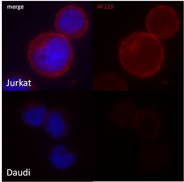
Application: ImmunocytochemistrySample Tested: Immersion-fixed Jurkat and Daudi cells.Species: HumanVerified Customer | Posted 05/13/2015CD30/TNFRSF8 Antibody [AF229] immunofluorescent staining in Jurkat and Daudi human cells.

There are no reviews that match your criteria.
Protocols
Find general support by application which include: protocols, troubleshooting, illustrated assays, videos and webinars.
- Appropriate Fixation of IHC/ICC Samples
- Cellular Response to Hypoxia Protocols
- ClariTSA™ Fluorophore Kits
- Detection & Visualization of Antibody Binding
- ELISA Sample Preparation & Collection Guide
- ELISA Troubleshooting Guide
- How to Run an R&D Systems DuoSet ELISA
- How to Run an R&D Systems Quantikine ELISA
- How to Run an R&D Systems Quantikine™ QuicKit™ ELISA
- ICC Cell Smear Protocol for Suspension Cells
- ICC Immunocytochemistry Protocol Videos
- ICC for Adherent Cells
- Immunocytochemistry (ICC) Protocol
- Immunocytochemistry Troubleshooting
- Immunofluorescence of Organoids Embedded in Cultrex Basement Membrane Extract
- Immunohistochemistry (IHC) and Immunocytochemistry (ICC) Protocols
- Preparing Samples for IHC/ICC Experiments
- Preventing Non-Specific Staining (Non-Specific Binding)
- Primary Antibody Selection & Optimization
- Protocol for VisUCyte™ HRP Polymer Detection Reagent
- Protocol for the Fluorescent ICC Staining of Cell Smears - Graphic
- Protocol for the Fluorescent ICC Staining of Cultured Cells on Coverslips - Graphic
- Protocol for the Preparation and Fluorescent ICC Staining of Cells on Coverslips
- Protocol for the Preparation and Fluorescent ICC Staining of Non-adherent Cells
- Protocol for the Preparation and Fluorescent ICC Staining of Stem Cells on Coverslips
- Protocol for the Preparation of a Cell Smear for Non-adherent Cell ICC - Graphic
- Quantikine HS ELISA Kit Assay Principle, Alkaline Phosphatase
- Quantikine HS ELISA Kit Principle, Streptavidin-HRP Polymer
- R&D Systems Quality Control Western Blot Protocol
- Sandwich ELISA (Colorimetric) – Biotin/Streptavidin Detection Protocol
- Sandwich ELISA (Colorimetric) – Direct Detection Protocol
- TUNEL and Active Caspase-3 Detection by IHC/ICC Protocol
- The Importance of IHC/ICC Controls
- Troubleshooting Guide: ELISA
- Troubleshooting Guide: Western Blot Figures
- Western Blot Conditions
- Western Blot Protocol
- Western Blot Protocol for Cell Lysates
- Western Blot Troubleshooting
- Western Blot Troubleshooting Guide
- View all Protocols, Troubleshooting, Illustrated assays and Webinars
Associated Pathways
