CD45, previously called LCA (leukocyte common antigen), T200, or Ly5 in mice, is member C of the class 1 (receptor‑like) protein tyrosine phosphatase family (PTPRC) (1, 2). It is a variably glycosylated 180‑220 kDa transmembrane protein that is abundantly expressed on all nucleated cells of hematopoietic origin (1‑3). CD45 has several isoforms, expressed according to cell type, developmental stage and antigenic exposure (1‑5). The longest form, CD45RABC (called B220 in mouse), is expressed on B lymphocytes (5). The CD45RABC cDNA encodes 1304 amino acids (aa), including a 23 aa signal sequence, a 552 aa extracellular domain containing the splicing region, a cysteine‑rich region and two fibronectin type III domains, a 22 aa transmembrane sequence, and a 707 aa cytoplasmic domain that contains two phosphatase domains, D1 and D2. Only D1 has phosphatase activity. CD45R0 is the shortest form, lacking exons 4, 5 and 6 which encode aa 32‑191. It is expressed on memory cells, while intermediate sizes are expressed on other T cells (3, 4, 6). CD45 has been best studied in T cells, where it determines T cell receptor signaling thresholds (3, 6‑8). CD45 is moved into or out of the immunological synapse (IS) membrane microdomain depending on the relative influence of interaction with the extracellular galectin lattice or the intracellular actin cytoskeleton (9, 10). Galectin interaction can be fine‑tuned by varying usage of the heavily O‑glycosylated spliced regions and sialylation of N‑linked carbohydrates (4, 9). Within the IS, CD45 dephosphorylates and negatively regulates the Src family kinase, Lck (8‑10). In other leukocytes, CD45 influences differentiation and links immunoreceptor signaling with cytokine secretion and cell survival, partially overlapping in function with DEP‑1/CD148 (11‑14). CD45 deletion causes in severe immunodeficiency, while point mutations may be associated with autoimmune disorders (6, 7).

Key Product Details
Validated by
Knockout/Knockdown
Species Reactivity
Validated:
Human
Cited:
Human, Rat
Applications
Validated:
Knockout Validated, Flow Cytometry, Immunocytochemistry, CyTOF-ready
Cited:
Immunohistochemistry, Immunohistochemistry-Paraffin, Flow Cytometry, Immunocytochemistry, ELISA Capture, Immunopanning
Label
Unconjugated
Antibody Source
Monoclonal Mouse IgG1 Clone # 2D1
Loading...
Product Specifications
Immunogen
Human peripheral blood mononuclear cells
Specificity
Detects human CD45. This antibody recognizes all isoforms of human CD45.
Clonality
Monoclonal
Host
Mouse
Isotype
IgG1
Scientific Data Images for Human CD45 Antibody (2D1)
Detection of CD45 in Human Blood Lymphocytes by Flow Cytometry.
Human peripheral blood lymphocytes were stained with Mouse Anti-Human CD45 Monoclonal Antibody (Catalog # MAB1430, filled histogram) or isotype control antibody (Catalog # MAB002, open histogram), followed by Phycoerythrin-conjugated Anti-Mouse IgG Secondary Antibody (Catalog # F0102B). View our protocol for Staining Membrane-associated Proteins.
CD45 in Human PBMCs.
CD45 was detected in immersion fixed human peripheral blood mononuclear cells (PBMCs) stimulated with PHA blast using Mouse Anti-Human CD45 Monoclonal Antibody (Catalog # MAB1430) at 10 µg/mL for 3 hours at room temperature. Cells were stained using the NorthernLights™ 557-conjugated Anti-Mouse IgG Secondary Antibody (yellow; Catalog # NL007) and counter-stained with DAPI (blue). View our protocol for Fluorescent ICC Staining of Non-adherent Cells.
Detection of CD45 in THP-1 Human Cell Line by Flow Cytometry.
THP-1 human acute monocytic leukemia cell line was stained with Mouse Anti-Human CD45 Monoclonal Antibody (Catalog # MAB1430, filled histogram) or isotype control antibody (MAB002), followed by Phycoerythrin-conjugated Anti-Mouse IgG Secondary Antibody (F0102B). Staining was performed using our Staining Membrane-associated Proteins protocol.
CD45 Specificity is Shown by Flow Cytometry in Knockout Cell Line.
CD45 knockout THP-1 human acute monocytic leukemia cell line was stained with Mouse Anti-Human CD45 Monoclonal Antibody (Catalog # MAB1430, filled histogram) or isotype control antibody (MAB002) followed by PE-conjugated anti-Mouse IgG Secondary Antibody (F0102B). No staining in the CD45 knockout THP-1 cell line was observed. Staining was performed using our Staining Membrane-associated Proteins protocol.Applications for Human CD45 Antibody (2D1)
Application
Recommended Usage
CyTOF-ready
Ready to be labeled using established conjugation methods. No BSA or other carrier proteins that could interfere with conjugation.
Flow Cytometry
0.25 µg/106 cells
Sample: Human peripheral blood lymphocytes and THP-1 human acute monocytic leukemia cell line
Sample: Human peripheral blood lymphocytes and THP-1 human acute monocytic leukemia cell line
Immunocytochemistry
8-25 µg/mL
Sample: Immersion fixed human peripheral blood mononuclear cells (PBMCs) stimulated with PHA blast
Sample: Immersion fixed human peripheral blood mononuclear cells (PBMCs) stimulated with PHA blast
Knockout Validated
Optimal dilutions of this antibody should be experimentally determined.
Reviewed Applications
Read 2 reviews rated 5 using MAB1430 in the following applications:
Flow Cytometry Panel Builder
Bio-Techne Knows Flow Cytometry
Save time and reduce costly mistakes by quickly finding compatible reagents using the Panel Builder Tool.
Advanced Features
- Spectra Viewer - Custom analysis of spectra from multiple fluorochromes
- Spillover Popups - Visualize the spectra of individual fluorochromes
- Antigen Density Selector - Match fluorochrome brightness with antigen density

Formulation, Preparation, and Storage
Purification
Protein A or G purified from hybridoma culture supernatant
Reconstitution
Reconstitute at 0.5 mg/mL in sterile PBS. For liquid material, refer to CoA for concentration.
Loading...
Formulation
Lyophilized from a 0.2 μm filtered solution in PBS with Trehalose. *Small pack size (SP) is supplied either lyophilized or as a 0.2 µm filtered solution in PBS.
Shipping
Lyophilized product is shipped at ambient temperature. Liquid small pack size (-SP) is shipped with polar packs. Upon receipt, store immediately at the temperature recommended below.
Stability & Storage
Use a manual defrost freezer and avoid repeated freeze-thaw cycles.
- 12 months from date of receipt, -20 to -70 °C as supplied.
- 1 month, 2 to 8 °C under sterile conditions after reconstitution.
- 6 months, -20 to -70 °C under sterile conditions after reconstitution.
Calculators
Background: CD45
References
- Anderson, J.N. et al. (2004) FASEB J. 18:8.
- Streuli, M. et al. (1987) J. Exp. Med. 166:1548.
- Hermiston, M.L. et al. (2003) Annu. Rev. Immunol. 21:107.
- Earl, L.A. and L.G. Baum (2008) Immunol. Cell Biol. 86:608.
- Ralph, S.J. et al. (1987) EMBO J. 6:1251.
- Falahti, R. and D. Leitenberg (2008) J. Immunol. 181:6082.
- Tchilian, E.Z. and P.C.L. Beverley (2006) Trends Immunol. 27:146.
- McNiell, L. et al. (2007) Immunity 27:425.
- Chen, I-J. et al. (2007) J. Biol. Chem. 282:35361.
- Freiberg, B.A. et al. (2002) Nat. Immunol. 3:911.
- Zhu, J.W. et al. (2008) Immunity 28:183.
- Huntington, N.D. et al. (2006) Nat. Immunol. 7:190.
- Hesslein, D.G. et al. (2006) Proc. Natl. Acad. Sci. USA 103:7012.
- Cross, J.L. et al. (2008) J. Immunol. 180:8020.
Long Name
Cluster of Differentiation 45
Alternate Names
CD45, LCA, PTPRC, T200 Glycoprotein
Gene Symbol
PTPRC
Additional CD45 Products
Product Documents for Human CD45 Antibody (2D1)
Certificate of Analysis
To download a Certificate of Analysis, please enter a lot or batch number in the search box below.
Note: Certificate of Analysis not available for kit components.
Product Specific Notices for Human CD45 Antibody (2D1)
For research use only
Related Research Areas
Citations for Human CD45 Antibody (2D1)
 Powered by Bioz
Customer Reviews for Human CD45 Antibody (2D1) (2)
5 out of 5
2 Customer Ratings
Have you used Human CD45 Antibody (2D1)?
Submit a review and receive an Amazon gift card!
$25/€18/£15/$25CAN/¥2500 Yen for a review with an image
$10/€7/£6/$10CAN/¥1110 Yen for a review without an image
Submit a review
Customer Images
Showing
1
-
2 of
2 reviews
Showing All
Filter By:
-
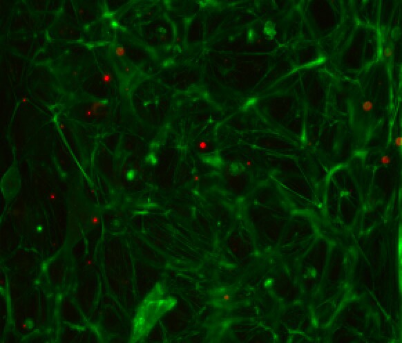
Application: Immunocytochemistry/ImmunofluorescenceSample Tested: Peripheral blood mononuclear cells (PBMCs)Species: HumanVerified Customer | Posted 09/08/2025

-
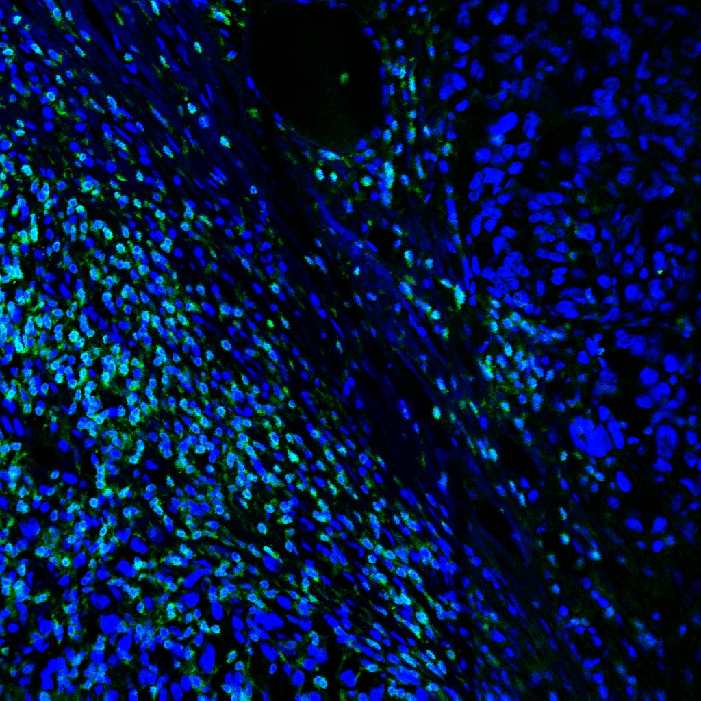
Application: Immunocytochemistry/ImmunofluorescenceSample Tested: Melanoma tissueSpecies: HumanVerified Customer | Posted 09/27/2023
There are no reviews that match your criteria.
Protocols
Find general support by application which include: protocols, troubleshooting, illustrated assays, videos and webinars.
- 7-Amino Actinomycin D (7-AAD) Cell Viability Flow Cytometry Protocol
- Appropriate Fixation of IHC/ICC Samples
- Cellular Response to Hypoxia Protocols
- ClariTSA™ Fluorophore Kits
- Detection & Visualization of Antibody Binding
- Extracellular Membrane Flow Cytometry Protocol
- Flow Cytometry Protocol for Cell Surface Markers
- Flow Cytometry Protocol for Staining Membrane Associated Proteins
- Flow Cytometry Staining Protocols
- Flow Cytometry Troubleshooting Guide
- ICC Cell Smear Protocol for Suspension Cells
- ICC Immunocytochemistry Protocol Videos
- ICC for Adherent Cells
- Immunocytochemistry (ICC) Protocol
- Immunocytochemistry Troubleshooting
- Immunofluorescence of Organoids Embedded in Cultrex Basement Membrane Extract
- Immunohistochemistry (IHC) and Immunocytochemistry (ICC) Protocols
- Intracellular Flow Cytometry Protocol Using Alcohol (Methanol)
- Intracellular Flow Cytometry Protocol Using Detergents
- Intracellular Nuclear Staining Flow Cytometry Protocol Using Detergents
- Intracellular Staining Flow Cytometry Protocol Using Alcohol Permeabilization
- Intracellular Staining Flow Cytometry Protocol Using Detergents to Permeabilize Cells
- Preparing Samples for IHC/ICC Experiments
- Preventing Non-Specific Staining (Non-Specific Binding)
- Primary Antibody Selection & Optimization
- Propidium Iodide Cell Viability Flow Cytometry Protocol
- Protocol for Liperfluo
- Protocol for VisUCyte™ HRP Polymer Detection Reagent
- Protocol for the Characterization of Human Th22 Cells
- Protocol for the Characterization of Human Th9 Cells
- Protocol for the Fluorescent ICC Staining of Cell Smears - Graphic
- Protocol for the Fluorescent ICC Staining of Cultured Cells on Coverslips - Graphic
- Protocol for the Preparation and Fluorescent ICC Staining of Cells on Coverslips
- Protocol for the Preparation and Fluorescent ICC Staining of Non-adherent Cells
- Protocol for the Preparation and Fluorescent ICC Staining of Stem Cells on Coverslips
- Protocol for the Preparation of a Cell Smear for Non-adherent Cell ICC - Graphic
- Protocol: Annexin V and PI Staining by Flow Cytometry
- Protocol: Annexin V and PI Staining for Apoptosis by Flow Cytometry
- TUNEL and Active Caspase-3 Detection by IHC/ICC Protocol
- The Importance of IHC/ICC Controls
- Troubleshooting Guide: Fluorokine Flow Cytometry Kits
- View all Protocols, Troubleshooting, Illustrated assays and Webinars
Loading...
Associated Pathways


