CHI3L1, also known as HCgp39 (human cartilage glycoprotein 39) and YKL40, is a secreted glycoprotein belonging to the family of chitinase-like lectins. These proteins are structurally related to the glycosylhydrolase family 18, but lack enzymatic activity. CHI3L1 is expressed by chondrocytes, synovial cells, macrophages and neutrophils. It has been found to play a role in down‑regulating cytokine-induced inflammatory responses. Human CHI3L1 shares 73%, 80%, 83% and 86% amino acid sequence identity with mouse, rat, bovine and porcine CHI3L1, respectively.
Human Chitinase 3-like 1/YKL-40 Antibody
R&D Systems | Catalog # AF2599

Key Product Details
Validated by
Biological Validation
Species Reactivity
Validated:
Human
Cited:
Human, Mouse, Primate
Applications
Validated:
Immunohistochemistry, Western Blot, Immunocytochemistry, Simple Western
Cited:
Immunohistochemistry, Immunohistochemistry-Paraffin, Immunohistochemistry-Frozen, Western Blot, Flow Cytometry, Simple Western
Label
Unconjugated
Antibody Source
Polyclonal Goat IgG
Loading...
Product Specifications
Immunogen
Mouse myeloma cell line NS0-derived recombinant human Chitinase 3-like 1/YKL-40
Tyr22-Thr383
Accession # P36222
Tyr22-Thr383
Accession # P36222
Specificity
Detects human Chitinase 3-like 1/YKL-40 in direct ELISAs and Western blots. In direct ELISAs, approximately 15% cross‑reactivity with recombinant mouse Chitinase 3-like 1/YKL-40 is observed.
Clonality
Polyclonal
Host
Goat
Isotype
IgG
Scientific Data Images for Human Chitinase 3-like 1/YKL-40 Antibody
Detection of Human Chitinase 3-like 1/YKL-40 by Western Blot.
Western blot shows lysates of THP-1 human acute monocytic leukemia cell line treated (+) with 50 ng/mL of PMA for 72 hours. PVDF membrane was probed with 1 µg/mL of Goat Anti-Human Chitinase 3-like 1/YKL-40 Antigen Affinity-purified Polyclonal Antibody (Catalog # AF2599) followed by HRP-conjugated Anti-Goat IgG Secondary Antibody (HAF109). A specific band was detected for Chitinase 3-like 1/YKL-40 at approximately 45 kDa (as indicated). This experiment was conducted under reducing conditions and using Immunoblot Buffer Group 8.
Chitinase 3‑like 1/YKL-40 in Human Ovarian Cancer.
Chitinase 3-like 1/YKL-40 was detected in immersion fixed paraffin-embedded sections of human ovarian cancer using Goat Anti-Human Chitinase 3-like 1/YKL-40 Antigen Affinity-purified Polyclonal Antibody (Catalog # AF2599) at 15 µg/mL overnight at 4 °C. Before incubation with the primary antibody, tissue was subjected to heat-induced epitope retrieval using Antigen Retrieval Reagent-Basic (CTS013). Tissue was stained using the Anti-Goat HRP-DAB Cell & Tissue Staining Kit (brown; CTS008) and counterstained with hematoxylin (blue). Specific staining was localized to epithelial cells. View our protocol for Chromogenic IHC Staining of Paraffin-embedded Tissue Sections.
Chitinase 3‑like 1/YKL-40 in THP‑1 Human Cell Line.
Chitinase 3‑like 1/YKL-40 was detected in immersion fixed THP‑1 human acute monocytic leukemia cell line treated with PMA (positive staining) and THP‑1 human acute monocytic leukemia cell line (untreated; negative staining) using Goat Anti-Human Chitinase 3‑like 1/YKL-40 Antigen Affinity-purified Polyclonal Antibody (Catalog # AF2599) at 1.7 µg/mL for 3 hours at room temperature. Cells were stained using the NorthernLights™ 557-conjugated Anti-Goat IgG Secondary Antibody (red; NL001) and counterstained with DAPI (blue). Specific staining was localized to cytoplasm. Staining was performed using our protocol for Fluorescent ICC Staining of Non-adherent Cells.
Chitinase 3‑like 1/YKL-40 in Human Cartilage.
Chitinase 3-like 1/YKL-40 was detected in immersion fixed paraffin-embedded sections of human cartilage using Goat Anti-Human Chitinase 3-like 1/YKL-40 Antigen Affinity-purified Polyclonal Antibody (Catalog # AF2599) at 15 µg/mL overnight at 4 °C. Tissue was stained using the Anti-Goat HRP-DAB Cell & Tissue Staining Kit (brown; CTS008) and counterstained with hematoxylin (blue). Specific staining was localized to chondrocytes. View our protocol for Chromogenic IHC Staining of Paraffin-embedded Tissue Sections.
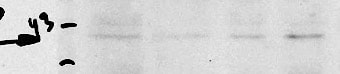
Detection of Human Chitinase 3‑like 1/YKL-40 by Western Blot.
Western blot shows lysates of human bone marrow, mouse bone marrow. PVDF membrane was probed with 0.5 µg/mL of Goat Anti-Human Chitinase 3‑like 1/YKL-40 Antigen Affinity-purified Polyclonal Antibody (Catalog # AF2599) followed by HRP-conjugated Anti-Goat IgG Secondary Antibody (Catalog # HAF017). A specific band was detected for Chitinase 3‑like 1/YKL-40 at approximately 40 kDa (as indicated). This experiment was conducted under reducing conditions and using Western Blot Buffer Group 1.
Detection of Human Chitinase 3‑like 1/YKL-40 by Simple WesternTM.
Simple Western lane view shows lysates of Human bone marrow, loaded at 0.2 mg/mL. A specific band was detected for Chitinase 3‑like 1 at approximately 50 kDa (as indicated) using 25 µg/mL of Goat Anti-Human Chitinase 3‑like 1/YKL-40 Antigen Affinity-purified Polyclonal Antibody (Catalog # AF2599). This experiment was conducted under reducing conditions and using the 12-230 kDa separation system.
Detection of Human Chitinase 3‑like 1/YKL-40 by Simple WesternTM.
Simple Western lane view shows lysates of THP-1 human acute monocytic leukemia cell line untreated (-) or treated (+) with 200 nM PMA for 24 hours and 10 ug/ml LPS for 3 hours, loaded at 0.2 mg/mL. A specific band was detected for Chitinase 3‑like 1/YKL-40 at approximately 50 kDa (as indicated) using 25 µg/mL of Goat Anti-Human Chitinase 3‑like 1/YKL-40 Antigen Affinity-purified Polyclonal Antibody (Catalog # AF2599). This experiment was conducted under reducing conditions and using the 12-230 kDa separation system.
Detection of Human Chitinase 3-like 1 by Western Blot
YKL-40 expression in AD brain tissue (a) RT-qPCR analysis of YKL-40 in the frontal cortex of control, AD (I-III), AD (IV-VI) and rpAD (IV-VI) samples. GAPDH was used for normalization. Kruskal-Wallis and Dunn’s post-hoc tests were used to estimate statistical differences. b Western blot analysis of YKL-40 in the frontal cortex of control, AD (IV-VI) and rpAD (IV-VI) samples. Normalization was based on GAPDH levels. Graphic summary of densitometry analyses performed on western blot results acquired from 8 control, 8 AD and 6 rpAD samples. c Immunohistochemical analysis of YKL-40 in the cerebral cortex, white matter, subpial layer and cerebellum in control and AD cases. d Immunohistochemical analysis of YKL-40 in the temporal cortex and hippocampus in AD cases. e Immunohistochemical analysis of YKL-40+ astrocytes surrounding beta -amyloid plaques (left) and in blood vessels with amyloid angiopathy (right) in the hippocampal region of AD cases Brown staining corresponds to YKL-40 staining and light blue to haematoxylin counterstaining. f Double-labeling immunofluorescence of YKL-40 (green) and amyloid beta (red) in the hippocampus of AD. g Double-labeling immunofluorescence of YKL-40 (green) and GFAP (red) in cerebral cortex and white matter in AD tissues. Fold changes in expression of mRNA and protein were determined relative to the control cases. *p < 0.05, ***p < 0.001 Image collected and cropped by CiteAb from the following publication (https://pubmed.ncbi.nlm.nih.gov/29126445), licensed under a CC-BY license. Not internally tested by R&D Systems.
Detection of Human Chitinase 3-like 1 by Western Blot
YKL-40 expression in DLB brain tissue. a RT-qPCR analysis of YKL-40 in the frontal cortex of control, DLB and rpDLB samples. Normalization was based on GAPDH levels. b Western blot analysis of YKL-40 in the frontal cortex of control, DLB and rpDLB samples. Normalization was carried out with GAPDH. Kruskal-Wallis and Dunn’s post-hoc tests were used for determination of statistical differences. c Immunohistochemical analysis of YKL-40 in the cerebral cortex, white matter and subpial layer in DLB cases. Brown staining corresponds to YKL-40 staining and light blue to haematoxylin counterstaining. Fold changes in expression (mRNA and protein) were determined relative to the control cases Image collected and cropped by CiteAb from the following publication (https://pubmed.ncbi.nlm.nih.gov/29126445), licensed under a CC-BY license. Not internally tested by R&D Systems.
Detection of Human Chitinase 3-like 1 by Western Blot
YKL-40 expression in the brain tissue of sCJD and related mouse model. a RT-qPCR analysis of YKL-40 in the frontal cortex (left panel) and cerebellum (right panel) of control, sCJD MM1 and sCJD VV2 samples. GAPDH was used for normalization. b Western blot analysis of YKL-40 in the frontal cortex (upper panel) and cerebellum (bottom panel) of control, sCJD MM1 and sCJD VV2 samples. For normalization GAPDH was used. Graphical representation of Western Blot data acquired from the analysis of eight samples per group. Fold changes in the expression of mRNA and protein were determined relative to the control cases. Kruskal-Wallis and Dunn’s post-hoc tests were used to determine statistical differences. ***p < 0.001 Image collected and cropped by CiteAb from the following publication (https://pubmed.ncbi.nlm.nih.gov/29126445), licensed under a CC-BY license. Not internally tested by R&D Systems.
Detection of Human Chitinase 3-like 1 by Western Blot
YKL-40 expression in experimental models of prion diseases. a RT-qPCR analysis of YKL-40 in the cortex of control and sCJD MM1 inoculated tg340PRNP129MM mice at 120 dpi (pre-clinical), 160 dpi (early clinical), 180 dpi (clinical) and 210 dpi (clinical with 10–1 diluted inoculum). Four animals per group were analyzed. Normalization was performed using Hprt. b Representative Western-blot analyses for YKL-40 immunodetection in the cortex of control and sCJD MM1-inoculated tg340PRNP129MM mice at 120 dpi (pre-clinical) and 180 dpi (clinical). Three animals per group were analyzed. Normalization was based on beta -actin levels. Numbers indicate densitometry results from three animals per group. Unpaired t-tests were performed to determine statistical differences. c RT-qPCR analysis of YKL-40 in the whole brain of control and RML scrapie-infected mice at pre-clinical (120 dpi) and clinical disease (180 dpi) stages. GAPDH was used for normalization. Similar results were acquired when normalization was based on Hprt expression levels (not shown). Unpaired t-tests were used for estimation of statistical differences. d Immunohistochemical analysis of YKL-40 expression in the cerebral cortex, hippocampus and thalamus of control and RML scrapie-infected mice at pre-clinical (60 and 90 dpi) and clinical (150 dpi) disease stages. Scale bar = 50 μm. Arrows indicate YKL-40 positive reactive astrocytes. Three animals per time point were used. Two sections were stained per animal (sagittal and coronal sections were used). Brown staining corresponds to YKL-40 staining and light blue to haematoxylin counterstaining. e RT-qPCR analysis of YKL-40 in the cerebral cortex of control and 22 L scrapie-infected mice at pre-clinical (60 dpi) and clinical (140 dpi) disease stages. f RT-qPCR analysis of YKL-40 in the cerebellum of control and 22 L scrapie-infected mice at clinical (140 dpi) disease stages. In all cases GAPDH was used for normalization. Similar results were obtained when Hprt was used
Human Chitinase 3-like 1 ELISA Standard Curve
Recombinant Human Chitinase 3‑like 1 (Catalog # 2599-CH) was serially diluted and captured by Rat Anti-Human/Primate Chitinase 3-like 1/YKL-40 Monoclonal Antibody (Catalog # MAB25991) coated on a Clear Polystyrene Microplate (Catalog # DY990). Goat Anti-Human Chitinase 3-like 1/YKL-40 Antigen Affinity-purified Polyclonal Antibody (Catalog # AF2599) was biotinylated and incubated with the protein captured on the plate. Detection of the standard curve was achieved by incubating Streptavidin-HRP (Catalog # DY998)Applications for Human Chitinase 3-like 1/YKL-40 Antibody
Application
Recommended Usage
Immunocytochemistry
1.5-15 µg/mL
Sample: THP‑1 human acute monocytic leukemia cell line treated with PMA
Sample: THP‑1 human acute monocytic leukemia cell line treated with PMA
Immunohistochemistry
5-15 µg/mL
Sample: Immersion fixed paraffin-embedded sections of human brain tissue and human cartilage
Sample: Immersion fixed paraffin-embedded sections of human brain tissue and human cartilage
Simple Western
10-25 µg/mL
Sample: THP‑1 human acute monocytic leukemia cell line treated with PMA/LPS and human bone marrow
Sample: THP‑1 human acute monocytic leukemia cell line treated with PMA/LPS and human bone marrow
Western Blot
0.5-1.0 µg/mL
Sample: THP‑1 human acute monocytic leukemia cell line, human bone marrow and mouse bone marrow
Sample: THP‑1 human acute monocytic leukemia cell line, human bone marrow and mouse bone marrow
Reviewed Applications
Read 2 reviews rated 3.5 using AF2599 in the following applications:
Formulation, Preparation, and Storage
Purification
Antigen Affinity-purified
Reconstitution
Reconstitute at 0.2 mg/mL in sterile PBS. For liquid material, refer to CoA for concentration.
Loading...
Formulation
Lyophilized from a 0.2 μm filtered solution in PBS with Trehalose. *Small pack size (SP) is supplied either lyophilized or as a 0.2 µm filtered solution in PBS.
Shipping
Lyophilized product is shipped at ambient temperature. Liquid small pack size (-SP) is shipped with polar packs. Upon receipt, store immediately at the temperature recommended below.
Stability & Storage
Use a manual defrost freezer and avoid repeated freeze-thaw cycles.
- 12 months from date of receipt, -20 to -70 °C as supplied.
- 1 month, 2 to 8 °C under sterile conditions after reconstitution.
- 6 months, -20 to -70 °C under sterile conditions after reconstitution.
Calculators
Background: Chitinase 3-like 1/YKL-40
Alternate Names
CHI3L1, Chitinase 3 like 1, HCgp39, YKL-40
Gene Symbol
CHI3L1
UniProt
Additional Chitinase 3-like 1/YKL-40 Products
- All Products for Chitinase 3-like 1/YKL-40
- Chitinase 3-like 1/YKL-40 cDNA Clones
- Chitinase 3-like 1/YKL-40 ELISA Kits
- Chitinase 3-like 1/YKL-40 Luminex Assays
- Chitinase 3-like 1/YKL-40 Lysates
- Chitinase 3-like 1/YKL-40 Primary Antibodies
- Chitinase 3-like 1/YKL-40 Proteins and Enzymes
- Chitinase 3-like 1/YKL-40 Simple Plex
Product Documents for Human Chitinase 3-like 1/YKL-40 Antibody
Certificate of Analysis
To download a Certificate of Analysis, please enter a lot or batch number in the search box below.
Note: Certificate of Analysis not available for kit components.
Product Specific Notices for Human Chitinase 3-like 1/YKL-40 Antibody
For research use only
Related Research Areas
Citations for Human Chitinase 3-like 1/YKL-40 Antibody
 Powered by Bioz
Customer Reviews for Human Chitinase 3-like 1/YKL-40 Antibody (2)
3.5 out of 5
2 Customer Ratings
Have you used Human Chitinase 3-like 1/YKL-40 Antibody?
Submit a review and receive an Amazon gift card!
$25/€18/£15/$25CAN/¥2500 Yen for a review with an image
$10/€7/£6/$10CAN/¥1110 Yen for a review without an image
Submit a review
Customer Images
Showing
1
-
2 of
2 reviews
Showing All
Filter By:
-
Application: Western BlotSample Tested: Primary glioblastoma stem cellsSpecies: HumanVerified Customer | Posted 10/04/2018

-
Application: ELISASample Tested: EDTA PlasmaSpecies: HumanVerified Customer | Posted 12/01/2017
There are no reviews that match your criteria.
Protocols
Find general support by application which include: protocols, troubleshooting, illustrated assays, videos and webinars.
- Antigen Retrieval Protocol (PIER)
- Antigen Retrieval for Frozen Sections Protocol
- Appropriate Fixation of IHC/ICC Samples
- Cellular Response to Hypoxia Protocols
- Chromogenic IHC Staining of Formalin-Fixed Paraffin-Embedded (FFPE) Tissue Protocol
- Chromogenic Immunohistochemistry Staining of Frozen Tissue
- ClariTSA™ Fluorophore Kits
- Detection & Visualization of Antibody Binding
- Fluorescent IHC Staining of Frozen Tissue Protocol
- Graphic Protocol for Heat-induced Epitope Retrieval
- Graphic Protocol for the Preparation and Fluorescent IHC Staining of Frozen Tissue Sections
- Graphic Protocol for the Preparation and Fluorescent IHC Staining of Paraffin-embedded Tissue Sections
- Graphic Protocol for the Preparation of Gelatin-coated Slides for Histological Tissue Sections
- ICC Cell Smear Protocol for Suspension Cells
- ICC Immunocytochemistry Protocol Videos
- ICC for Adherent Cells
- IHC Sample Preparation (Frozen sections vs Paraffin)
- Immunocytochemistry (ICC) Protocol
- Immunocytochemistry Troubleshooting
- Immunofluorescence of Organoids Embedded in Cultrex Basement Membrane Extract
- Immunofluorescent IHC Staining of Formalin-Fixed Paraffin-Embedded (FFPE) Tissue Protocol
- Immunohistochemistry (IHC) and Immunocytochemistry (ICC) Protocols
- Immunohistochemistry Frozen Troubleshooting
- Immunohistochemistry Paraffin Troubleshooting
- Preparing Samples for IHC/ICC Experiments
- Preventing Non-Specific Staining (Non-Specific Binding)
- Primary Antibody Selection & Optimization
- Protocol for Heat-Induced Epitope Retrieval (HIER)
- Protocol for Making a 4% Formaldehyde Solution in PBS
- Protocol for VisUCyte™ HRP Polymer Detection Reagent
- Protocol for the Fluorescent ICC Staining of Cell Smears - Graphic
- Protocol for the Fluorescent ICC Staining of Cultured Cells on Coverslips - Graphic
- Protocol for the Preparation & Fixation of Cells on Coverslips
- Protocol for the Preparation and Chromogenic IHC Staining of Frozen Tissue Sections
- Protocol for the Preparation and Chromogenic IHC Staining of Frozen Tissue Sections - Graphic
- Protocol for the Preparation and Chromogenic IHC Staining of Paraffin-embedded Tissue Sections
- Protocol for the Preparation and Chromogenic IHC Staining of Paraffin-embedded Tissue Sections - Graphic
- Protocol for the Preparation and Fluorescent ICC Staining of Cells on Coverslips
- Protocol for the Preparation and Fluorescent ICC Staining of Non-adherent Cells
- Protocol for the Preparation and Fluorescent ICC Staining of Stem Cells on Coverslips
- Protocol for the Preparation and Fluorescent IHC Staining of Frozen Tissue Sections
- Protocol for the Preparation and Fluorescent IHC Staining of Paraffin-embedded Tissue Sections
- Protocol for the Preparation of Gelatin-coated Slides for Histological Tissue Sections
- Protocol for the Preparation of a Cell Smear for Non-adherent Cell ICC - Graphic
- R&D Systems Quality Control Western Blot Protocol
- TUNEL and Active Caspase-3 Detection by IHC/ICC Protocol
- The Importance of IHC/ICC Controls
- Troubleshooting Guide: Immunohistochemistry
- Troubleshooting Guide: Western Blot Figures
- Western Blot Conditions
- Western Blot Protocol
- Western Blot Protocol for Cell Lysates
- Western Blot Troubleshooting
- Western Blot Troubleshooting Guide
- View all Protocols, Troubleshooting, Illustrated assays and Webinars
Loading...