cIAP-1 (also known as BIR2, MIHB and HIAP-2) is a member of the inhibitor of apoptosis (IAP) family of proteins that inhibit the proteolytic activity of mature caspases. cIAP-1 has 3 BIR (baculovirus inhibitor of apoptosis) domains, a RING finger domain, and a caspase recruitment domain (CARD). cIAP-1 inhibits caspases by interaction of the BIR domain with the active caspase. Caspase activity may be restored through interactions with the Reaper like motif on mitochondrial proteins such as SMAC/Diablo or HTRA-2/Omi. cIAP-1 is reported to be cleaved by caspases in fetal rat hepatocytes treated with TGF-beta.

Key Product Details
Validated by
Knockout/Knockdown, Biological Validation
Species Reactivity
Validated:
Human
Cited:
Human, Mouse, Feline
Applications
Validated:
Knockout Validated, Immunohistochemistry, Western Blot, Simple Western
Cited:
Immunohistochemistry, Western Blot, Flow Cytometry, Immunocytochemistry, Immunoprecipitation, Proximity Ligation Assay (PLA)
Label
Unconjugated
Antibody Source
Polyclonal Goat IgG
Loading...
Product Specifications
Immunogen
E. coli-derived recombinant human cIAP-1/HIAP-2
His2-Ser618
Accession # Q13490
His2-Ser618
Accession # Q13490
Specificity
Detects human cIAP-1/HIAP-2. Does not cross-react with recombinant human cIAP-2 or XIAP.
Clonality
Polyclonal
Host
Goat
Isotype
IgG
Scientific Data Images for Human cIAP-1/HIAP-2 Antibody
Detection of Human cIAP‑1/HIAP‑2 by Western Blot.
Western blot shows lysates of HEK293 human embryonic kidney cell line transfected with human cIAP-1 (lane 1), human cIAP-2 (lane 2), or non-transfected (lane 3). PVDF membrane was probed with 0.5 µg/mL Goat Anti-Human cIAP-1/HIAP-2 Antigen Affinity-purified Polyclonal Antibody (Catalog # AF8181) followed by HRP-conjugated Anti-Goat IgG Secondary Antibody (Catalog # HAF017). For additional reference, lystates of HepG2 human hepatocellular carcinoma cell line and Jurkat human acute T cell leukemia cell line were included. A specific band for cIAP-1/HIAP-2 was detected at approximately 65 kDa (as indicated). This experiment was conducted under reducing conditions and using Immunoblot Buffer Group 2.
cIAP‑1/HIAP‑2 in Human Lymph Node.
cIAP-1/HIAP-2 was detected in immersion fixed paraffin-embedded sections of human lymph node using Goat Anti-Human cIAP-1/HIAP-2 Antigen Affinity-purified Polyclonal Antibody (Catalog # AF8181) at 10 µg/mL overnight at 4 °C. Tissue was stained using the Anti-Goat HRP-DAB Cell & Tissue Staining Kit (brown; Catalog # CTS008) and counterstained with hematoxylin (blue). Specific staining was localized to lymphocytes. View our protocol for Chromogenic IHC Staining of Paraffin-embedded Tissue Sections.
cIAP‑1/HIAP‑2 in Human Lymphoma.
cIAP-1/HIAP-2 was detected in immersion fixed paraffin-embedded sections of human lymphoma using Goat Anti-Human cIAP-1/HIAP-2 Antigen Affinity-purified Polyclonal Antibody (Catalog # AF8181) at 15 µg/mL overnight at 4 °C. Tissue was stained using the Anti-Goat HRP-DAB Cell & Tissue Staining Kit (brown; Catalog # CTS008) and counterstained with hematoxylin (blue). Lower panel shows a lack of labeling if primary antibodies are omitted and tissue is stained only with secondary antibody followed by incubation with detection reagents. View our protocol for Chromogenic IHC Staining of Paraffin-embedded Tissue Sections.
Detection of Human cIAP‑1/HIAP‑2 by Simple WesternTM.
Simple Western lane view shows lysates of HepG2 human hepatocellular carcinoma cell line, loaded at 0.5 mg/mL. A specific band was detected for cIAP-1/HIAP-2 at approximately 66 kDa (as indicated) using 5 µg/mL of Goat Anti-Human cIAP-1/HIAP-2 Antigen Affinity-purified Polyclonal Antibody (Catalog # AF8181) followed by 1:50 dilution of HRP-conjugated Anti-Goat IgG Secondary Antibody (Catalog # HAF019). This experiment was conducted under reducing conditions and using the 12-230 kDa separation system.
Western Blot Shows Human cIAP‑1/HIAP‑2 Specificity by Using Knockout Cell Line.
Western blot shows lysates of HeLa human cervical epithelial carcinoma parental cell line and cIAP-1/HIAP-2 knockout HeLa cell line (KO). PVDF membrane was probed with 0.5 µg/mL of Goat Anti-Human cIAP-1/HIAP-2 Antigen Affinity-purified Polyclonal Antibody (Catalog # AF8181) followed by HRP-conjugated Anti-Goat IgG Secondary Antibody (Catalog # HAF017). A specific band was detected for cIAP-1/HIAP-2 at approximately 68 kDa (as indicated) in the parental HeLa cell line, but is not detectable in knockout HeLa cell line. GAPDH (Catalog # AF5718) is shown as a loading control. This experiment was conducted under reducing conditions and using Immunoblot Buffer Group 1.
Specificity of Human cIAP‑1/HIAP‑2 by Simple WesternTM.
Simple Western lane view shows lysates of HeLa human cervical epithelial carcinoma parental cell line and cIAP-1/HIAP-2 knockout HeLa cell line (KO), loaded at 0.2 mg/mL. A specific band was detected for cIAP-1/HIAP-2 at approximately 66 kDa (as indicated) in the parental HeLa cell line, but is not detectable in knockout HeLa cell line. Goat Anti-Human cIAP-1/HIAP-2 Antigen Affinity-purified Polyclonal Antibody (Catalog # AF8181) was used at 5 µg/mL followed by 1:50 dilution of HRP-conjugated Anti-Goat IgG Secondary Antibody (Catalog # HAF019). This experiment was conducted under reducing conditions and using the 12-230 kDa separation system.
Detection of Human cIAP-1/HIAP-2 by Western Blot
IAP expression in glioblastoma cell lines. Expression levels of cIAP1, cIAP2, XIAP and ML-IAP were analyzed by western blotting and quantified in U87MG and GL261 adherent GBM cell lines, and in GBM6 and GBM9 spheres. Expression level of beta -actin served as loading control. The four GBM cell lines expressed heterogeneously cIAP1, cIAP2, XIAP and ML-IAP. A representative experiment of four experiments is shown. Quantification was performed using ImageJ software (National Institutes of Health, Bethesda, MD, USA) and data presented were normalized to beta -actin expression Image collected and cropped by CiteAb from the following publication (https://pubmed.ncbi.nlm.nih.gov/27490930), licensed under a CC-BY license. Not internally tested by R&D Systems.
Detection of Human cIAP-1/HIAP-2 by Western Blot
cIAP-1 and cIAP-2 are downregulated in cholangiocytes of PSC patients. (a) Representative images of liver sections stained for cIAP-1 (left panel) and cIAP-2 (right panel) in patients with normal, NASH and PSC (stage IV) liver histology. Photomicrographs of small bile ducts (SBD) and large bile ducts (LBD) taken at × 20 magnification. The arrows point to the bile ducts. (b) Histological scoring for cIAP-1 in normal (5, 31), NASH (12, 120) and PSC (16, 131) patients and for cIAP-2 in normal (5, 31), NASH (12, 118) and PSC (16, 123) patients. Numbers in parentheses indicate total number of patients and total number of small and large bile ducts evaluated, respectively. Grade 0=no protein expression; grade 3=high protein expression. **P<0.01, ***P<0.001. (c) Representative images of liver sections stained for TWEAK (left panel) and Fn14 (right panel) from patients with normal or PSC (stage IV) liver histology. Photomicrographs of small bile ducts and large bile ducts taken at × 40 and × 20 magnification, respectively. The arrows point to the bile ducts. (d) Immunoblot analysis showing expression of cIAP-1, cIAP-2 and actin (loading control) in H69 cells treated with human recombinant TWEAK (100 ng/ml) for the indicated times Image collected and cropped by CiteAb from the following publication (https://www.nature.com/articles/cddis2016459), licensed under a CC-BY license. Not internally tested by R&D Systems.
Detection of Human cIAP-1/HIAP-2 by Immunocytochemistry/Immunofluorescence
Prognostic value of cIAP1, cIAP2, XIAP and ML-IAP protein expression in human glioblastomas (cohorts 1 and 2). (a) cIAP1-, cIAP2-, XIAP- and ML-IAP-positive stainings in GBM. IAPs were heterogeneously expressed by tumor cells in GBM samples (Table 1). Stainings were diffused with a stronger punctuated positivity into the cytoplasm. Black arrows highlight cIAP2-positive nuclei. Scale bars, 50 μm. (b) Correlation of ML-IAP protein expression with PFS and OS in cohort 1. The cutoff was 35% and was determined by performing a ROC curve. ML-IAP expression of ⩾35% was correlated with a poor prognosis. (c) Correlation of ML-IAP protein expression with PFS and OS in cohort 2. The cutoff was the same as that for cohort 1 analysis (35%). ML-IAP expression of ⩾35% was correlated with a poor prognosis Image collected and cropped by CiteAb from the following publication (https://pubmed.ncbi.nlm.nih.gov/27490930), licensed under a CC-BY license. Not internally tested by R&D Systems.
Detection of Human cIAP-1/HIAP-2 by Western Blot
Apoptosis and IAP expression upon SMAC mimetic GDC-0152 treatment in glioblastoma cell lines. (a) Apoptosis (SubG0/G1) of DMSO control and GDC-0152-treated cells was determined by flow cytometry of propidium iodide-stained nuclei and percentage of apoptosis is shown. U87MG and GL261 cell lines were treated for 72 h and GBM6 and GBM9 cell lines were treated for 8 days at the indicated concentrations. At these respective time points, percentage of U87MG cells dead by apoptosis, percentage of GL261 cells, percentage of GBM6 cells and percentage of GBM9 cells. Data are expressed as mean+S.E.M. Three independent experiments were performed for the GL261 cell lines and five for the U87MG, GBM6 and GBM9 cell lines. *P<0.05; **P<0.01; ***P<0.005. (b) Expression levels of cIAP1, cIAP2, XIAP and ML-IAP were analyzed by western blotting. Cell lines were treated with 1 μM of GDC-0152. U87MG and GL261 were treated for 72 h and GBM6 and GBM9 cell lines for 8 days. In all GBM cell lines GDC-0152 decreased IAP expression. Expression level of beta -actin served as loading control. A representative experiment of three experiments is shown Image collected and cropped by CiteAb from the following publication (https://pubmed.ncbi.nlm.nih.gov/27490930), licensed under a CC-BY license. Not internally tested by R&D Systems.
Detection of Human cIAP-1/HIAP-2 by Western Blot
BV6 considerably reduces expression of IAP proteins. SW480, HT-29 and HCT-15 cells were plated in a 3D laminin-rich extracellular matrix and treated with 1 μM BV6 or DMSO control 4 h before irradiation with 4 Gy. At different time points, expression of indicated proteins was analyzed by Western blotting, while beta -actin served as loading control. Two independent experiments were performed with similar results Image collected and cropped by CiteAb from the following open publication (https://pubmed.ncbi.nlm.nih.gov/26383618), licensed under a CC-BY license. Not internally tested by R&D Systems.
Detection of Human cIAP-1/HIAP-2 by Western Blot
cIAP-1 and cIAP-2 are downregulated in cholangiocytes of PSC patients. (a) Representative images of liver sections stained for cIAP-1 (left panel) and cIAP-2 (right panel) in patients with normal, NASH and PSC (stage IV) liver histology. Photomicrographs of small bile ducts (SBD) and large bile ducts (LBD) taken at × 20 magnification. The arrows point to the bile ducts. (b) Histological scoring for cIAP-1 in normal (5, 31), NASH (12, 120) and PSC (16, 131) patients and for cIAP-2 in normal (5, 31), NASH (12, 118) and PSC (16, 123) patients. Numbers in parentheses indicate total number of patients and total number of small and large bile ducts evaluated, respectively. Grade 0=no protein expression; grade 3=high protein expression. **P<0.01, ***P<0.001. (c) Representative images of liver sections stained for TWEAK (left panel) and Fn14 (right panel) from patients with normal or PSC (stage IV) liver histology. Photomicrographs of small bile ducts and large bile ducts taken at × 40 and × 20 magnification, respectively. The arrows point to the bile ducts. (d) Immunoblot analysis showing expression of cIAP-1, cIAP-2 and actin (loading control) in H69 cells treated with human recombinant TWEAK (100 ng/ml) for the indicated times Image collected and cropped by CiteAb from the following open publication (https://pubmed.ncbi.nlm.nih.gov/28055006), licensed under a CC-BY license. Not internally tested by R&D Systems.Applications for Human cIAP-1/HIAP-2 Antibody
Application
Recommended Usage
Immunohistochemistry
5-15 µg/mL
Sample: Immersion fixed paraffin-embedded sections of human lymph node and human lymphoma
Sample: Immersion fixed paraffin-embedded sections of human lymph node and human lymphoma
Knockout Validated
cIAP‑1/HIAP‑2
is specifically detected in HeLa human cervical epithelial carcinoma parental cell line but is not detectable in
cIAP‑1/HIAP‑2 knockout HeLa cell line.
Simple Western
5 µg/mL
Sample: HepG2 human hepatocellular carcinoma cell line
Sample: HepG2 human hepatocellular carcinoma cell line
Western Blot
0.5 µg/mL
Sample: HEK293 human embryonic kidney cell line transfected with human cIAP-1, HepG2 human hepatocellular carcinoma cell line, and Jurkat human acute T cell leukemia cell line
Sample: HEK293 human embryonic kidney cell line transfected with human cIAP-1, HepG2 human hepatocellular carcinoma cell line, and Jurkat human acute T cell leukemia cell line
Reviewed Applications
Read 8 reviews rated 4 using AF8181 in the following applications:
Formulation, Preparation, and Storage
Purification
Antigen Affinity-purified
Reconstitution
Reconstitute at 0.2 mg/mL in sterile PBS. For liquid material, refer to CoA for concentration.
Loading...
Formulation
Lyophilized from a 0.2 μm filtered solution in PBS with Trehalose. *Small pack size (SP) is supplied either lyophilized or as a 0.2 µm filtered solution in PBS.
Shipping
Lyophilized product is shipped at ambient temperature. Liquid small pack size (-SP) is shipped with polar packs. Upon receipt, store immediately at the temperature recommended below.
Stability & Storage
Use a manual defrost freezer and avoid repeated freeze-thaw cycles.
- 12 months from date of receipt, -20 to -70 °C as supplied.
- 1 month, 2 to 8 °C under sterile conditions after reconstitution.
- 6 months, -20 to -70 °C under sterile conditions after reconstitution.
Calculators
Background: cIAP-1/HIAP-2
References
- Roy, N. et al. (1997) EMBO J. 23:6914.
- Deveraux, Q. et al. (1997) Nature 388:300.
- Deveraux, Q. and J. Reed (1999) Genes & Develop. 13:239.
- Herrera, B. et al. (2002) FEBS Letters 520:93.
Long Name
Cellular Inhibitor of Apoptosis Protein 1
Alternate Names
BIRC2, cIAP1, HIAP-2, MIHB
Gene Symbol
BIRC2
UniProt
Additional cIAP-1/HIAP-2 Products
Product Documents for Human cIAP-1/HIAP-2 Antibody
Certificate of Analysis
To download a Certificate of Analysis, please enter a lot or batch number in the search box below.
Note: Certificate of Analysis not available for kit components.
Product Specific Notices for Human cIAP-1/HIAP-2 Antibody
For research use only
Related Research Areas
Citations for Human cIAP-1/HIAP-2 Antibody
 Powered by Bioz
Customer Reviews for Human cIAP-1/HIAP-2 Antibody (8)
4 out of 5
8 Customer Ratings
Have you used Human cIAP-1/HIAP-2 Antibody?
Submit a review and receive an Amazon gift card!
$25/€18/£15/$25CAN/¥2500 Yen for a review with an image
$10/€7/£6/$10CAN/¥1110 Yen for a review without an image
Submit a review
Customer Images
Showing
1
-
5 of
8 reviews
Showing All
Filter By:
-
Application: MicroarraysSample Tested: EDTA PlasmaSpecies: HumanVerified Customer | Posted 01/03/2020

-
Application: MicroarraySample Tested: EDTA PlasmaSpecies: HumanVerified Customer | Posted 01/09/2019
-
Application: ELISASample Tested: PlasmaSpecies: HumanVerified Customer | Posted 11/10/2018
-
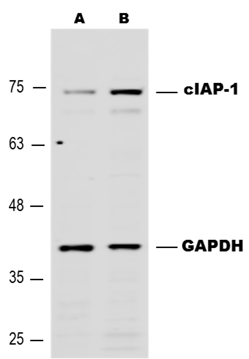
Application: Western BlotSample Tested: MCF-7 human breast cancer cell lineSpecies: HumanVerified Customer | Posted 03/27/2017Expression of hcIAP-1/HIAP-2 in MCF-7 cells untreated (A) and after treatment with 10 uM Paclitaxel for 4 hours (B) using Goat anti-Human cIAP-1/HIAP-2 antibody (#AF8181) at 0.5 ug/mL, followed by Donkey anti-Goat secondary antibody at 1:5,000 dilution.
-
Application: Western BlotSample Tested: See PMID 22553342Species: HumanVerified Customer | Posted 01/09/2015
-
Application: Western BlotSample Tested: See PMID 22212761Species: HumanVerified Customer | Posted 01/09/2015
-
Application: Western BlotSample Tested: See PMID 20074295Species: HumanVerified Customer | Posted 01/09/2015
-
Application: Western BlotSample Tested: See PMID 22711539Species: HumanVerified Customer | Posted 01/09/2015
There are no reviews that match your criteria.
Protocols
Find general support by application which include: protocols, troubleshooting, illustrated assays, videos and webinars.
- Antigen Retrieval Protocol (PIER)
- Antigen Retrieval for Frozen Sections Protocol
- Appropriate Fixation of IHC/ICC Samples
- Cellular Response to Hypoxia Protocols
- Chromogenic IHC Staining of Formalin-Fixed Paraffin-Embedded (FFPE) Tissue Protocol
- Chromogenic Immunohistochemistry Staining of Frozen Tissue
- ClariTSA™ Fluorophore Kits
- Detection & Visualization of Antibody Binding
- Fluorescent IHC Staining of Frozen Tissue Protocol
- Graphic Protocol for Heat-induced Epitope Retrieval
- Graphic Protocol for the Preparation and Fluorescent IHC Staining of Frozen Tissue Sections
- Graphic Protocol for the Preparation and Fluorescent IHC Staining of Paraffin-embedded Tissue Sections
- Graphic Protocol for the Preparation of Gelatin-coated Slides for Histological Tissue Sections
- IHC Sample Preparation (Frozen sections vs Paraffin)
- Immunofluorescent IHC Staining of Formalin-Fixed Paraffin-Embedded (FFPE) Tissue Protocol
- Immunohistochemistry (IHC) and Immunocytochemistry (ICC) Protocols
- Immunohistochemistry Frozen Troubleshooting
- Immunohistochemistry Paraffin Troubleshooting
- Preparing Samples for IHC/ICC Experiments
- Preventing Non-Specific Staining (Non-Specific Binding)
- Primary Antibody Selection & Optimization
- Protocol for Heat-Induced Epitope Retrieval (HIER)
- Protocol for Making a 4% Formaldehyde Solution in PBS
- Protocol for VisUCyte™ HRP Polymer Detection Reagent
- Protocol for the Preparation & Fixation of Cells on Coverslips
- Protocol for the Preparation and Chromogenic IHC Staining of Frozen Tissue Sections
- Protocol for the Preparation and Chromogenic IHC Staining of Frozen Tissue Sections - Graphic
- Protocol for the Preparation and Chromogenic IHC Staining of Paraffin-embedded Tissue Sections
- Protocol for the Preparation and Chromogenic IHC Staining of Paraffin-embedded Tissue Sections - Graphic
- Protocol for the Preparation and Fluorescent IHC Staining of Frozen Tissue Sections
- Protocol for the Preparation and Fluorescent IHC Staining of Paraffin-embedded Tissue Sections
- Protocol for the Preparation of Gelatin-coated Slides for Histological Tissue Sections
- R&D Systems Quality Control Western Blot Protocol
- TUNEL and Active Caspase-3 Detection by IHC/ICC Protocol
- The Importance of IHC/ICC Controls
- Troubleshooting Guide: Immunohistochemistry
- Troubleshooting Guide: Western Blot Figures
- Western Blot Conditions
- Western Blot Protocol
- Western Blot Protocol for Cell Lysates
- Western Blot Troubleshooting
- Western Blot Troubleshooting Guide
- View all Protocols, Troubleshooting, Illustrated assays and Webinars
Loading...
Associated Pathways


