Cerbeblon (CRBN) is a substrate recognition component of an E3 protein ligase complex which mediates ubiquitination and proteasomal degradation of target proteins. In embryonic development, degradation of regulatory proteins is required for normal limb outgrowth. Thalidomide, a teratogenic drug prescribed to pregnant women in the 1950s, binds to CRBN in the E3 protein ligase complex. Despite its teratogenic effect, CRBN is used to treat multiple myeloma and complications of leprosy. Widely expressed, CRBN may play a role in memory and learning by regulating assembly and surface expression of large-conductance calcium-activated potassium channels in hippocampal neurons. Mutations in the CRBN gene is linked to the Mental Retardation, Autosomal Recessive2a (MR2A) form of mental retardation.

Key Product Details
Species Reactivity
Human
Applications
Immunohistochemistry, Immunocytochemistry
Label
Unconjugated
Antibody Source
Monoclonal Mouse IgG2B Clone # 978015
Loading...
Product Specifications
Immunogen
Synthetic peptide Human CRBN
Accession # Q96SW2
Accession # Q96SW2
Specificity
Detects human CRBN in immunocytochemistry and immunohistochemistry.
Clonality
Monoclonal
Host
Mouse
Isotype
IgG2B
Scientific Data Images for Human CRBN Antibody (978015)
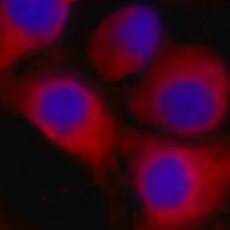
CRBN in RAW 264.7 Mouse Cell Line.
CRBN was detected in immersion fixed RAW 264.7 mouse monocyte/macrophage cell line using Mouse Anti-Human CRBN Polyclonal Antibody (Catalog # MAB9574) at 1 µg/mL for 3 hours at room temperature. Cells were stained using the NorthernLights™ 557-conjugated Anti-Mouse IgG Secondary Antibody (red; Catalog # NL007) and counterstained with DAPI (blue). Specific staining was localized to cytoplasm. View our protocol for Fluorescent ICC Staining of Non-adherent Cells.
CRBN in Human Testis.
CRBN was detected in immersion fixed paraffin-embedded sections of human testis using Mouse Anti-Human CRBN Polyclonal Antibody (Catalog # MAB9574) at 5 µg/mL for 1 hour at room temperature followed by incubation with the Anti-Mouse IgG VisUCyte™ HRP Polymer Antibody (Catalog # VC001). Tissue was stained using DAB (brown) and counterstained with hematoxylin (blue). Specific staining was localized to cytoplasm. View our protocol for IHC Staining with VisUCyte HRP Polymer Detection Reagents.Applications for Human CRBN Antibody (978015)
Application
Recommended Usage
Immunocytochemistry
5-25 µg/mL
Sample: Immersion fixed RAW 264.7 mouse monocyte/macrophage cell line
Sample: Immersion fixed RAW 264.7 mouse monocyte/macrophage cell line
Immunohistochemistry
1-25 µg/mL
Sample: Immersion fixed paraffin-embedded sections of human testis
Sample: Immersion fixed paraffin-embedded sections of human testis
Reviewed Applications
Read 1 review rated 5 using MAB9574 in the following applications:
Formulation, Preparation, and Storage
Purification
Protein A or G purified from hybridoma culture supernatant
Reconstitution
Reconstitute at 0.5 mg/mL in sterile PBS. For liquid material, refer to CoA for concentration.
Loading...
Formulation
Lyophilized from a 0.2 μm filtered solution in PBS with Trehalose. *Small pack size (SP) is supplied either lyophilized or as a 0.2 µm filtered solution in PBS.
Shipping
Lyophilized product is shipped at ambient temperature. Liquid small pack size (-SP) is shipped with polar packs. Upon receipt, store immediately at the temperature recommended below.
Stability & Storage
Use a manual defrost freezer and avoid repeated freeze-thaw cycles.
- 12 months from date of receipt, -20 to -70 °C as supplied.
- 1 month, 2 to 8 °C under sterile conditions after reconstitution.
- 6 months, -20 to -70 °C under sterile conditions after reconstitution.
Calculators
Background: CRBN
Long Name
Cereblon
Alternate Names
Cereblon, MRT2, MRT2A, Protein X 0001
Gene Symbol
CRBN
UniProt
Additional CRBN Products
Product Documents for Human CRBN Antibody (978015)
Certificate of Analysis
To download a Certificate of Analysis, please enter a lot or batch number in the search box below.
Note: Certificate of Analysis not available for kit components.
Product Specific Notices for Human CRBN Antibody (978015)
For research use only
Citations for Human CRBN Antibody (978015)
 Powered by Bioz
Customer Reviews for Human CRBN Antibody (978015) (1)
5 out of 5
1 Customer Rating
Have you used Human CRBN Antibody (978015)?
Submit a review and receive an Amazon gift card!
$25/€18/£15/$25CAN/¥2500 Yen for a review with an image
$10/€7/£6/$10CAN/¥1110 Yen for a review without an image
Submit a review
Customer Images
Showing
1
-
1 of
1 review
Showing All
Filter By:
-
Application: Immunocytochemistry/ImmunofluorescenceSample Tested: macrophagesSpecies: HumanVerified Customer | Posted 07/25/2022

There are no reviews that match your criteria.
Protocols
Find general support by application which include: protocols, troubleshooting, illustrated assays, videos and webinars.
- Antigen Retrieval Protocol (PIER)
- Antigen Retrieval for Frozen Sections Protocol
- Appropriate Fixation of IHC/ICC Samples
- Cellular Response to Hypoxia Protocols
- Chromogenic IHC Staining of Formalin-Fixed Paraffin-Embedded (FFPE) Tissue Protocol
- Chromogenic Immunohistochemistry Staining of Frozen Tissue
- ClariTSA™ Fluorophore Kits
- Detection & Visualization of Antibody Binding
- Fluorescent IHC Staining of Frozen Tissue Protocol
- Graphic Protocol for Heat-induced Epitope Retrieval
- Graphic Protocol for the Preparation and Fluorescent IHC Staining of Frozen Tissue Sections
- Graphic Protocol for the Preparation and Fluorescent IHC Staining of Paraffin-embedded Tissue Sections
- Graphic Protocol for the Preparation of Gelatin-coated Slides for Histological Tissue Sections
- ICC Cell Smear Protocol for Suspension Cells
- ICC Immunocytochemistry Protocol Videos
- ICC for Adherent Cells
- IHC Sample Preparation (Frozen sections vs Paraffin)
- Immunocytochemistry (ICC) Protocol
- Immunocytochemistry Troubleshooting
- Immunofluorescence of Organoids Embedded in Cultrex Basement Membrane Extract
- Immunofluorescent IHC Staining of Formalin-Fixed Paraffin-Embedded (FFPE) Tissue Protocol
- Immunohistochemistry (IHC) and Immunocytochemistry (ICC) Protocols
- Immunohistochemistry Frozen Troubleshooting
- Immunohistochemistry Paraffin Troubleshooting
- Preparing Samples for IHC/ICC Experiments
- Preventing Non-Specific Staining (Non-Specific Binding)
- Primary Antibody Selection & Optimization
- Protocol for Heat-Induced Epitope Retrieval (HIER)
- Protocol for Making a 4% Formaldehyde Solution in PBS
- Protocol for VisUCyte™ HRP Polymer Detection Reagent
- Protocol for the Fluorescent ICC Staining of Cell Smears - Graphic
- Protocol for the Fluorescent ICC Staining of Cultured Cells on Coverslips - Graphic
- Protocol for the Preparation & Fixation of Cells on Coverslips
- Protocol for the Preparation and Chromogenic IHC Staining of Frozen Tissue Sections
- Protocol for the Preparation and Chromogenic IHC Staining of Frozen Tissue Sections - Graphic
- Protocol for the Preparation and Chromogenic IHC Staining of Paraffin-embedded Tissue Sections
- Protocol for the Preparation and Chromogenic IHC Staining of Paraffin-embedded Tissue Sections - Graphic
- Protocol for the Preparation and Fluorescent ICC Staining of Cells on Coverslips
- Protocol for the Preparation and Fluorescent ICC Staining of Non-adherent Cells
- Protocol for the Preparation and Fluorescent ICC Staining of Stem Cells on Coverslips
- Protocol for the Preparation and Fluorescent IHC Staining of Frozen Tissue Sections
- Protocol for the Preparation and Fluorescent IHC Staining of Paraffin-embedded Tissue Sections
- Protocol for the Preparation of Gelatin-coated Slides for Histological Tissue Sections
- Protocol for the Preparation of a Cell Smear for Non-adherent Cell ICC - Graphic
- TUNEL and Active Caspase-3 Detection by IHC/ICC Protocol
- The Importance of IHC/ICC Controls
- Troubleshooting Guide: Immunohistochemistry
- View all Protocols, Troubleshooting, Illustrated assays and Webinars
Loading...