The G protein-coupled receptor, RDC1, belongs to a subgroup of chemokine receptors and has been designated CXCR7. CXCR7 can bind with high-affinity to CXCL12/SDF-1 and CXCL11/I-TAC. It is also a co-receptor for several HIV and SIV strains. In their N-termini and extracellular loops 1, 2, and 3, human and mouse CXCR7 share 84%, 100%, 96% and 86% amino acid sequence identity, respectively. Reports of mRNA levels and/or protein expression (as assessed using anti‑CXCR7, clone 9C4) (1, 2) indicate that CXCR7 occurs on a wide variety of tissues and cells including monocytes, B cells, T cells and mature dendritic cells. In contrast, based on ligand binding analysis and receptor level (as assessed using anti‑CXCR7, clone 11G8), surface expression of CXCR7 was reported to be restricted to tumor cells, activated endothelial cells, fetal liver cells, and few other cell types (3). The basis of these inconsistent observations is not known but may be attributed to cell context and the use of different antibodies that may recognize different epitopes.
Human CXCR7/RDC-1 Antibody (358440)
R&D Systems | Catalog # MAB42274

Key Product Details
Species Reactivity
Human
Applications
Immunohistochemistry, Immunocytochemistry
Label
Unconjugated
Antibody Source
Monoclonal Mouse IgG2A Clone # 358440
Loading...
Product Specifications
Immunogen
NS0 mouse myeloma cell line transfected with human CXCR7/RDC‑1
Met1-Lys362
Accession # AAA62370
Met1-Lys362
Accession # AAA62370
Specificity
Detects human CXCR7/RDC-1 in direct ELISAs.
Clonality
Monoclonal
Host
Mouse
Isotype
IgG2A
Scientific Data Images for Human CXCR7/RDC-1 Antibody (358440)
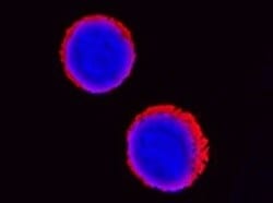
CXCR7/RDC‑1 in MCF‑7 Human Cell Line.
CXCR7/RDC‑1 was detected in immersion fixed MCF‑7 human breast cancer cell line using Mouse Anti-Human CXCR7/RDC‑1 Monoclonal Antibody (Catalog # MAB42274) at 8 µg/mL for 3 hours at room temperature. Cells were stained using the NorthernLights™ 557-conjugated Anti-Mouse IgG Secondary Antibody (red; NL007) and counterstained with DAPI (blue). Specific staining was localized to cell surface and cytoplasm. Staining was performed using our protocol for Fluorescent ICC Staining of Non-adherent Cells.
CXCR7/RDC‑1 in Human Breast Cancer Tissue.
CXCR7/RDC‑1 was detected in immersion fixed paraffin-embedded sections of human breast cancer tissue using Mouse Anti-Human CXCR7/RDC‑1 Monoclonal Antibody (Catalog # MAB42274) at 0.5 µg/mL for 1 hour at room temperature followed by incubation with the Anti-Mouse IgG VisUCyte™ HRP Polymer Antibody (VC001). Before incubation with the primary antibody, tissue was subjected to heat-induced epitope retrieval using Antigen Retrieval Reagent-Basic (CTS013). Tissue was stained using DAB (brown) and counterstained with hematoxylin (blue). Specific staining was localized to endothelial cells. Staining was performed using our protocol for IHC Staining with VisUCyte HRP Polymer Detection Reagents.Applications for Human CXCR7/RDC-1 Antibody (358440)
Application
Recommended Usage
Immunocytochemistry
8-25 µg/mL
Sample: Immersion fixed MCF‑7 human breast cancer cell line
Sample: Immersion fixed MCF‑7 human breast cancer cell line
Immunohistochemistry
0.5-25 µg/mL
Sample: Immersion fixed paraffin-embedded sections of human breast cancer tissue
Sample: Immersion fixed paraffin-embedded sections of human breast cancer tissue
Reviewed Applications
Read 1 review rated 5 using MAB42274 in the following applications:
Formulation, Preparation, and Storage
Purification
Protein A or G purified from hybridoma culture supernatant
Reconstitution
Reconstitute at 0.5 mg/mL in sterile PBS. For liquid material, refer to CoA for concentration.
Loading...
Formulation
Lyophilized from a 0.2 μm filtered solution in PBS with Trehalose. *Small pack size (SP) is supplied either lyophilized or as a 0.2 µm filtered solution in PBS.
Shipping
Lyophilized product is shipped at ambient temperature. Liquid small pack size (-SP) is shipped with polar packs. Upon receipt, store immediately at the temperature recommended below.
Stability & Storage
Use a manual defrost freezer and avoid repeated freeze-thaw cycles.
- 12 months from date of receipt, -20 to -70 °C as supplied.
- 1 month, 2 to 8 °C under sterile conditions after reconstitution.
- 6 months, -20 to -70 °C under sterile conditions after reconstitution.
Calculators
Background: CXCR7/RDC-1
References
- Balabanian, K. et al. (2005) J. Biol. Chem. 280:35760.
- Infantino, S. et al. (2006) J. Immunol. 176:2197.
- Burns, J.M. et al. (2006) J. Exp. Med. 203:2201.
Alternate Names
ACKR3, CMKOR1, CXCR7, GPR159, RDC-1
Gene Symbol
ACKR3
UniProt
Additional CXCR7/RDC-1 Products
Product Documents for Human CXCR7/RDC-1 Antibody (358440)
Certificate of Analysis
To download a Certificate of Analysis, please enter a lot or batch number in the search box below.
Note: Certificate of Analysis not available for kit components.
Product Specific Notices for Human CXCR7/RDC-1 Antibody (358440)
For research use only
Citations for Human CXCR7/RDC-1 Antibody (358440)
 Powered by Bioz
Customer Reviews for Human CXCR7/RDC-1 Antibody (358440) (1)
5 out of 5
1 Customer Rating
Have you used Human CXCR7/RDC-1 Antibody (358440)?
Submit a review and receive an Amazon gift card!
$25/€18/£15/$25CAN/¥2500 Yen for a review with an image
$10/€7/£6/$10CAN/¥1110 Yen for a review without an image
Submit a review
Customer Images
Showing
1
-
1 of
1 review
Showing All
Filter By:
-
Application: Immunocytochemistry/ImmunofluorescenceSample Tested: Blood mononuclear cells (PBMCs)Species: HumanVerified Customer | Posted 02/01/2022

There are no reviews that match your criteria.
Protocols
Find general support by application which include: protocols, troubleshooting, illustrated assays, videos and webinars.
- Antigen Retrieval Protocol (PIER)
- Antigen Retrieval for Frozen Sections Protocol
- Appropriate Fixation of IHC/ICC Samples
- Cellular Response to Hypoxia Protocols
- Chromogenic IHC Staining of Formalin-Fixed Paraffin-Embedded (FFPE) Tissue Protocol
- Chromogenic Immunohistochemistry Staining of Frozen Tissue
- ClariTSA™ Fluorophore Kits
- Detection & Visualization of Antibody Binding
- Fluorescent IHC Staining of Frozen Tissue Protocol
- Graphic Protocol for Heat-induced Epitope Retrieval
- Graphic Protocol for the Preparation and Fluorescent IHC Staining of Frozen Tissue Sections
- Graphic Protocol for the Preparation and Fluorescent IHC Staining of Paraffin-embedded Tissue Sections
- Graphic Protocol for the Preparation of Gelatin-coated Slides for Histological Tissue Sections
- ICC Cell Smear Protocol for Suspension Cells
- ICC Immunocytochemistry Protocol Videos
- ICC for Adherent Cells
- IHC Sample Preparation (Frozen sections vs Paraffin)
- Immunocytochemistry (ICC) Protocol
- Immunocytochemistry Troubleshooting
- Immunofluorescence of Organoids Embedded in Cultrex Basement Membrane Extract
- Immunofluorescent IHC Staining of Formalin-Fixed Paraffin-Embedded (FFPE) Tissue Protocol
- Immunohistochemistry (IHC) and Immunocytochemistry (ICC) Protocols
- Immunohistochemistry Frozen Troubleshooting
- Immunohistochemistry Paraffin Troubleshooting
- Preparing Samples for IHC/ICC Experiments
- Preventing Non-Specific Staining (Non-Specific Binding)
- Primary Antibody Selection & Optimization
- Protocol for Heat-Induced Epitope Retrieval (HIER)
- Protocol for Making a 4% Formaldehyde Solution in PBS
- Protocol for VisUCyte™ HRP Polymer Detection Reagent
- Protocol for the Fluorescent ICC Staining of Cell Smears - Graphic
- Protocol for the Fluorescent ICC Staining of Cultured Cells on Coverslips - Graphic
- Protocol for the Preparation & Fixation of Cells on Coverslips
- Protocol for the Preparation and Chromogenic IHC Staining of Frozen Tissue Sections
- Protocol for the Preparation and Chromogenic IHC Staining of Frozen Tissue Sections - Graphic
- Protocol for the Preparation and Chromogenic IHC Staining of Paraffin-embedded Tissue Sections
- Protocol for the Preparation and Chromogenic IHC Staining of Paraffin-embedded Tissue Sections - Graphic
- Protocol for the Preparation and Fluorescent ICC Staining of Cells on Coverslips
- Protocol for the Preparation and Fluorescent ICC Staining of Non-adherent Cells
- Protocol for the Preparation and Fluorescent ICC Staining of Stem Cells on Coverslips
- Protocol for the Preparation and Fluorescent IHC Staining of Frozen Tissue Sections
- Protocol for the Preparation and Fluorescent IHC Staining of Paraffin-embedded Tissue Sections
- Protocol for the Preparation of Gelatin-coated Slides for Histological Tissue Sections
- Protocol for the Preparation of a Cell Smear for Non-adherent Cell ICC - Graphic
- TUNEL and Active Caspase-3 Detection by IHC/ICC Protocol
- The Importance of IHC/ICC Controls
- Troubleshooting Guide: Immunohistochemistry
- View all Protocols, Troubleshooting, Illustrated assays and Webinars
Loading...
Associated Pathways
