Human DC-Sign (dendritic cell-specific ICAM-3 grabbing nonintegrin; also CD209) is a member of the chromosome 19 C-type lectin family that includes DC-SIGN, DC-SIGN-related protein, CD23 and LSECtin (1). DC-SIGN was initially reported to be a 46 kDa, 404 amino acid (aa) type II transmembrane protein that contained a 40 aa cytoplasmic N-terminus, a 21 aa transmembrane segment, and a 343 aa extracellular C-terminus (2). The extracellular region contains a distal, 115 aa Ca++-dependent carbohydrate-binding lectin domain and a membrane-proximal linker segment that is composed of seven 23 aa repeats (2, 3). The lectin domain is believed to preferably bind mannose, either within the context of ICAM-3 (on T cells) or ICAM-2 (on endothelial cells) (2, 4, 5). DC-SIGN expression appears to be limited to dendritic cells (DC) and macrophages (6), and DC interaction with the ICAMs both aids DC cell trafficking and immunological synapse formation (7). Since the original report on DC-SIGN, multiple splice forms have been discovered, generating both membrane-bound and soluble forms (3). There are eight type A isoforms, all of which begin with the same 15 aa of exon 1a. Four contain the transmembrane region of exon II, and four do not (i.e., are soluble). Among these eight type A isoforms, only three retain the entire 343 aa found in the full length form described in reference #2 (the full length form is referred to as type I mDC-SIGN1A) (3). Five additional isoforms utilize an alternate start site, and these are referred to as type B isoforms. These all show a 35 aa cytoplasmic domain. One also has a transmembrane segment; four do not. Two of the five contain full, unspliced extracellular regions (3). All of this suggests enormous complexity in DC-SIGN biology. DC-SIGN is not well conserved across species. Human and mouse show little overall aa identity. In the lectin domain, however, human DC-SIGN shares 68% aa identity with mouse DC-SIGN (8). Human and rhesus monkey DC-SIGN share 91% aa identity over the entire extracellular region (8). A detailed description of the additional properties of this monoclonal antibody (MAB161) have been published (9, 10).
Human DC-SIGN/CD209 Antibody (120507)
R&D Systems | Catalog # MAB161
Clone 120507 was used by HLDA to establish CD designation

Key Product Details
Validated by
Biological Validation
Species Reactivity
Validated:
Human
Cited:
Human, Mouse, Rat, Hamster, Insect - Mosquito, Macaca fascicularis fascicularis, Primate - Cercopithecus aethiops (African Green Monkey), Primate - Macaca fascicularis (Crab-eating Monkey or Cynomolgus Macaque), Primate - Macaca mulatta (Rhesus Macaque)
Applications
Validated:
Western Blot, Adhesion Blockade, Flow Cytometry, COMET, CyTOF-ready
Cited:
Immunohistochemistry, Immunohistochemistry-Paraffin, Immunohistochemistry-Frozen, Western Blot, Neutralization, Flow Cytometry, Immunocytochemistry, Blocking, ELISA Detection, ELISA Development, Functional Assay
Label
Unconjugated
Antibody Source
Monoclonal Mouse IgG2B Clone # 120507
Loading...
Product Specifications
Immunogen
NIH-3T3 mouse embryonic fibroblast cell line transfected with human DC‑SIGN/CD209
Specificity
Detects human DC‑SIGN/CD209 on transfected NIH/3T3 cells and on monocyte derived dendritic cells. Does not react with parental mouse cells or irrelevant transfectants, such as human DC-SIGN2.
Clonality
Monoclonal
Host
Mouse
Isotype
IgG2B
Endotoxin Level
<0.10 EU per 1 μg of the antibody by the LAL method.
Scientific Data Images for Human DC-SIGN/CD209 Antibody (120507)
Detection of DC-SIGN in Human Colon via seqIF™ staining on COMET™
DC-SIGN was detected in immersion fixed paraffin-embedded sections of human colon using Mouse Anti-Human DC-SIGN Monoclonal Antibody (Catalog # MAB161) at 0.5 µg/mL at 37 ° Celsius for 2 minutes. Before incubation with the primary antibody, tissue underwent an all-in-one dewaxing and antigen retrieval preprocessing using PreTreatment Module (PT Module) and Dewax and HIER Buffer H (pH 9; Epredia Catalog # TA-999-DHBH). Tissue was stained using the Alexa Fluor™ 647 Goat anti-Mouse IgG Secondary Antibody at 1:200 at 37 ° Celsius for 2 minutes. (Yellow; Lunaphore Catalog # DR647MS) and counterstained with DAPI (blue; Lunaphore Catalog # DR100). Specific staining was localized to the membrane and cyoplasm. Protocol available in COMET™ Panel Builder.
Detection of DC-SIGN in Human Liver via seqIF™ staining on COMET™
DC-SIGN was detected in immersion fixed paraffin-embedded sections of human liver using Mouse Anti-Human DC-SIGN Monoclonal Antibody (Catalog # MAB161) at 0.5 µg/mL at 37 ° Celsius for 2 minutes. Before incubation with the primary antibody, tissue underwent an all-in-one dewaxing and antigen retrieval preprocessing using PreTreatment Module (PT Module) and Dewax and HIER Buffer H (pH 9; Epredia Catalog # TA-999-DHBH). Tissue was stained using the Alexa Fluor™ 647 Goat anti-Mouse IgG Secondary Antibody at 1:200 at 37 ° Celsius for 2 minutes. (Yellow; Lunaphore Catalog # DR647MS) and counterstained with DAPI (blue; Lunaphore Catalog # DR100). Specific staining was localized to the membrane and cyoplasm. Protocol available in COMET™ Panel Builder.
Detection of DC-SIGN in Human Lymph Node via seqIF™ staining on COMET™
DC-SIGN was detected in immersion fixed paraffin-embedded sections of human lymph node using Mouse Anti-Human DC-SIGN Monoclonal Antibody (Catalog # MAB161) at 0.5 µg/mL at 37 ° Celsius for 2 minutes. Before incubation with the primary antibody, tissue underwent an all-in-one dewaxing and antigen retrieval preprocessing using PreTreatment Module (PT Module) and Dewax and HIER Buffer H (pH 9; Epredia Catalog # TA-999-DHBH). Tissue was stained using the Alexa Fluor™ 647 Goat anti-Mouse IgG Secondary Antibody at 1:200 at 37 ° Celsius for 2 minutes. (Yellow; Lunaphore Catalog # DR647MS) and counterstained with DAPI (blue; Lunaphore Catalog # DR100). Specific staining was localized to the membrane and cyoplasm. Protocol available in COMET™ Panel Builder.
Detection of DC‑SIGN in Human DC‑SIGN Transfected 3T3 Mouse Cell Line by Flow Cytometry.
Human DC-SIGN and DC-SIGN2 transfected 3T3 mouse embryonic fibroblast cell line were stained with Mouse Anti-Human DC-SIGN Monoclonal Antibody (Catalog # MAB161, filled histograms) or isotype control antibody (Catalog # MAB0041, open histogram), followed by Phycoerythrin-conjugated Anti-Mouse IgG F(ab')2Secondary Antibody (Catalog # F0102B).
Detection of DC‑SIGN in Human Monocyte Derived Dendritic Cells by Flow Cytometry.
Human monocyte derived dendritic cells were stained with Mouse Anti-Human DC-SIGN Monoclonal Antibody (Catalog # MAB161) followed by PE-conjugated anti-mouse IgG (Catalog # F0102B) and Anti-Human B7-2/CD86 Fluorescein-conjugated Monoclonal Antibody (Catalog # FAB141F). Quadrant markers were set based on control antibody staining (Catalog # MAB0041).
Detection of Human DC-SIGN/CD209 by Flow Cytometry
Infectivity of KSHV is enhanced in the presence of DC-SIGN and DC-SIGNR.A) 293T cells were transfected with empty pcDNA3 vector or expression constructs for DC-SIGN or DC-SIGNR. After 24 hours, cells were infected with 20 µl Bac16 delta K3 delta K5 or left uninfected as controls. Cells were harvested after additional 24 hours and surface stained with a DC-SIGN/R antibody (H-200) and analyzed by flow cytometry. Top three panels show transfected cells stained for DC-SIGN/R followed by PE- (DC-SIGN), FITC- (DC-SIGNR) or both (vector) conjugated secondary antibodies. Bottom panels shows KSHV infection of 293T cells transiently expressing DC-SIGN or DC-SIGNR. B, left panels) 293 cell lines stably expressing a vector construct, DC-SIGN or DC-SIGNR were fluorescently stained for surface expression of DC-SIGN or DC-SIGNR. The mean channel fluorescence is indicated in the upper right hand corner. Open histograms – secondary antibody alone; shaded histograms – DC-SIGN or DC-SIGNR staining. B, right panel) 293 pcDNA3, DC-SIGN or DC-SIGNR stable cell lines were pre-incubated with a control antibody (anti-ICAM1, 7 µg/ml), with mannan (100 µg/ml) or a monoclonal antibody specific for DC-SIGN (MAB161; 7 µg/ml) for 30 minutes on ice. These cells were then infected with wild type KSHV (Bac16 or rKSHV.219) at an MOI of 0.01. After 72 hours cells were harvested and evaluated for infection by flow cytometry measuring GFP expression. Infection rates were normalized to 293 pcDNA3 cells treated with the control antibody. The fold increase in relative infectivity is indicated. Data are representative of four independent experiments with two performed in triplicate. C) 293 pcDNA3, DC-SIGN or DC-SIGNR stable lines were infected with 50 µl of concentrated Bac16 wildtype (wt), or mutants with deletion of K3 only ( delta K3), K5 only ( delta K5), or deletion of both K3 and K5 ( delta K3 delta K5) as indicated. At 72 hours post-infection, the cells were stained for surface expression of DC-SIGN, DC-SIGNR or MHC class I. GFP fluore
Detection of Cynomolgus Monkey DC-SIGN/CD209 by Immunohistochemistry
Rapid induction and persistence of DC-SIGN, S100 and CD68 post-vaccination in lymphoid tissue.Galleries of representative fields of A DC-SIGN and B CD68 staining in MLN at 3, 7, 10, 21, 125 d.p.i. C Representative fields of DC-SIGN signal tracking through the spleen 3–125 d.p.i. maximal from d3 in red pulp (rp) and marginal zones (mg) D Representative fields of splenic S100 levels illustrate similar staining intensity 3–125 d.p.i. predominantly in the germinal centres and red pulp. Staining intensities scoring were; pale yellow (no staining, −); yellow (very low, +); dark yellow (low, ++), red (medium, +++) and magenta (high, ++++). Image collected and cropped by CiteAb from the following publication (https://pubmed.ncbi.nlm.nih.gov/25162725), licensed under a CC-BY license. Not internally tested by R&D Systems.
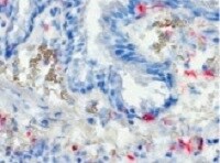
Detection of DC-SIGN/CD209 by Immunohistochemistry
Infection of DC-SIGN+ cells in the lung.(A) Infection of the DC-SIGNhi, DC-SIGNlo and DC-SIGN- cells in BAL from MV-infected macaques, determined by detection of EGFP in flow cytometry at day 2–5 d.p.i. Each dot represents an individual animal. Lines indicate geometric means. (B) Macroscopic images from EGFP+ lung slices collected 3 d.p.i., cultured for additional 3,5,7 or 10 days. (C) Phenotype of cells migrating from the ex vivo cultured lung slice, collected from supernatant after 5 days of culturing (D) Phenotype of EGFP+ cells collected from lung slice medium. (E-F) DC-SIGN expression on lung sections from uninfected macaques (E) or 2.d.p.i. (F) Asterisks indicate DC-SIGN reactivity. Image collected and cropped by CiteAb from the following open publication (https://pubmed.ncbi.nlm.nih.gov/23227146), licensed under a CC-BY license. Not internally tested by R&D Systems.
Detection of DC-SIGN/CD209 by Western Blot
K3 and K5 affect the stability of DC-SIGN and DC-SIGNR in a RING-CH domain-dependent mechanism.A) 293 cells stably expressing wild-type K3 or K5, or the RING-CH mutant of either viral protein were transiently transfected with 2 µg DC-SIGN or DC-SIGNR constructs. At ∼48 hpt,cells were lysed in RIPA buffer and 30 µg of normalized lysate were loaded per sample. Protein levels of DC-SIGN or DC-SIGNR were determined by WB, and then blots were reprobed for lamin B as a loading control. Data is representative of at least three independent experiments. B) THP-1 cell stably expressing the indicated K5 constructs or empty vector were stained for cell surface levels of DC-SIGN. Solid histogram, empty vector; grey histogram, K5 construct; dotted histogram, isotype control. C) The same THP-1 cell lines used in Panel B, were lysed and subjected to western blotting with either a DC-SIGN (H-200) or GAPDH (O411) antibody, as loading control. Data is representative of at least three independent experiments. Image collected and cropped by CiteAb from the following open publication (https://pubmed.ncbi.nlm.nih.gov/23460925), licensed under a CC-BY license. Not internally tested by R&D Systems.
Detection of DC-SIGN/CD209 by Immunocytochemistry/ Immunofluorescence
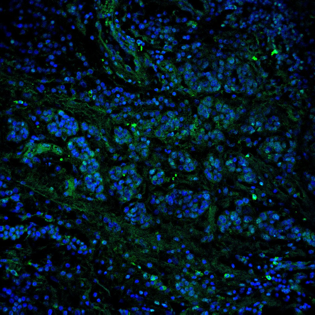
DC-SIGN is expressed by DCs and a subset of macrophages in lymph nodes.(A) TBLN cells were stained for DC-SIGN in combination with DC and macrophage markers and analyzed by flow cytometry. Gray areas show negative controls (DC-SIGN single staining). Percentages of positive cells expressing the markers are annotated in the upper right corner. (B) DC-SIGN expression of CD11c+ and CD83+ cells in TBLNs. (C-D) Dual immunofluorescence staining of DC-SIGN (green) and MAC387 (red) in lung sections 2 or 3 d.p.i. (C i-iv) and axillary lymphoid tissue 4 d.p.i. (D). Nuclei are stained blue with Hoechst. Image collected and cropped by CiteAb from the following open publication (https://pubmed.ncbi.nlm.nih.gov/23227146), licensed under a CC-BY license. Not internally tested by R&D Systems.Applications for Human DC-SIGN/CD209 Antibody (120507)
Application
Recommended Usage
Adhesion Blockade
The adhesion of NIH-3T3 mouse embryonic fibroblast cells (5 x 10⁴ cells/well) to immobilized Recombinant Human ICAM-3/CD50 Fc Chimera (Catalog # 715-IC, 5 µg/mL, 100 µL/well) was maximally inhibited (80-100%) by 5 µg/mL of the antibody.
COMET
0.5 µg/mL
Sample: Immersion fixed paraffin-embedded sections of human colon, liver and lymph node
Sample: Immersion fixed paraffin-embedded sections of human colon, liver and lymph node
CyTOF-ready
Ready to be labeled using established conjugation methods. No BSA or other carrier proteins that could interfere with conjugation.
Flow Cytometry
2.5 µg/106 cells
Sample: Human DC‑SIGN transfected 3T3 mouse embryonic fibroblast cell line and human monocyte derived dendritic cells
Sample: Human DC‑SIGN transfected 3T3 mouse embryonic fibroblast cell line and human monocyte derived dendritic cells
Western Blot
1 µg/mL
Sample: Recombinant Human DC-SIGN Fc Chimera (Catalog # 161-DC)
Sample: Recombinant Human DC-SIGN Fc Chimera (Catalog # 161-DC)
Reviewed Applications
Read 4 reviews rated 4.3 using MAB161 in the following applications:
Flow Cytometry Panel Builder
Bio-Techne Knows Flow Cytometry
Save time and reduce costly mistakes by quickly finding compatible reagents using the Panel Builder Tool.
Advanced Features
- Spectra Viewer - Custom analysis of spectra from multiple fluorochromes
- Spillover Popups - Visualize the spectra of individual fluorochromes
- Antigen Density Selector - Match fluorochrome brightness with antigen density

Formulation, Preparation, and Storage
Purification
Protein A or G purified from hybridoma culture supernatant
Reconstitution
Reconstitute at 0.5 mg/mL in sterile PBS. For liquid material, refer to CoA for concentration.
Loading...
Formulation
Lyophilized from a 0.2 μm filtered solution in PBS with Trehalose. See Certificate of Analysis for details.
*Small pack size (-SP) is supplied either lyophilized or as a 0.2 µm filtered solution in PBS.
*Small pack size (-SP) is supplied either lyophilized or as a 0.2 µm filtered solution in PBS.
Shipping
Lyophilized product is shipped at ambient temperature. Liquid small pack size (-SP) is shipped with polar packs. Upon receipt, store immediately at the temperature recommended below.
Stability & Storage
Use a manual defrost freezer and avoid repeated freeze-thaw cycles.
- 12 months from date of receipt, -20 to -70 °C as supplied.
- 1 month, 2 to 8 °C under sterile conditions after reconstitution.
- 6 months, -20 to -70 °C under sterile conditions after reconstitution.
Calculators
Background: DC-SIGN/CD209
References
- Liu, W. et al. (2004) J. Biol. Chem. 279:18748.
- Curtis, B.M. et al. (1992) Proc. Natl. Acad. Sci. USA 89:8356.
- Mummidi, S. et al. (2001) J. Biol. Chem. 276:33196.
- Su, S.V. et al. (2004) J. Biol. Chem. 279:19122.
- Cambi, A. et al. (2005) Cell. Microbiol. 7:481.
- Serrano-Gomez, D. et al. (2004) J. Immunol. 173:5635.
- Geijtenbeek, T.B.H. and Y. van Kooyk (2003) Curr. Top. Microbiol. Immunol. 276:32.
- Baribaud, F. et al. (2001) J. Virol. 75:10281.
- Wu, L. et al. (2002) J. Virol. 76:5905.
- Baribaud, F. et al. (2002) J. Virol.76:9135.
Long Name
Dendritic Cell-specific ICAM-3-grabbing Non-integrin 1
Alternate Names
CD209, CLEC4L, DC-SIGN1, DCSIGN
Gene Symbol
CD209
Additional DC-SIGN/CD209 Products
Product Documents for Human DC-SIGN/CD209 Antibody (120507)
Certificate of Analysis
To download a Certificate of Analysis, please enter a lot or batch number in the search box below.
Note: Certificate of Analysis not available for kit components.
Product Specific Notices for Human DC-SIGN/CD209 Antibody (120507)
For research use only
Related Research Areas
Citations for Human DC-SIGN/CD209 Antibody (120507)
 Powered by Bioz
Customer Reviews for Human DC-SIGN/CD209 Antibody (120507) (4)
4.3 out of 5
4 Customer Ratings
Have you used Human DC-SIGN/CD209 Antibody (120507)?
Submit a review and receive an Amazon gift card!
$25/€18/£15/$25CAN/¥2500 Yen for a review with an image
$10/€7/£6/$10CAN/¥1110 Yen for a review without an image
Submit a review
Customer Images
Showing
1
-
4 of
4 reviews
Showing All
Filter By:
-
Application: Immunocytochemistry/ImmunofluorescenceSample Tested: Melanoma tissueSpecies: HumanVerified Customer | Posted 11/04/2021

-
Application: ImmunohistochemistrySample Tested: Lung tissueSpecies: HumanVerified Customer | Posted 08/09/2021
-
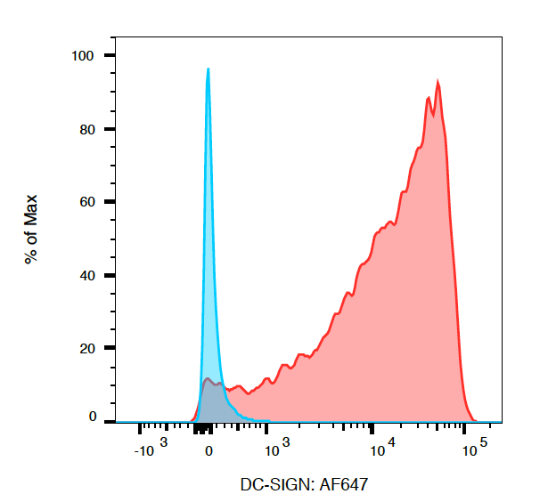
Application: Flow CytometrySample Tested: DCSIGN Transfected HEK293TSpecies: HumanVerified Customer | Posted 01/05/2018DC-SIGN transfected cells are seen in red and stained, untransfected cells are in blue. DC-SIGN AF647 conjugated was used, see other experimental details for more info.HEK293T cells were transfected with pcDNA3.DC-SIGN, 48hrs post-transfection cells were analyzed by flow cytometry. A 1/500 dilution of antibody was used to stain transfected cells (~2E6 cells).
-
Application: Flow CytometrySample Tested: Dendritic cellsSpecies: HumanVerified Customer | Posted 07/27/2016
There are no reviews that match your criteria.
Protocols
Find general support by application which include: protocols, troubleshooting, illustrated assays, videos and webinars.
- 7-Amino Actinomycin D (7-AAD) Cell Viability Flow Cytometry Protocol
- Cellular Response to Hypoxia Protocols
- Extracellular Membrane Flow Cytometry Protocol
- Flow Cytometry Protocol for Cell Surface Markers
- Flow Cytometry Protocol for Staining Membrane Associated Proteins
- Flow Cytometry Staining Protocols
- Flow Cytometry Troubleshooting Guide
- Intracellular Flow Cytometry Protocol Using Alcohol (Methanol)
- Intracellular Flow Cytometry Protocol Using Detergents
- Intracellular Nuclear Staining Flow Cytometry Protocol Using Detergents
- Intracellular Staining Flow Cytometry Protocol Using Alcohol Permeabilization
- Intracellular Staining Flow Cytometry Protocol Using Detergents to Permeabilize Cells
- Propidium Iodide Cell Viability Flow Cytometry Protocol
- Protocol for Liperfluo
- Protocol for the Characterization of Human Th22 Cells
- Protocol for the Characterization of Human Th9 Cells
- Protocol: Annexin V and PI Staining by Flow Cytometry
- Protocol: Annexin V and PI Staining for Apoptosis by Flow Cytometry
- R&D Systems Quality Control Western Blot Protocol
- Troubleshooting Guide: Fluorokine Flow Cytometry Kits
- Troubleshooting Guide: Western Blot Figures
- Western Blot Conditions
- Western Blot Protocol
- Western Blot Protocol for Cell Lysates
- Western Blot Troubleshooting
- Western Blot Troubleshooting Guide
- View all Protocols, Troubleshooting, Illustrated assays and Webinars


