Epithelial Cellular Adhesion Molecule (EpCAM), also known as KS1/4, gp40, GA733-2, 17-1A, and TROP-1, is a 40 kDa transmembrane glycoprotein composed of a 242 amino acid (aa) extracellular domain with two epidermal-growth-factor-like (EGF-like) repeats within the cysteine-rich N-terminal region, a 23 aa transmembrane domain, and a 26 aa cytoplasmic domain. Human and mouse EpCAM share 82% aa sequence identity. In human, EpCAM also shares 49% aa sequence homology with Trop-2/EGP-1. During embryonic development, EpCAM is detected in fetal lung, kidney, liver, pancreas, skin, and germ cells. In adults, human EpCAM is detected in basolateral cell membranes of all simple, pseudo-stratified, and transitional epithelia, but is not detected in normal squamous stratified epithelia, mesenchymal tissue, muscular tissue, neuro-endocrine tissue, or lymphoid tissue (1). EpCAM expression has been found to increase in actively proliferating epithelia tissues and during adult liver regeneration (1, 2). EpCAM expression is also found to increase in human malignant neoplasias, with most carcinoma expressing EpCAM including those of arising from squamousal epithelia (1). EpCAM has been shown function as a homophilic Ca2+ independent adhesion molecule (3). Homophilic adhesion via EpCAM requires the interaction of both EGF-like repeats, with the first EGF-like repeat mediating reciprocal interaction between EpCAM molecules on opposing cells, while the second repeat is involved in lateral interaction of EpCAM. Lateral interaction of EpCAM lead to the formation of dimers and tetramers (4). During homophilic adhesion the cytoplasmic tail of EpCAM interacts with the actin cytoskeleton via a direct association alpha -actinin (5).
Human EpCAM/TROP1 Antibody (158210)
R&D Systems | Catalog # MAB960

Key Product Details
Species Reactivity
Validated:
Human
Cited:
Human, Mouse, Plant - Brassica Rapa, Plant - Nicotiana benthamiana, Xenograft
Applications
Validated:
Multiplex Immunofluorescence, Immunohistochemistry, Western Blot, Immunocytochemistry, COMET
Cited:
Immunohistochemistry, Immunohistochemistry-Paraffin, Western Blot, Neutralization, Flow Cytometry, Immunocytochemistry, Adhesion Assay, Cell Capture, ELISA Capture, Phagocytosis Assay
Label
Unconjugated
Antibody Source
Monoclonal Mouse IgG2A Clone # 158210
Loading...
Product Specifications
Immunogen
Mouse myeloma cell line NS0-derived recombinant human EpCAM/TROP-1
Gln24-Lys265
Accession # P16422
Gln24-Lys265
Accession # P16422
Specificity
Detects human EpCAM/TROP-1 in direct ELISAs and Western blots. This antibody detects an epitope found in the extracellular domain between amino acids 136 and 265. In direct ELISAs and Western blots, no cross-reactivity with recombinant human (rh) ALCAM, rhBCAM, rhMCAM, rhNCAM-L1, or recombinant mouse (rm) OCAM is observed. In direct ELISAs, less than 5% cross-reactivity with rmEpCAM/TROP-1 is observed.
Clonality
Monoclonal
Host
Mouse
Isotype
IgG2A
Scientific Data Images for Human EpCAM/TROP1 Antibody (158210)
Detection of EpCAM/TROP-1 in Human Colon via seqIF™ staining on COMET™
EpCAM/TROP-1 was detected in immersion fixed paraffin-embedded sections of human Colon using Mouse Anti-Human EpCAM/TROP-1, Monoclonal Antibody (Catalog # MAB960) at 1ug/mL at 37° Celsius for 4 minutes. Before incubation with the primary antibody, tissue underwent an all-in-one dewaxing and antigen retrieval preprocessing using PreTreatment Module (PT Module) and Dewax and HIER Buffer H (pH 9; Epredia Catalog # TA-999-DHBH). Tissue was stained using the Alexa Fluor™ 555 Goat anti-Mouse IgG Secondary Antibody at 1:100 at 37° Celsius for 2 minutes. (Yellow; Lunaphore Catalog # DR555MS) and counterstained with DAPI (blue; Lunaphore Catalog # DR100). Specific staining was localized to the membrane. Protocol available in COMET™ Panel Builder.
Detection of EpCAM/TROP-1 in Human Lung Cancer via seqIF™ staining on COMET™
EpCAM/TROP-1 was detected in immersion fixed paraffin-embedded sections of human Lung Cancer using Mouse Anti-Human EpCAM/TROP-1, Monoclonal Antibody (Catalog # MAB960) at 1ug/mL at 37° Celsius for 4 minutes. Before incubation with the primary antibody, tissue underwent an all-in-one dewaxing and antigen retrieval preprocessing using PreTreatment Module (PT Module) and Dewax and HIER Buffer H (pH 9; Epredia Catalog # TA-999-DHBH). Tissue was stained using the Alexa Fluor™ 555 Goat anti-Mouse IgG Secondary Antibody at 1:100 at 37 ° Celsius for 2 minutes. (Yellow; Lunaphore Catalog # DR555MS) and counterstained with DAPI (blue; Lunaphore Catalog # DR100). Specific staining was localized to the membrane. Protocol available in COMET™ Panel Builder.
Detection of EpCAM/TROP-1 in Human Kidney via seqIF™ staining on COMET™
EpCAM/TROP-1 was detected in immersion fixed paraffin-embedded sections of human Kidney using Mouse Anti-Human EpCAM/TROP-1, Monoclonal Antibody (Catalog # MAB960) at 1ug/mL at 37° Celsius for 4 minutes. Before incubation with the primary antibody, tissue underwent an all-in-one dewaxing and antigen retrieval preprocessing using PreTreatment Module (PT Module) and Dewax and HIER Buffer H (pH 9; Epredia Catalog # TA-999-DHBH). Tissue was stained using the Alexa Fluor™ 555 Goat anti-Mouse IgG Secondary Antibody at 1:100 at 37 ° Celsius for 2 minutes. (Yellow; Lunaphore Catalog # DR555MS) and counterstained with DAPI (blue; Lunaphore Catalog # DR100). Specific staining was localized to the membrane. Protocol available in COMET™ Panel Builder.
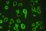
EpCAM/TROP‑1 in HT-29 Human Cell Line.
EpCAM/TROP-1 was detected in immersion fixed HT-29 human colon adenocarcinoma cell line using Mouse Anti-Human EpCAM/TROP-1 Monoclonal Antibody (Catalog # MAB960) at 10 µg/mL for 3 hours at room temperature. Cells were stained using the NorthernLights™ 493-conjugated Anti-Mouse IgG Secondary Antibody (green; NL009) and counterstained with DAPI (blue). View our protocol for Fluorescent ICC Staining of Cells on Coverslips.
EpCAM/TROP‑1 in HT-29 Human Cell Line.
EpCAM/TROP-1 was detected in immersion fixed HT-29 human colon adenocarcinoma cell line using Mouse Anti-Human EpCAM/TROP-1 Monoclonal Antibody (Catalog # MAB960) at 10 µg/mL for 3 hours at room temperature. Cells were stained using the NorthernLights™ 557-conjugated Anti-Mouse IgG Secondary Antibody (yellow; NL007) and counterstained with DAPI (blue). View our protocol for Fluorescent ICC Staining of Cells on Coverslips.
EpCAM/TROP‑1 in Human Adenocarcinoma.
EpCAM/TROP-1 was detected in immersion fixed paraffin-embedded sections of human adenocarcinoma using Mouse Anti-Human EpCAM/TROP-1 Monoclonal Antibody (Catalog # MAB960) at 15 µg/mL overnight at 4 °C. Tissue was stained using the Anti-Mouse HRP-DAB Cell & Tissue Staining Kit (brown; CTS002) and counterstained with hematoxylin (blue). Specific staining was localized to cancer cells. View our protocol for Chromogenic IHC Staining of Paraffin-embedded Tissue Sections. and Daudi Human Burkitt's Lymphoma Cell Line (Negative) Cells.")
Detection of EpCAM/TROP‑1 in HT‑29 Human Colon Adenocarcinoma Cell Line (Positive) and Daudi Human Burkitt's Lymphoma Cell Line (Negative) Cells.
EpCAM/TROP‑1 was detected in immersion fixed HT‑29 human colon adenocarcinoma cell line (positive) and Daudi human Burkitt's lymphoma cell line (negative) cells using Mouse Anti-Human EpCAM/TROP‑1 Monoclonal Antibody (Catalog # MAB960) at 8 µg/mL for 3 hours at room temperature. Cells were stained using the NorthernLights™ 557-conjugated Anti-Mouse IgG Secondary Antibody (red; Catalog # NL007) and counterstained with DAPI (blue). Specific staining was localized to cell surface and cytoplasm. View our protocol for Fluorescent ICC Staining of Cells on Coverslips.Applications for Human EpCAM/TROP1 Antibody (158210)
Application
Recommended Usage
COMET
Optimal dilutions of this antibody should be experimentally determined.
Immunocytochemistry
8-25 µg/mL
Sample: Immersion fixed HT‑29 human colon adenocarcinoma cell line (positive) and Daudi human Burkitt's lymphoma cell line (negative) cells
Sample: Immersion fixed HT‑29 human colon adenocarcinoma cell line (positive) and Daudi human Burkitt's lymphoma cell line (negative) cells
Immunohistochemistry
8-25 µg/mL
Sample: Immersion fixed paraffin-embedded sections of human adenocarcinoma
Sample: Immersion fixed paraffin-embedded sections of human adenocarcinoma
Multiplex Immunofluorescence
3-25 µg/mL
Sample: Immersion fixed paraffin-embedded sections of human colon
Sample: Immersion fixed paraffin-embedded sections of human colon
Western Blot
1 µg/mL
Sample: Recombinant Human EpCAM/TROP‑1 Fc Chimera (Catalog # 960-EP)
Sample: Recombinant Human EpCAM/TROP‑1 Fc Chimera (Catalog # 960-EP)
Reviewed Applications
Read 2 reviews rated 4.5 using MAB960 in the following applications:
Formulation, Preparation, and Storage
Purification
Protein A or G purified from hybridoma culture supernatant
Reconstitution
Reconstitute at 0.5 mg/mL in sterile PBS. For liquid material, refer to CoA for concentration.
Loading...
Formulation
Lyophilized from a 0.2 μm filtered solution in PBS with Trehalose. See Certificate of Analysis for details.
*Small pack size (-SP) is supplied either lyophilized or as a 0.2 µm filtered solution in PBS.
*Small pack size (-SP) is supplied either lyophilized or as a 0.2 µm filtered solution in PBS.
Shipping
Lyophilized product is shipped at ambient temperature. Liquid small pack size (-SP) is shipped with polar packs. Upon receipt, store immediately at the temperature recommended below.
Stability & Storage
Use a manual defrost freezer and avoid repeated freeze-thaw cycles.
- 12 months from date of receipt, -20 to -70 °C as supplied.
- 1 month, 2 to 8 °C under sterile conditions after reconstitution.
- 6 months, -20 to -70 °C under sterile conditions after reconstitution.
Calculators
Background: EpCAM/TROP1
References
- Balzar, M. et al. (1999) J. Mol. Med. 77:699.
- Boer, C.J. et al. (1999) J. Pathol. 188:201.
- Litvinow, S.V. et al. (1994) J. Cell Biol. 125:437.
- Balzar, M. et al. (2001) Mol. Cell. Biol. 21:2570.
- Balzar, M. et al. (1998) Mol. Cell. Biol. 18:4388.
Long Name
Epithelial Cell Adhesion Molecule
Alternate Names
17-1A, CD326, GA733-2, gp40, KS1/4, M4S1, TACSTD1, TROP1
Gene Symbol
EPCAM
UniProt
Additional EpCAM/TROP1 Products
Product Documents for Human EpCAM/TROP1 Antibody (158210)
Certificate of Analysis
To download a Certificate of Analysis, please enter a lot or batch number in the search box below.
Note: Certificate of Analysis not available for kit components.
Product Specific Notices for Human EpCAM/TROP1 Antibody (158210)
For research use only
Citations for Human EpCAM/TROP1 Antibody (158210)
 Powered by Bioz
Customer Reviews for Human EpCAM/TROP1 Antibody (158210) (2)
4.5 out of 5
2 Customer Ratings
Have you used Human EpCAM/TROP1 Antibody (158210)?
Submit a review and receive an Amazon gift card!
$25/€18/£15/$25CAN/¥2500 Yen for a review with an image
$10/€7/£6/$10CAN/¥1110 Yen for a review without an image
Submit a review
Customer Images
Showing
1
-
2 of
2 reviews
Showing All
Filter By:
-
Application: ImmunohistochemistrySample Tested: Lung tissueSpecies: HumanVerified Customer | Posted 10/13/2021

-
Application: Western BlotSample Tested: NCI-N87 human gastric carcinoma cell line, HT-29 human colon adenocarcinoma cell line and LoVo human colorectal adenocarcinoma cell lineSpecies: HumanVerified Customer | Posted 07/16/2018
There are no reviews that match your criteria.
Protocols
Find general support by application which include: protocols, troubleshooting, illustrated assays, videos and webinars.
- Antigen Retrieval Protocol (PIER)
- Antigen Retrieval for Frozen Sections Protocol
- Appropriate Fixation of IHC/ICC Samples
- Cellular Response to Hypoxia Protocols
- Chromogenic IHC Staining of Formalin-Fixed Paraffin-Embedded (FFPE) Tissue Protocol
- Chromogenic Immunohistochemistry Staining of Frozen Tissue
- ClariTSA™ Fluorophore Kits
- Detection & Visualization of Antibody Binding
- Fluorescent IHC Staining of Frozen Tissue Protocol
- Graphic Protocol for Heat-induced Epitope Retrieval
- Graphic Protocol for the Preparation and Fluorescent IHC Staining of Frozen Tissue Sections
- Graphic Protocol for the Preparation and Fluorescent IHC Staining of Paraffin-embedded Tissue Sections
- Graphic Protocol for the Preparation of Gelatin-coated Slides for Histological Tissue Sections
- ICC Cell Smear Protocol for Suspension Cells
- ICC Immunocytochemistry Protocol Videos
- ICC for Adherent Cells
- IHC Sample Preparation (Frozen sections vs Paraffin)
- Immunocytochemistry (ICC) Protocol
- Immunocytochemistry Troubleshooting
- Immunofluorescence of Organoids Embedded in Cultrex Basement Membrane Extract
- Immunofluorescent IHC Staining of Formalin-Fixed Paraffin-Embedded (FFPE) Tissue Protocol
- Immunohistochemistry (IHC) and Immunocytochemistry (ICC) Protocols
- Immunohistochemistry Frozen Troubleshooting
- Immunohistochemistry Paraffin Troubleshooting
- Preparing Samples for IHC/ICC Experiments
- Preventing Non-Specific Staining (Non-Specific Binding)
- Primary Antibody Selection & Optimization
- Protocol for Heat-Induced Epitope Retrieval (HIER)
- Protocol for Making a 4% Formaldehyde Solution in PBS
- Protocol for VisUCyte™ HRP Polymer Detection Reagent
- Protocol for the Fluorescent ICC Staining of Cell Smears - Graphic
- Protocol for the Fluorescent ICC Staining of Cultured Cells on Coverslips - Graphic
- Protocol for the Preparation & Fixation of Cells on Coverslips
- Protocol for the Preparation and Chromogenic IHC Staining of Frozen Tissue Sections
- Protocol for the Preparation and Chromogenic IHC Staining of Frozen Tissue Sections - Graphic
- Protocol for the Preparation and Chromogenic IHC Staining of Paraffin-embedded Tissue Sections
- Protocol for the Preparation and Chromogenic IHC Staining of Paraffin-embedded Tissue Sections - Graphic
- Protocol for the Preparation and Fluorescent ICC Staining of Cells on Coverslips
- Protocol for the Preparation and Fluorescent ICC Staining of Non-adherent Cells
- Protocol for the Preparation and Fluorescent ICC Staining of Stem Cells on Coverslips
- Protocol for the Preparation and Fluorescent IHC Staining of Frozen Tissue Sections
- Protocol for the Preparation and Fluorescent IHC Staining of Paraffin-embedded Tissue Sections
- Protocol for the Preparation of Gelatin-coated Slides for Histological Tissue Sections
- Protocol for the Preparation of a Cell Smear for Non-adherent Cell ICC - Graphic
- R&D Systems Quality Control Western Blot Protocol
- TUNEL and Active Caspase-3 Detection by IHC/ICC Protocol
- The Importance of IHC/ICC Controls
- Troubleshooting Guide: Immunohistochemistry
- Troubleshooting Guide: Western Blot Figures
- Western Blot Conditions
- Western Blot Protocol
- Western Blot Protocol for Cell Lysates
- Western Blot Troubleshooting
- Western Blot Troubleshooting Guide
- View all Protocols, Troubleshooting, Illustrated assays and Webinars

