Fascin (that which creates fascicles [bundles] of actin; also known as 55 kDa actin-bundling protein, p55 and singed-like protein) is an intracellular 55-58 kDa member of the fascin family of proteins. It has a restricted expression pattern, being found in oligodendrocytes, select endothelium, cerebellar stellate neurons and blood, interdigitating, and thymic medullary dendritic cells. Fascin is found associated with actin in filopodia, and serves to coordinate and stabilize actin bundle formation, both in normal cells and tumor cells. In the latter cell type, filopodia have been renamed invadopodia, and their appearance is crucial for the creation of a stable platform that coordinates local matrix degradation. Human fascin is 493 amino acids (aa) in length. It contains an N-terminal fascin-like domain (aa 139-256) that contains part of one of two actin-binding sequences (aa 136-143), followed by two additional fascin-like domains (aa 260-378 and 383-493), the latter of which contains the second actin-binding sequence (aa 386-395). There are also two acetylation sites and two utilized phosphorylation sites at Ser38 and Ser39. Phosphorylation of the latter site inhibits fascin interaction with actin. At least two isoform variants may exist. One contains a 12 aa substitution for aa 427-493, while a second shows a deletion of aa 371-426. Full-length human fascin shares 97% aa sequence identity with mouse fascin. Two additional human fascins termed retinal and testis fascin have been identified. They are products of distinct genes and share 56% and 27% aa sequence identity with the standard (p55) fascin, respectively.

Key Product Details
Validated by
Knockout/Knockdown
Species Reactivity
Human
Applications
Knockout Validated, Immunohistochemistry, Western Blot, Simple Western
Label
Unconjugated
Antibody Source
Monoclonal Mouse IgG2A Clone # 833223
Loading...
Product Specifications
Immunogen
E. coli-derived recombinant human Fascin
Met1-Tyr493
Accession # Q16658
Met1-Tyr493
Accession # Q16658
Specificity
Detects human Fascin in ELISAs and Western blots. Detects mouse and rat Fascin in Western blots. In direct ELISAs, no cross-reactivity with recombinant
human Fascin-2 is observed.
Clonality
Monoclonal
Host
Mouse
Isotype
IgG2A
Scientific Data Images for Human Fascin Antibody (833223)
Detection of Human, Mouse, and Rat Fascin by Western Blot.
Western blot shows lysates of HeLa human cervical epithelial carcinoma cell line, K562 human chronic myelogenous leukemia cell line, Neuro-2A mouse neuroblastoma cell line, and Rat-2 rat embryonic fibroblast cell line. PVDF membrane was probed with 1 µg/mL of Mouse Anti-Human Fascin Monoclonal Antibody (Catalog # MAB7745) followed by HRP-conjugated Anti-Mouse IgG Secondary Antibody (Catalog # HAF018). A specific band was detected for Fascin at approximately 55 kDa (as indicated). This experiment was conducted under reducing conditions and using Immunoblot Buffer Group 1.
Fascin in Human Liver.
Fascin was detected in immersion fixed paraffin-embedded sections of human liver using Mouse Anti-Human Fascin Monoclonal Antibody (Catalog # MAB7745) at 15 µg/mL overnight at 4 °C. Before incubation with the primary antibody, tissue was subjected to heat-induced epitope retrieval using Antigen Retrieval Reagent-Basic (Catalog # CTS013). Tissue was stained using the Anti-Mouse HRP-DAB Cell & Tissue Staining Kit (brown; Catalog # CTS002) and counterstained with hematoxylin (blue). Specific staining was localized to synusoids. View our protocol for Chromogenic IHC Staining of Paraffin-embedded Tissue Sections.
Detection of Human Fascin by Simple WesternTM.
Simple Western lane view shows lysates of HeLa human cervical epithelial carcinoma cell line, loaded at 0.5 mg/mL. A specific band was detected for Fascin at approximately 61 kDa (as indicated) using 10 µg/mL of Mouse Anti-Human Fascin Monoclonal Antibody (Catalog # MAB7745). This experiment was conducted under reducing conditions and using the 12-230 kDa separation system.
Western Blot Shows Human Fascin Specificity by Using Knockout Cell Line.
Western blot shows lysates of HeLa human cervical epithelial carcinoma parental cell line and Fascin knockout HeLa cell line (KO). PVDF membrane was probed with 1 µg/mL of Mouse Anti-Human Fascin Monoclonal Antibody (Catalog # MAB7745) followed by HRP-conjugated Anti-Mouse IgG Secondary Antibody (Catalog # HAF018). A specific band was detected for Fascin at approximately 55 kDa (as indicated) in the parental HeLa cell line, but is not detectable in knockout HeLa cell line. GAPDH (Catalog # MAB5718) is shown as a loading control. This experiment was conducted under reducing conditions and using Immunoblot Buffer Group 1. New adjunct appears with knockout cell line.Applications for Human Fascin Antibody (833223)
Application
Recommended Usage
Immunohistochemistry
8-25 µg/mL
Sample: Immersion fixed paraffin-embedded sections of human liver
Sample: Immersion fixed paraffin-embedded sections of human liver
Knockout Validated
Fascin
is specifically detected in HeLa human cervical epithelial carcinoma parental cell line but is not detectable in
Fascin knockout HeLa cell line.
Simple Western
10 µg/mL
Sample: HeLa human cervical epithelial carcinoma cell line
Sample: HeLa human cervical epithelial carcinoma cell line
Western Blot
1 µg/mL
Sample: HeLa human cervical epithelial carcinoma cell line, K562 human chronic myelogenous leukemia cell line, Neuro‑2A mouse neuroblastoma cell line, and Rat‑2 rat embryonic fibroblast cell line
Sample: HeLa human cervical epithelial carcinoma cell line, K562 human chronic myelogenous leukemia cell line, Neuro‑2A mouse neuroblastoma cell line, and Rat‑2 rat embryonic fibroblast cell line
Reviewed Applications
Read 2 reviews rated 5 using MAB7745 in the following applications:
Formulation, Preparation, and Storage
Purification
Protein A or G purified from hybridoma culture supernatant
Reconstitution
Sterile PBS to a final concentration of 0.5 mg/mL. For liquid material, refer to CoA for concentration.
Loading...
Formulation
Lyophilized from a 0.2 μm filtered solution in PBS with Trehalose. *Small pack size (SP) is supplied either lyophilized or as a 0.2 µm filtered solution in PBS.
Shipping
Lyophilized product is shipped at ambient temperature. Liquid small pack size (-SP) is shipped with polar packs. Upon receipt, store immediately at the temperature recommended below.
Stability & Storage
Use a manual defrost freezer and avoid repeated freeze-thaw cycles.
- 12 months from date of receipt, -20 to -70 °C as supplied.
- 1 month, 2 to 8 °C under sterile conditions after reconstitution.
- 6 months, -20 to -70 °C under sterile conditions after reconstitution.
Calculators
Background: Fascin
Alternate Names
FAN1, FSCN1, HSN, Singed-like Protein, SNL
Gene Symbol
FSCN1
UniProt
Additional Fascin Products
Product Documents for Human Fascin Antibody (833223)
Certificate of Analysis
To download a Certificate of Analysis, please enter a lot or batch number in the search box below.
Note: Certificate of Analysis not available for kit components.
Product Specific Notices for Human Fascin Antibody (833223)
For research use only
Related Research Areas
Customer Reviews for Human Fascin Antibody (833223) (2)
5 out of 5
2 Customer Ratings
Have you used Human Fascin Antibody (833223)?
Submit a review and receive an Amazon gift card!
$25/€18/£15/$25CAN/¥2500 Yen for a review with an image
$10/€7/£6/$10CAN/¥1110 Yen for a review without an image
Submit a review
Customer Images
Showing
1
-
2 of
2 reviews
Showing All
Filter By:
-
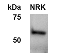
Application: Western BlotSample Tested: NRK whole cell lysateSpecies: RatVerified Customer | Posted 06/05/2015Western Blot of Fascin1 in NRK cells

-
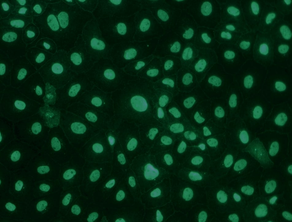
Application: ImmunofluorescenceSample Tested: NRK cellsSpecies: RatVerified Customer | Posted 05/22/2015immunofluorescence of Fascin in NRK cells
There are no reviews that match your criteria.
Protocols
Find general support by application which include: protocols, troubleshooting, illustrated assays, videos and webinars.
- Antigen Retrieval Protocol (PIER)
- Antigen Retrieval for Frozen Sections Protocol
- Appropriate Fixation of IHC/ICC Samples
- Cellular Response to Hypoxia Protocols
- Chromogenic IHC Staining of Formalin-Fixed Paraffin-Embedded (FFPE) Tissue Protocol
- Chromogenic Immunohistochemistry Staining of Frozen Tissue
- ClariTSA™ Fluorophore Kits
- Detection & Visualization of Antibody Binding
- Fluorescent IHC Staining of Frozen Tissue Protocol
- Graphic Protocol for Heat-induced Epitope Retrieval
- Graphic Protocol for the Preparation and Fluorescent IHC Staining of Frozen Tissue Sections
- Graphic Protocol for the Preparation and Fluorescent IHC Staining of Paraffin-embedded Tissue Sections
- Graphic Protocol for the Preparation of Gelatin-coated Slides for Histological Tissue Sections
- IHC Sample Preparation (Frozen sections vs Paraffin)
- Immunofluorescent IHC Staining of Formalin-Fixed Paraffin-Embedded (FFPE) Tissue Protocol
- Immunohistochemistry (IHC) and Immunocytochemistry (ICC) Protocols
- Immunohistochemistry Frozen Troubleshooting
- Immunohistochemistry Paraffin Troubleshooting
- Preparing Samples for IHC/ICC Experiments
- Preventing Non-Specific Staining (Non-Specific Binding)
- Primary Antibody Selection & Optimization
- Protocol for Heat-Induced Epitope Retrieval (HIER)
- Protocol for Making a 4% Formaldehyde Solution in PBS
- Protocol for VisUCyte™ HRP Polymer Detection Reagent
- Protocol for the Preparation & Fixation of Cells on Coverslips
- Protocol for the Preparation and Chromogenic IHC Staining of Frozen Tissue Sections
- Protocol for the Preparation and Chromogenic IHC Staining of Frozen Tissue Sections - Graphic
- Protocol for the Preparation and Chromogenic IHC Staining of Paraffin-embedded Tissue Sections
- Protocol for the Preparation and Chromogenic IHC Staining of Paraffin-embedded Tissue Sections - Graphic
- Protocol for the Preparation and Fluorescent IHC Staining of Frozen Tissue Sections
- Protocol for the Preparation and Fluorescent IHC Staining of Paraffin-embedded Tissue Sections
- Protocol for the Preparation of Gelatin-coated Slides for Histological Tissue Sections
- R&D Systems Quality Control Western Blot Protocol
- TUNEL and Active Caspase-3 Detection by IHC/ICC Protocol
- The Importance of IHC/ICC Controls
- Troubleshooting Guide: Immunohistochemistry
- Troubleshooting Guide: Western Blot Figures
- Western Blot Conditions
- Western Blot Protocol
- Western Blot Protocol for Cell Lysates
- Western Blot Troubleshooting
- Western Blot Troubleshooting Guide
- View all Protocols, Troubleshooting, Illustrated assays and Webinars
Loading...