
Key Product Details
Validated by
Knockout/Knockdown
Species Reactivity
Validated:
Human
Cited:
Human
Applications
Validated:
Knockout Validated, Western Blot, ELISA Capture (Matched Antibody Pair), Flow Cytometry, Immunocytochemistry, CyTOF-ready
Cited:
Western Blot, Flow Cytometry, Immunocytochemistry, ELISA Capture, ELISA Detection, ELISA Development, ELISA Development (Capture)
Label
Unconjugated
Antibody Source
Monoclonal Mouse IgG1 Clone # 548908
Loading...
Product Specifications
Immunogen
Chinese hamster ovary cell line CHO-derived recombinant human FOLR1
Arg25-Met233
Accession # P15328
Arg25-Met233
Accession # P15328
Specificity
Detects human FOLR1 in direct ELISAs and Western blots. In direct ELISAs, no cross-reactivity with recombinant human FOLR2, 3 or 4 is observed.
Clonality
Monoclonal
Host
Mouse
Isotype
IgG1
Scientific Data Images for Human FOLR1 Antibody (548908)
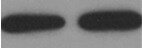
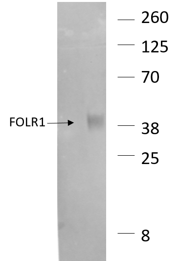
Detection of Human FOLR1 by Western Blot.
Western blot shows lysates of human cortex tissue and HeLa human cervical epithelial carcinoma cell line. PVDF membrane was probed with 2 µg/mL of Mouse Anti-Human FOLR1 Monoclonal Antibody (Catalog # MAB5646) followed by HRP-conjugated Anti-Mouse IgG Secondary Antibody (HAF007). A specific band was detected for FOLR1 at approximately 40 kDa (as indicated). This experiment was conducted using Immunoblot Buffer Group 1. Use under non-reducing conditions only.
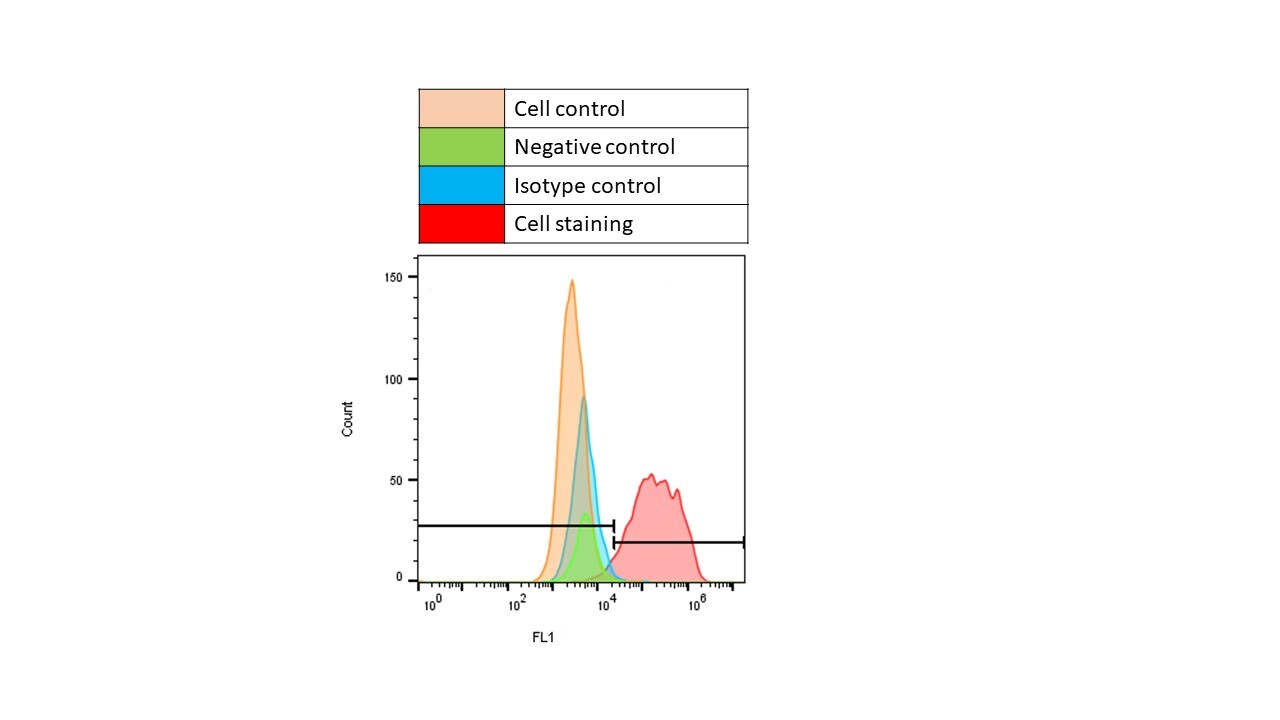
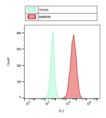
Detection of FOLR1 in MCF‑7 Human Cell Line by Flow Cytometry.
MCF-7 human breast cancer cell line was stained with Mouse Anti-Human FOLR1 Monoclonal Antibody (Catalog # MAB5646, filled histogram) or isotype control antibody (MAB002, open histogram), followed by Phycoerythrin-conjugated Anti-Mouse IgG Secondary Antibody (F0102B). View our protocol for Staining Membrane-associated Proteins.
FOLR1 in MCF-7 Human Cell Line.
FOLR1 was detected in immersion fixed MCF-7 human breast cancer cell line using 10 µg/mL Mouse Anti-Human FOLR1 Monoclonal Antibody (Catalog # MAB5646) for 3 hours at room temperature. Cells were stained with the NorthernLights™ 557-conjugated Anti-Mouse IgG Secondary Antibody (red; NL007) and counterstained with DAPI (blue). View our protocol for Fluorescent ICC Staining of Cells on Coverslips.
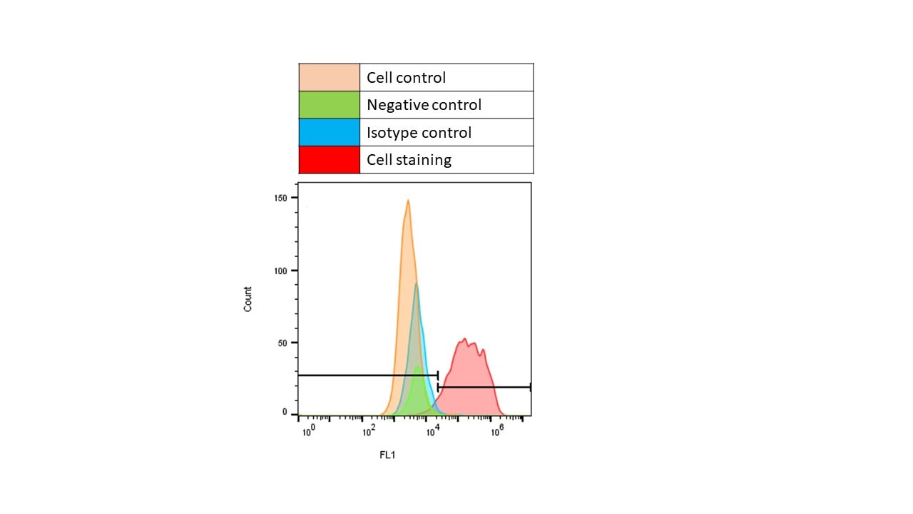
FOLR1 Specificity is Shown by Flow Cytometry in Knockout Cell Line.
FOLR1 knockout MCF-7 human breast cancer cell line was stained with Mouse Anti-Human FOLR1 Monoclonal Antibody (Catalog # MAB5646, filled histogram) or isotype control antibody (MAB002, open histogram) followed by anti-Mouse IgG PE-conjugated secondary antibody (F0102B). No staining in the FOLR1 knockout MCF-7 cell line was observed. View our protocol for Staining Membrane-associated Proteins.
Detection of Human FOLR1 by Immunocytochemistry/Immunofluorescence
(A) Immunofluorescent (left) and flow cytometry (right) detection of the EpCAM expression in MCF7 cells and the FR alpha expression in A2780 cells. EpCAM and FR alpha stained with Alexa Fluor® 488 are green at an excitation of 488 nm, and the nuclei stained with DAPI are blue at an excitation of 405 nm. As a negative control, we used EpCAM to stain Jurkat cells, which are EpCAM negatively expressed, and we used FR alpha to stain A549 cells, which are FR alpha negatively expressed. Histograms of flow cytometric analysis: MCF7 cells (top) were stained with anti-EpCAM antibodies (red) and A2780 cells (bottom) were stained with anti-FR alpha antibodies (red), the negative control was autofluorescent (orange), and the isotype was mouse IgG plus secondary antibody (blue). (B) Schematic of our CTCs enrichment strategy. Anti-EpCAM-MNs and Anti-FR alpha -MNs was added to the whole blood and after incubation, magnetic separation and fluorescence identification, the cells of DAPI+/CK+/CD45− were defined as CTC. Image collected and cropped by CiteAb from the following publication (https://pubmed.ncbi.nlm.nih.gov/29352248), licensed under a CC-BY license. Not internally tested by R&D Systems.
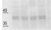
Detection of FOLR1 in HeLa cells by Flow Cytometry
HeLa cells were stained with Mouse Anti-Human FOLR1 Monoclonal Antibody (Catalog # MAB5646, filled histogram) or isotype control antibody (Catalog # MAB002, open histogram) followed by Phycoerythrin-conjugated Anti-Mouse IgG Secondary Antibody (Catalog # F0102B). View our protocol for Staining Membrane-associated Proteins.
Human FOLR1 ELISA Standard Curve
Recombinant Human FOLR1 (Catalog # 5646-FR) was serially diluted and captured by Mouse Anti-Human FOLR1 Monoclonal Antibody (Catalog # MAB5646) coated on a Clear Polystyrene Microplate (Catalog # DY990). Goat Anti-Human FOLR1 Antigen Affinity-purified Polyclonal Antibody (Catalog # AF5646) was biotinylated and incubated with the protein captured on the plate. Detection of the standard curve was achieved by incubating Streptavidin-HRP (Catalog # DY998)Applications for Human FOLR1 Antibody (548908)
Application
Recommended Usage
CyTOF-ready
Ready to be labeled using established conjugation methods. No BSA or other carrier proteins that could interfere with conjugation.
Flow Cytometry
0.25 µg/106 cells
Sample: MCF‑7 human breast cancer cell line; HeLa human cervical epithelial carcinoma cell line
Sample: MCF‑7 human breast cancer cell line; HeLa human cervical epithelial carcinoma cell line
Immunocytochemistry
8-25 µg/mL
Sample: Immersion fixed MCF-7 human breast cancer cell line
Sample: Immersion fixed MCF-7 human breast cancer cell line
Knockout Validated
FOLR1 is specifically detected in MCF-7 human breast cancer parental cell line but is not detectable in FOLR1 knockout MCF-7 cell line.
Western Blot
2 µg/mL
Sample: Human cortex tissue and HeLa human cervical epithelial carcinoma cell line under non-reducing conditions only
Sample: Human cortex tissue and HeLa human cervical epithelial carcinoma cell line under non-reducing conditions only
Human FOLR1 Sandwich Immunoassay
Please Note: Optimal dilutions of this antibody should be experimentally determined.
Reviewed Applications
Read 8 reviews rated 3.9 using MAB5646 in the following applications:
Flow Cytometry Panel Builder
Bio-Techne Knows Flow Cytometry
Save time and reduce costly mistakes by quickly finding compatible reagents using the Panel Builder Tool.
Advanced Features
- Spectra Viewer - Custom analysis of spectra from multiple fluorochromes
- Spillover Popups - Visualize the spectra of individual fluorochromes
- Antigen Density Selector - Match fluorochrome brightness with antigen density

Formulation, Preparation, and Storage
Purification
Protein A or G purified from hybridoma culture supernatant
Reconstitution
Reconstitute at 0.5 mg/mL in sterile PBS. For liquid material, refer to CoA for concentration.
Loading...
Formulation
Lyophilized from a 0.2 μm filtered solution in PBS with Trehalose. *Small pack size (SP) is supplied either lyophilized or as a 0.2 µm filtered solution in PBS.
Shipping
Lyophilized product is shipped at ambient temperature. Liquid small pack size (-SP) is shipped with polar packs. Upon receipt, store immediately at the temperature recommended below.
Stability & Storage
Use a manual defrost freezer and avoid repeated freeze-thaw cycles.
- 12 months from date of receipt, -20 to -70 °C as supplied.
- 1 month, 2 to 8 °C under sterile conditions after reconstitution.
- 6 months, -20 to -70 °C under sterile conditions after reconstitution.
Calculators
Background: FOLR1
References
- Kelemen, L.E. (2006) Int. J. Cancer 119:243.
- Fowler, B. et al. (2001) Kidney Int. 59:S-221.
- Luhrs, C.A. et al. (1989) J. Biol. Chem. 264:21446.
- Lacey, S.W. et al. (1989) J. Clin. Invest. 84:715.
- Elwood, P.C. (1989) J. Biol. Chem. 264:14893.
- Rijnboutt, S. et al. (1996) J. Cell Biol. 132:35.
- Ross, J.F. et al. (1994) Cancer 73:2432.
- Parker, N. et al. (2005) Anal. Biochem. 338:284.
- Piedrahita, J.A. et al. (1999) Nat. Genet. 23:228.
- Paulos, C.M. et al. (2004) Mol. Pharmacol. 66:1406.
- Elwood, P.C. et al. (1991) J. Biol. Chem. 26:2346.
Long Name
Folate Receptor 1
Alternate Names
FBP, Folbp1, FR-alpha, MOv18
Gene Symbol
FOLR1
UniProt
Additional FOLR1 Products
Product Documents for Human FOLR1 Antibody (548908)
Certificate of Analysis
To download a Certificate of Analysis, please enter a lot or batch number in the search box below.
Note: Certificate of Analysis not available for kit components.
Product Specific Notices for Human FOLR1 Antibody (548908)
For research use only
Related Research Areas
Citations for Human FOLR1 Antibody (548908)
 Powered by Bioz
Customer Reviews for Human FOLR1 Antibody (548908) (8)
3.9 out of 5
8 Customer Ratings
Have you used Human FOLR1 Antibody (548908)?
Submit a review and receive an Amazon gift card!
$25/€18/£15/$25CAN/¥2500 Yen for a review with an image
$10/€7/£6/$10CAN/¥1110 Yen for a review without an image
Submit a review
Customer Images

Showing
1
-
5 of
8 reviews
Showing All
Filter By:
-
Application: Western BlotSample Tested: Human cancer cell lineSpecies: HumanVerified Customer | Posted 08/26/2021

-
Application: Flow CytometrySample Tested: IGROV1 cell lineSpecies: HumanVerified Customer | Posted 04/10/2019
-
Application: Flow CytometrySample Tested: IGROV cell line and IGROV1 cell lineSpecies: HumanVerified Customer | Posted 04/07/2019
-
Application: Western BlotSample Tested: Human cancer cell lineSpecies: HumanVerified Customer | Posted 10/04/2018After very long exposure gave a band of an expected size. Very weak signal even with ECL Plus.
-
Application: Western BlotSample Tested: SK-OV-3 human ovarian adenocarcinoma cell line, igrov-1, NCI-H292, HT-29 human colon adenocarcinoma cell line and CAOV3Species: HumanVerified Customer | Posted 05/08/2018
-
Application: Flow CytometrySample Tested: human cancer cell lineSpecies: HumanVerified Customer | Posted 04/03/2018
-
Application: Flow CytometrySample Tested: Human cancer cell lineSpecies: HumanVerified Customer | Posted 03/21/2018
-
Application: Western BlotSample Tested: Ovarian cancer tissueSpecies: HumanVerified Customer | Posted 02/23/2016
Bio-Techne ResponseR&D Systems Technical Service will investigate.
There are no reviews that match your criteria.
Protocols
Find general support by application which include: protocols, troubleshooting, illustrated assays, videos and webinars.
- 7-Amino Actinomycin D (7-AAD) Cell Viability Flow Cytometry Protocol
- Appropriate Fixation of IHC/ICC Samples
- Cellular Response to Hypoxia Protocols
- ClariTSA™ Fluorophore Kits
- Detection & Visualization of Antibody Binding
- Extracellular Membrane Flow Cytometry Protocol
- Flow Cytometry Protocol for Cell Surface Markers
- Flow Cytometry Protocol for Staining Membrane Associated Proteins
- Flow Cytometry Staining Protocols
- Flow Cytometry Troubleshooting Guide
- ICC Cell Smear Protocol for Suspension Cells
- ICC Immunocytochemistry Protocol Videos
- ICC for Adherent Cells
- Immunocytochemistry (ICC) Protocol
- Immunocytochemistry Troubleshooting
- Immunofluorescence of Organoids Embedded in Cultrex Basement Membrane Extract
- Immunohistochemistry (IHC) and Immunocytochemistry (ICC) Protocols
- Intracellular Flow Cytometry Protocol Using Alcohol (Methanol)
- Intracellular Flow Cytometry Protocol Using Detergents
- Intracellular Nuclear Staining Flow Cytometry Protocol Using Detergents
- Intracellular Staining Flow Cytometry Protocol Using Alcohol Permeabilization
- Intracellular Staining Flow Cytometry Protocol Using Detergents to Permeabilize Cells
- Preparing Samples for IHC/ICC Experiments
- Preventing Non-Specific Staining (Non-Specific Binding)
- Primary Antibody Selection & Optimization
- Propidium Iodide Cell Viability Flow Cytometry Protocol
- Protocol for Liperfluo
- Protocol for VisUCyte™ HRP Polymer Detection Reagent
- Protocol for the Characterization of Human Th22 Cells
- Protocol for the Characterization of Human Th9 Cells
- Protocol for the Fluorescent ICC Staining of Cell Smears - Graphic
- Protocol for the Fluorescent ICC Staining of Cultured Cells on Coverslips - Graphic
- Protocol for the Preparation and Fluorescent ICC Staining of Cells on Coverslips
- Protocol for the Preparation and Fluorescent ICC Staining of Non-adherent Cells
- Protocol for the Preparation and Fluorescent ICC Staining of Stem Cells on Coverslips
- Protocol for the Preparation of a Cell Smear for Non-adherent Cell ICC - Graphic
- Protocol: Annexin V and PI Staining by Flow Cytometry
- Protocol: Annexin V and PI Staining for Apoptosis by Flow Cytometry
- R&D Systems Quality Control Western Blot Protocol
- TUNEL and Active Caspase-3 Detection by IHC/ICC Protocol
- The Importance of IHC/ICC Controls
- Troubleshooting Guide: Fluorokine Flow Cytometry Kits
- Troubleshooting Guide: Western Blot Figures
- Western Blot Conditions
- Western Blot Protocol
- Western Blot Protocol for Cell Lysates
- Western Blot Troubleshooting
- Western Blot Troubleshooting Guide
- View all Protocols, Troubleshooting, Illustrated assays and Webinars
Loading...