Galectin-10 (also eosinophil lysophospholipase and Charcot-Leyden Crystal protein) is a 16 kDa member of the lectin family of proteins. It is expressed intracellularly by eosinophils, basophils and CD25+ Treg cells. Although originally believed to possess lysophospholipase activity, this has been shown to be incorrect. It is known to bind lysophospholipase and its inhibitors, and to bind mannose in a very unusual manner. Human Galectin-10 is 142 amino acids (aa) in length. There is one galectin domain (aa 6-138) that contains two dimerization motifs (aa 6-10 and 131-135). Two molecular weight isoforms of 15 and 14 kDa have been described. Human Galectin-10 has no known structural counterpart in rodents.
Human Galectin-10 Antibody (561603)
R&D Systems | Catalog # MAB5447

Key Product Details
Species Reactivity
Human
Applications
Western Blot, Flow Cytometry, Immunocytochemistry, CyTOF-ready
Label
Unconjugated
Antibody Source
Monoclonal Mouse IgG2B Clone # 561603
Loading...
Product Specifications
Immunogen
E. coli-derived recombinant human Galectin-10
Met1-Arg142
Accession # Q05315
Met1-Arg142
Accession # Q05315
Specificity
Detects human Galectin-10 in direct ELISAs and Western blots. In direct ELISAs, no cross-reactivity with recombinant human Galectin-1, -2, -3, -4, -7, -8, or -9/Ecalectin is observed.
Clonality
Monoclonal
Host
Mouse
Isotype
IgG2B
Scientific Data Images for Human Galectin-10 Antibody (561603)
Detection of Galectin‑10 in Human PBMCs by Flow Cytometry.
Human peripheral blood lymphocytes were stained with Human Galectin-10 Monoclonal Antibody (Catalog # MAB5447) followed by Fluorescein-conjugated Anti-Mouse IgG Secondary Antibody (Catalog # F0103B) and Human IL-2 Ra APC-conjugated Monoclonal Antibody (Catalog # FAB1020A). Quadrant markers were set based on control antibody staining (Catalog # IC0041Fand IC003A).
Detection of Human Galectin‑10 by Western Blot.
Western blot shows lysates of human eosinophils (enriched, approximately 60%). PVDF Membrane was probed with 1 µg/mL of Human Galectin-10 Monoclonal Antibody (Catalog # MAB5447) followed by HRP-conjugated Anti-Mouse IgG Secondary Antibody (Catalog # HAF007). A specific band was detected for Galectin-10 at approximately 16 kDa (as indicated). This experiment was conducted under reducing conditions and using Immunoblot Buffer Group 1.
Galectin‑10 in HL‑60 Human Cell Line.
Galectin-10 was detected in immersion fixed HL-60 human acute promyelocytic leukemia cell line using Mouse Anti-Human Galectin-10 Monoclonal Antibody (Catalog # MAB5447) at 8 µg/mL for 3 hours at room temperature. Cells were stained using the NorthernLights™ 557-conjugated Anti-Mouse IgG Secondary Antibody (red; Catalog # NL007) and counterstained with DAPI (blue). Specific staining was localized to cytoplasm. View our protocol for Fluorescent ICC Staining of Non-adherent Cells.Applications for Human Galectin-10 Antibody (561603)
Application
Recommended Usage
CyTOF-ready
Ready to be labeled using established conjugation methods. No BSA or other carrier proteins that could interfere with conjugation.
Flow Cytometry
0.25 µg/106 cells
Sample: Human peripheral blood lymphocytes
Sample: Human peripheral blood lymphocytes
Immunocytochemistry
5-25 µg/mL
Sample: Immersion fixed HL‑60 human acute promyelocytic leukemia cell line
Sample: Immersion fixed HL‑60 human acute promyelocytic leukemia cell line
Western Blot
1 µg/mL
Sample: Human eosinophils (enriched, approximately 60%)
Sample: Human eosinophils (enriched, approximately 60%)
Reviewed Applications
Read 1 review rated 4 using MAB5447 in the following applications:
Flow Cytometry Panel Builder
Bio-Techne Knows Flow Cytometry
Save time and reduce costly mistakes by quickly finding compatible reagents using the Panel Builder Tool.
Advanced Features
- Spectra Viewer - Custom analysis of spectra from multiple fluorochromes
- Spillover Popups - Visualize the spectra of individual fluorochromes
- Antigen Density Selector - Match fluorochrome brightness with antigen density

Formulation, Preparation, and Storage
Purification
Protein A or G purified from hybridoma culture supernatant
Reconstitution
Sterile PBS to a final concentration of 0.5 mg/mL. For liquid material, refer to CoA for concentration.
Loading...
Formulation
Lyophilized from a 0.2 μm filtered solution in PBS with Trehalose. *Small pack size (SP) is supplied either lyophilized or as a 0.2 µm filtered solution in PBS.
Shipping
Lyophilized product is shipped at ambient temperature. Liquid small pack size (-SP) is shipped with polar packs. Upon receipt, store immediately at the temperature recommended below.
Stability & Storage
Use a manual defrost freezer and avoid repeated freeze-thaw cycles.
- 12 months from date of receipt, -20 to -70 °C as supplied.
- 1 month, 2 to 8 °C under sterile conditions after reconstitution.
- 6 months, -20 to -70 °C under sterile conditions after reconstitution.
Calculators
Background: Galectin-10
Alternate Names
CLC, GAL10, Galectin10, LGALS10
Entrez Gene IDs
1178 (Human)
Gene Symbol
CLC
UniProt
Additional Galectin-10 Products
Product Documents for Human Galectin-10 Antibody (561603)
Certificate of Analysis
To download a Certificate of Analysis, please enter a lot or batch number in the search box below.
Note: Certificate of Analysis not available for kit components.
Product Specific Notices for Human Galectin-10 Antibody (561603)
For research use only
Related Research Areas
Citations for Human Galectin-10 Antibody (561603)
 Powered by Bioz
Customer Reviews for Human Galectin-10 Antibody (561603) (1)
4 out of 5
1 Customer Rating
Have you used Human Galectin-10 Antibody (561603)?
Submit a review and receive an Amazon gift card!
$25/€18/£15/$25CAN/¥2500 Yen for a review with an image
$10/€7/£6/$10CAN/¥1110 Yen for a review without an image
Submit a review
Customer Images
Showing
1
-
1 of
1 review
Showing All
Filter By:
-
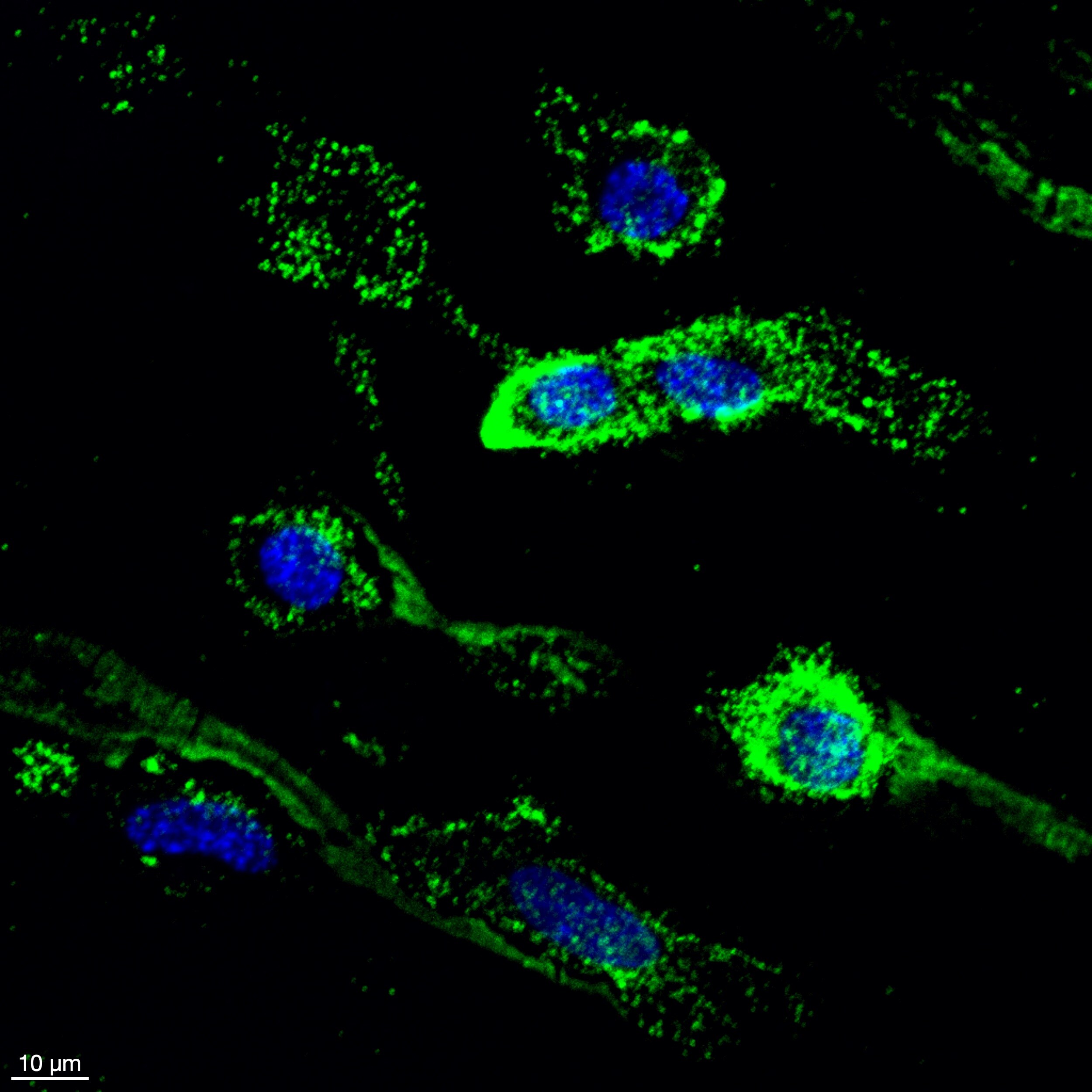
Application: ImmunocytochemistrySample Tested: ccd-841-conSpecies: HumanVerified Customer | Posted 04/18/2026Dapi (Blue) and Galectin-10 (Green)In vitro immunostaining with galectin-10 (1:1000)

There are no reviews that match your criteria.
Protocols
Find general support by application which include: protocols, troubleshooting, illustrated assays, videos and webinars.
- 7-Amino Actinomycin D (7-AAD) Cell Viability Flow Cytometry Protocol
- Appropriate Fixation of IHC/ICC Samples
- Cellular Response to Hypoxia Protocols
- ClariTSA™ Fluorophore Kits
- Detection & Visualization of Antibody Binding
- Extracellular Membrane Flow Cytometry Protocol
- Flow Cytometry Protocol for Cell Surface Markers
- Flow Cytometry Protocol for Staining Membrane Associated Proteins
- Flow Cytometry Staining Protocols
- Flow Cytometry Troubleshooting Guide
- ICC Cell Smear Protocol for Suspension Cells
- ICC Immunocytochemistry Protocol Videos
- ICC for Adherent Cells
- Immunocytochemistry (ICC) Protocol
- Immunocytochemistry Troubleshooting
- Immunofluorescence of Organoids Embedded in Cultrex Basement Membrane Extract
- Immunohistochemistry (IHC) and Immunocytochemistry (ICC) Protocols
- Intracellular Flow Cytometry Protocol Using Alcohol (Methanol)
- Intracellular Flow Cytometry Protocol Using Detergents
- Intracellular Nuclear Staining Flow Cytometry Protocol Using Detergents
- Intracellular Staining Flow Cytometry Protocol Using Alcohol Permeabilization
- Intracellular Staining Flow Cytometry Protocol Using Detergents to Permeabilize Cells
- Preparing Samples for IHC/ICC Experiments
- Preventing Non-Specific Staining (Non-Specific Binding)
- Primary Antibody Selection & Optimization
- Propidium Iodide Cell Viability Flow Cytometry Protocol
- Protocol for Liperfluo
- Protocol for VisUCyte™ HRP Polymer Detection Reagent
- Protocol for the Characterization of Human Th22 Cells
- Protocol for the Characterization of Human Th9 Cells
- Protocol for the Fluorescent ICC Staining of Cell Smears - Graphic
- Protocol for the Fluorescent ICC Staining of Cultured Cells on Coverslips - Graphic
- Protocol for the Preparation and Fluorescent ICC Staining of Cells on Coverslips
- Protocol for the Preparation and Fluorescent ICC Staining of Non-adherent Cells
- Protocol for the Preparation and Fluorescent ICC Staining of Stem Cells on Coverslips
- Protocol for the Preparation of a Cell Smear for Non-adherent Cell ICC - Graphic
- Protocol: Annexin V and PI Staining by Flow Cytometry
- Protocol: Annexin V and PI Staining for Apoptosis by Flow Cytometry
- R&D Systems Quality Control Western Blot Protocol
- TUNEL and Active Caspase-3 Detection by IHC/ICC Protocol
- The Importance of IHC/ICC Controls
- Troubleshooting Guide: Fluorokine Flow Cytometry Kits
- Troubleshooting Guide: Western Blot Figures
- Western Blot Conditions
- Western Blot Protocol
- Western Blot Protocol for Cell Lysates
- Western Blot Troubleshooting
- Western Blot Troubleshooting Guide
- View all Protocols, Troubleshooting, Illustrated assays and Webinars
Loading...