Interleukin 15 receptor alpha (IL-15 R alpha ) is a high affinity receptor that specifically binds IL-15 with high affinity and associates as a heterotrimer with the IL-2 receptors beta and gamma subunits to initiate signal transduction. IL-15 R alpha is expressed on a wide variety of T cells and B cells as well as non-lymphoid cells.
IL‑15 R alpha is a 58-60 kDa protein that shares structural similarities to the IL-2 R alpha protein. IL-15 R alpha and IL-2 R alpha genes also share similar intron-exon organization and are closely linked on human chromosome 10p14-p15. Human IL-15 R alpha shares 45% amino acid (aa) homology with the mouse form of the receptor. Eight isoforms of
IL‑15 R alpha mRNA have been identified resulting from alternative splicing events involving different exons. The exclusion of exon 2 results in an IL-15 R alpha isoform that does not bind IL-15. Human IL-15 R alpha DE3 cDNA encodes a 267 amino acid (aa) protein that contains a 30 aa signal sequence, a 175 aa extracellular region containing one N-linked glycosylation site, a 21 aa transmembrane domain, and a 41 aa cytoplasmic tail. Signaling of IL-15 can occur in one of three ways; through the heterotrimeric complex of IL-15 R alpha, IL-2 R beta, and IL-2 R gamma c, through the heterodimeric complex of IL-2 receptors beta and gamma common, through a novel 60-65 kDa IL-15 RX subunit found on mast cells. The binding of IL-15 to IL-15 R alpha has been reported to antagonize the TNF-alpha -mediated apoptosis in fibroblasts by competing with TNF RI for TRAF2 binding.

Key Product Details
Species Reactivity
Validated:
Human
Cited:
Human, Mouse, Primate - Macaca mulatta (Rhesus Macaque)
Applications
Validated:
Immunohistochemistry, Western Blot, Neutralization, Flow Cytometry, Dual RNAscope ISH-IHC Compatible, Immunocytochemistry, CyTOF-ready
Cited:
Immunohistochemistry, Immunohistochemistry-Paraffin, Western Blot, Neutralization, Flow Cytometry, Immunocytochemistry, Bioassay, ELISA Capture, ELISA Development, CyTOF-reported
Label
Unconjugated
Antibody Source
Polyclonal Goat IgG
Loading...
Product Specifications
Immunogen
S. frugiperda insect ovarian cell line Sf 21-derived recombinant human
IL‑15 R alpha
Ile31-Thr172
Accession # EAW86418
IL‑15 R alpha
Ile31-Thr172
Accession # EAW86418
Specificity
Detects human IL-15 R alpha in direct ELISAs and Western blots. In direct ELISAs, less than 10% cross-reactivity with recombinant humanIL‑2 R gamma is observed.
Clonality
Polyclonal
Host
Goat
Isotype
IgG
Endotoxin Level
<0.10 EU per 1 μg of the antibody by the LAL method.
Scientific Data Images for Human IL-15R alpha Antibody
IL‑15 R alpha in HDLM‑2 Human Cell Line.
IL-15 Ra was detected in immersion fixed HDLM-2 human Hodgkin's lymphoma cell line using Goat Anti-Human IL-15 Ra Antigen Affinity-purified Polyclonal Antibody (Catalog # AF247) at 1.7 µg/mL for 3 hours at room temperature. Cells were stained using the NorthernLights™ 557-conjugated Anti-Goat IgG Secondary Antibody (red; Catalog # NL001) and counterstained with DAPI (blue). Specific staining was localized to cytoplasm. View our protocol for Fluorescent ICC Staining of Non-adherent Cells.
IL‑15 R alpha in Human Prostate Cancer Tissue.
IL‑15 Ra was detected in immersion fixed paraffin-embedded sections of human prostate cancer tissue using 15 µg/mL Goat Anti-Human IL‑15 Ra Antigen Affinity-purified Polyclonal Antibody (Catalog # AF247) overnight at 4 °C. Tissue was stained with the Anti-Goat HRP-DAB Cell & Tissue Staining Kit (brown; Catalog # CTS008) and counterstained with hematoxylin (blue). View our protocol for Chromogenic IHC Staining of Paraffin-embedded Tissue Sections.
IL‑15 R alpha Inhibition of IL‑15-dependent Cell Proliferation and Neutralization by Human IL‑15 R alpha Antibody.
Recombinant Human IL-15 Ra Fc Chimera (Catalog # 147-IR) inhibits Recombinant Human IL-15 (Catalog # 247-IL) induced proliferation in the CTLL-2 mouse cytotoxic T cell line in a dose-dependent manner (orange line). Inhibition of Recombinant Human IL-15 (2 ng/mL) activity elicited by Recombinant Human IL-15 Ra Fc Chimera (30 ng/mL) is neutralized (green line) by increasing concentrations of Goat Anti-Human IL-15 Ra Antigen Affinity-purified Polyclonal Antibody (Catalog # AF247). The ND50 is typically 1-4 µg/mL.
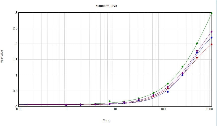
Detection of Human IL-15R alpha by ELISA
Measured levels of sIL-15Ralpha, IL-15, IL-6 and TNFalpha in synovial fluids from RA and OA patients. Cytokine concentration of IL-15Ralpha (A), IL-15 (B), TNFalpha (C) and IL-6 (D) were measured in in duplicate SF samples from RA (n = 30) and OA (n = 30) by ELISA. Box plot represents media ± SD. Differences between two groups were performed by Mann–Whitney U test for nonparametric data. Image collected and cropped by CiteAb from the following open publication (https://pubmed.ncbi.nlm.nih.gov/25879761), licensed under a CC-BY license. Not internally tested by R&D Systems.
Detection of IL-15R alpha in Human Pancreas.
Formalin-fixed paraffin-embedded tissue sections of human pancreas were probed for IL15Ra mRNA (ACD RNAScope Probe, catalog #532368; Fast Red chromogen, ACD catalog # 322750). Adjacent tissue section was processed for immunohistochemistry using goat anti-human IL15Ra polyclonal antibody (R&D Systems catalog # AF247) at 3ug/mL with overnight incubation at 4 degrees Celsius followed by incubation with anti-goat IgG VisUCyte HRP Polymer Antibody (Catalog # VC004) and DAB chromogen (yellow-brown). Tissue was counterstained with hematoxylin (blue). Specific staining was localized to exocrine glands. and IFNg(10ng/mL) for 24h by Flow Cytometry")
Detection of IL-15R alpha in human monocytes treated with LPS(100ng/mL) and IFNg(10ng/mL) for 24h by Flow Cytometry
Human monocytes treated with LPS(100ng/mL) and IFNg(10ng/mL) for 24h (filled histogram) or resting human monocytes (open histogram) were stained with Goat Anti-Human IL-15R alpha Antigen Affinity-purified Polyclonal Antibody (Catalog # AF247) followed by Allophycocyanin-conjugated Anti-Goat IgG Secondary Antibody (Catalog # F0108). View our protocol for Staining Membrane-associated Proteins.Applications for Human IL-15R alpha Antibody
Application
Recommended Usage
CyTOF-ready
Ready to be labeled using established conjugation methods. No BSA or other carrier proteins that could interfere with conjugation.
Dual RNAscope ISH-IHC Compatible
5-15 µg/mL
Sample: Immersion fixed paraffin-embedded sections of human pancreas
Sample: Immersion fixed paraffin-embedded sections of human pancreas
Flow Cytometry
0.25 µg/106 cells
Sample: Human monocytes treated with LPS and IFNg
Sample: Human monocytes treated with LPS and IFNg
Immunocytochemistry
1-15 µg/mL
Sample: Immersion fixed human Hodgkin's lymphoma cell line
Sample: Immersion fixed human Hodgkin's lymphoma cell line
Immunohistochemistry
5-15 µg/mL
Sample: Immersion fixed paraffin-embedded sections of human prostate cancer tissue
Sample: Immersion fixed paraffin-embedded sections of human prostate cancer tissue
Western Blot
0.1 µg/mL
Sample: Recombinant Human IL‑15 R alpha Fc Chimera (Catalog # 147-IR)
Sample: Recombinant Human IL‑15 R alpha Fc Chimera (Catalog # 147-IR)
Neutralization
Measured by its ability to neutralize IL‑15 R alpha -mediated inhibition of proliferation in the CTLL‑2 mouse cytotoxic T cell line. The Neutralization Dose (ND50) is typically 1-4 µg/mL in the presence of 30 ng/mL Recombinant Human IL‑15 R alpha Fc Chimera and 2 ng/mL Recombinant Human IL‑15.
Reviewed Applications
Read 2 reviews rated 4 using AF247 in the following applications:
Flow Cytometry Panel Builder
Bio-Techne Knows Flow Cytometry
Save time and reduce costly mistakes by quickly finding compatible reagents using the Panel Builder Tool.
Advanced Features
- Spectra Viewer - Custom analysis of spectra from multiple fluorochromes
- Spillover Popups - Visualize the spectra of individual fluorochromes
- Antigen Density Selector - Match fluorochrome brightness with antigen density

Formulation, Preparation, and Storage
Purification
Antigen Affinity-purified
Reconstitution
Reconstitute at 0.2 mg/mL in sterile PBS. For liquid material, refer to CoA for concentration.
Loading...
Formulation
Lyophilized from a 0.2 μm filtered solution in PBS with Trehalose. See Certificate of Analysis for details.
*Small pack size (-SP) is supplied either lyophilized or as a 0.2 µm filtered solution in PBS.
*Small pack size (-SP) is supplied either lyophilized or as a 0.2 µm filtered solution in PBS.
Shipping
Lyophilized product is shipped at ambient temperature. Liquid small pack size (-SP) is shipped with polar packs. Upon receipt, store immediately at the temperature recommended below.
Stability & Storage
Use a manual defrost freezer and avoid repeated freeze-thaw cycles.
- 12 months from date of receipt, -20 to -70 °C as supplied.
- 1 month, 2 to 8 °C under sterile conditions after reconstitution.
- 6 months, -20 to -70 °C under sterile conditions after reconstitution.
Calculators
Background: IL-15R alpha
References
- Anderson, D.M. et al. (1995) J. Biol. Chem. 270:29862.
- Bulfone-Paus, S. et al. (1999) FASEB 13:1575.
- Waldemann, T.A. and Y. Tagaya (1999) Ann. Rev. Immunol. 17:19.
- Dubois, S. et al. (1999) J. Biol. Chem. 274:26978.
Long Name
Interleukin 15 Receptor alpha
Alternate Names
CD215, IL-15 R alpha, IL-15Ra, IL15 R alpha, IL15RA
Gene Symbol
IL15RA
UniProt
Additional IL-15R alpha Products
Product Documents for Human IL-15R alpha Antibody
Certificate of Analysis
To download a Certificate of Analysis, please enter a lot or batch number in the search box below.
Note: Certificate of Analysis not available for kit components.
Product Specific Notices for Human IL-15R alpha Antibody
For research use only
Citations for Human IL-15R alpha Antibody
 Powered by Bioz
Customer Reviews for Human IL-15R alpha Antibody (2)
4 out of 5
2 Customer Ratings
Have you used Human IL-15R alpha Antibody?
Submit a review and receive an Amazon gift card!
$25/€18/£15/$25CAN/¥2500 Yen for a review with an image
$10/€7/£6/$10CAN/¥1110 Yen for a review without an image
Submit a review
Customer Images
Showing
1
-
2 of
2 reviews
Showing All
Filter By:
-
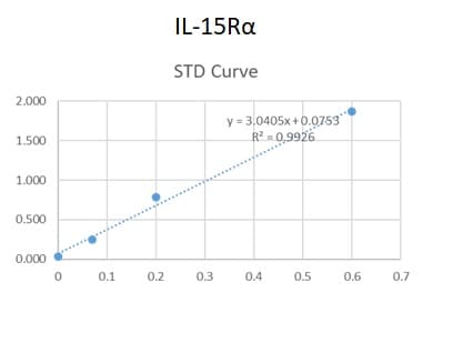
Application: ELISASample Tested: Serum and PlasmaSpecies: HumanVerified Customer | Posted 06/09/2020We used this antibody in an in-house ELISA along with mAb and protein standard (147-IR-100) to quantify IL-15R alpha in human serum and plasma. This combination could not detect IL-15R alpha in our samples but generated a good standard curve.

-
Application: ELISASample Tested: SerumSpecies: HumanVerified Customer | Posted 05/01/2020Ab was used as capture antibody in human serum ELISA
There are no reviews that match your criteria.
Protocols
Find general support by application which include: protocols, troubleshooting, illustrated assays, videos and webinars.
- 7-Amino Actinomycin D (7-AAD) Cell Viability Flow Cytometry Protocol
- Antigen Retrieval Protocol (PIER)
- Antigen Retrieval for Frozen Sections Protocol
- Appropriate Fixation of IHC/ICC Samples
- Cellular Response to Hypoxia Protocols
- Chromogenic IHC Staining of Formalin-Fixed Paraffin-Embedded (FFPE) Tissue Protocol
- Chromogenic Immunohistochemistry Staining of Frozen Tissue
- ClariTSA™ Fluorophore Kits
- Detection & Visualization of Antibody Binding
- Extracellular Membrane Flow Cytometry Protocol
- Flow Cytometry Protocol for Cell Surface Markers
- Flow Cytometry Protocol for Staining Membrane Associated Proteins
- Flow Cytometry Staining Protocols
- Flow Cytometry Troubleshooting Guide
- Fluorescent IHC Staining of Frozen Tissue Protocol
- Graphic Protocol for Heat-induced Epitope Retrieval
- Graphic Protocol for the Preparation and Fluorescent IHC Staining of Frozen Tissue Sections
- Graphic Protocol for the Preparation and Fluorescent IHC Staining of Paraffin-embedded Tissue Sections
- Graphic Protocol for the Preparation of Gelatin-coated Slides for Histological Tissue Sections
- ICC Cell Smear Protocol for Suspension Cells
- ICC Immunocytochemistry Protocol Videos
- ICC for Adherent Cells
- IHC Sample Preparation (Frozen sections vs Paraffin)
- ISH-IHC Protocol for Chromogenic Detection on Formalin Fixed Paraffin Embedded (FFPE) Tissue
- Immunocytochemistry (ICC) Protocol
- Immunocytochemistry Troubleshooting
- Immunofluorescence of Organoids Embedded in Cultrex Basement Membrane Extract
- Immunofluorescent IHC Staining of Formalin-Fixed Paraffin-Embedded (FFPE) Tissue Protocol
- Immunohistochemistry (IHC) and Immunocytochemistry (ICC) Protocols
- Immunohistochemistry Frozen Troubleshooting
- Immunohistochemistry Paraffin Troubleshooting
- Intracellular Flow Cytometry Protocol Using Alcohol (Methanol)
- Intracellular Flow Cytometry Protocol Using Detergents
- Intracellular Nuclear Staining Flow Cytometry Protocol Using Detergents
- Intracellular Staining Flow Cytometry Protocol Using Alcohol Permeabilization
- Intracellular Staining Flow Cytometry Protocol Using Detergents to Permeabilize Cells
- Preparing Samples for IHC/ICC Experiments
- Preventing Non-Specific Staining (Non-Specific Binding)
- Primary Antibody Selection & Optimization
- Propidium Iodide Cell Viability Flow Cytometry Protocol
- Protocol for Heat-Induced Epitope Retrieval (HIER)
- Protocol for Liperfluo
- Protocol for Making a 4% Formaldehyde Solution in PBS
- Protocol for VisUCyte™ HRP Polymer Detection Reagent
- Protocol for the Characterization of Human Th22 Cells
- Protocol for the Characterization of Human Th9 Cells
- Protocol for the Fluorescent ICC Staining of Cell Smears - Graphic
- Protocol for the Fluorescent ICC Staining of Cultured Cells on Coverslips - Graphic
- Protocol for the Preparation & Fixation of Cells on Coverslips
- Protocol for the Preparation and Chromogenic IHC Staining of Frozen Tissue Sections
- Protocol for the Preparation and Chromogenic IHC Staining of Frozen Tissue Sections - Graphic
- Protocol for the Preparation and Chromogenic IHC Staining of Paraffin-embedded Tissue Sections
- Protocol for the Preparation and Chromogenic IHC Staining of Paraffin-embedded Tissue Sections - Graphic
- Protocol for the Preparation and Fluorescent ICC Staining of Cells on Coverslips
- Protocol for the Preparation and Fluorescent ICC Staining of Non-adherent Cells
- Protocol for the Preparation and Fluorescent ICC Staining of Stem Cells on Coverslips
- Protocol for the Preparation and Fluorescent IHC Staining of Frozen Tissue Sections
- Protocol for the Preparation and Fluorescent IHC Staining of Paraffin-embedded Tissue Sections
- Protocol for the Preparation of Gelatin-coated Slides for Histological Tissue Sections
- Protocol for the Preparation of a Cell Smear for Non-adherent Cell ICC - Graphic
- Protocol: Annexin V and PI Staining by Flow Cytometry
- Protocol: Annexin V and PI Staining for Apoptosis by Flow Cytometry
- R&D Systems Quality Control Western Blot Protocol
- TUNEL and Active Caspase-3 Detection by IHC/ICC Protocol
- The Importance of IHC/ICC Controls
- Troubleshooting Guide: Fluorokine Flow Cytometry Kits
- Troubleshooting Guide: Immunohistochemistry
- Troubleshooting Guide: Western Blot Figures
- Western Blot Conditions
- Western Blot Protocol
- Western Blot Protocol for Cell Lysates
- Western Blot Troubleshooting
- Western Blot Troubleshooting Guide
- View all Protocols, Troubleshooting, Illustrated assays and Webinars


