CXCL8, also known as IL-8, is a CXC chemokine that is a potent chemoattractant for neutrophils and has a wide range of other pro-inflammatory effects.
Human IL-8/CXCL8 Antibody (6217)
R&D Systems | Catalog # MAB208

Key Product Details
Validated by
Biological Validation
Species Reactivity
Validated:
Human
Cited:
Human, Mouse, Rat, Bacteria - Escherichia coli, Bacteria - Porphyromonas gingivalis, Bovine, Complex Species Category, Guinea Pig, Rabbit, Xenograft
Applications
Validated:
Western Blot, ELISA Capture (Matched Antibody Pair), Neutralization, Intracellular Staining by Flow Cytometry, Immunocytochemistry, Simple Western, CyTOF-ready
Cited:
Immunohistochemistry, Immunohistochemistry-Paraffin, Immunohistochemistry-Frozen, Western Blot, Neutralization, Flow Cytometry, Immunocytochemistry, Immunoprecipitation, Chromatin Immunoprecipitation (ChIP), Affinity Chromatography, Antibody Array Development, Array Development, Binding Assay, Bioassay, Blocking, Co-Immunoprecipitation, Electrochemiluminescent Assay, ELISA Capture, ELISA Development, ELISA Development (Capture), ELISA Development (Detection), In vivo assay, Functional Assay, Inhibition, Luminex Development
Label
Unconjugated
Antibody Source
Monoclonal Mouse IgG1 Clone # 6217
Loading...
Product Specifications
Immunogen
E. coli-derived recombinant human IL-8/CXCL8
Ser28-Ser99
Accession # P10145
Ser28-Ser99
Accession # P10145
Specificity
Detects human IL-8/CXCL8 in ELISAs and Western blots. In Western blots, 100% cross-reactivity with recombinant porcine IL-8/CXCL8 and no cross-reactivity with recombinant rat CXCL3/CINC-2 alpha is observed.
Clonality
Monoclonal
Host
Mouse
Isotype
IgG1
Endotoxin Level
<0.10 EU per 1 μg of the antibody by the LAL method.
Scientific Data Images for Human IL-8/CXCL8 Antibody (6217)
Detection of Human IL-8/CXCL8 by Western Blot.
Western blot shows lysates of THP‑1 human acute monocytic leukemia cell line untreated (-) or treated (+) with 200 nM PMA and 10 ug/mL LPS for 24 hours and 3 hours. PVDF membrane was probed with 3 µg/mL of Mouse Anti-Human IL-8/CXCL8 Monoclonal Antibody (Catalog # MAB208) followed by HRP-conjugated Anti-Mouse IgG Secondary Antibody (HAF018). A specific band was detected for IL-8/CXCL8 at approximately 10 kDa (as indicated). This experiment was conducted under reducing conditions and using Western Blot Buffer Group 1.
IL-8/CXCL8 in Human PBMCs.
IL-8/CXCL8 was detected in immersion fixed human peripheral blood mononuclear cells (PBMCs) using Mouse Anti-Human IL-8/CXCL8 Monoclonal Antibody (Catalog # MAB208) at 10 µg/mL for 3 hours at room temperature. Cells were stained using the NorthernLights™ 557-conjugated Anti-Mouse IgG Secondary Antibody (red; Catalog # NL007) and counterstained with DAPI (blue). View our protocol for Fluorescent ICC Staining of Non-adherent Cells.
Detection of Human IL-8/CXCL8 by Simple WesternTM.
Simple Western lane view shows human PBMC conditioned media untreated (-) or treated (+) with 1 μg/mL LPS for 24 hrs, loaded at 0.2 mg/mL. A specific band was detected for IL-8/CXCL8 at approximately 8 kDa (as indicated) using 50 µg/mL of Mouse Anti-Human IL-8/CXCL8 Monoclonal Antibody (Catalog # MAB208). This experiment was conducted under reducing conditions and using the 2-40 kDa separation system.
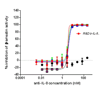
Chemotaxis Induced by IL-8/CXCL8 and Neutralization by Human IL-8/CXCL8 Antibody.
Recombinant Human IL-8/CXCL8 (Catalog # 208-IL) chemoattracts the BaF3 mouse pro-B cell line transfected with human CXCR2 in a dose-dependent manner (orange line). The amount of cells that migrated through to the lower chemotaxis chamber was measured by Resazurin (Catalog # AR002). Chemotaxis elicited by Recombinant Human IL-8/CXCL8 (20 ng/mL) is neutralized (green line) by increasing concentrations of Mouse Anti-Human IL-8/CXCL8 Monoclonal Antibody (Catalog # MAB208). The ND50 is typically 0.08-0.4 µg/mL.
Detection of IL-8/CXCL8 by Western Blot
CDK1/CDC14B-dependent phosphorylation of USP9X at serine 2563 promotes mitotic survival via WT1 and IL-8. e Immunoblot analysis confirming reversal of mitotic apoptosis following exogenous reconstitution of IL-8 in CXCL8-depleted cells. Experiment was performed as in a, with addition of exogenous IL-8 for the last 48 h. Cells were treated with nocodazole for 8 h. Image collected and cropped by CiteAb from the following open publication (https://pubmed.ncbi.nlm.nih.gov/32152317), licensed under a CC-BY license. Not internally tested by R&D Systems.
Detection of IL-8/CXCL8 by Western Blot
CDK1/CDC14B-dependent phosphorylation of USP9X at serine 2563 promotes mitotic survival via WT1 and IL-8. f Immunoblot analysis detecting induction of mitotic apoptosis in response to WT1, CXCL8, or WT1 and CXCL8 knockdown compared to control knockdown in U2OS cells that were arrested in mitosis using nocodazole (8 h). Image collected and cropped by CiteAb from the following open publication (https://pubmed.ncbi.nlm.nih.gov/32152317), licensed under a CC-BY license. Not internally tested by R&D Systems.
Detection of IL-8/CXCL8 by Western Blot
CDK1/CDC14B-dependent phosphorylation of USP9X at serine 2563 promotes mitotic survival via WT1 and IL-8.a. i Immunoblot analysis revealing decreased mitotic apoptosis after CDC14B knockdown in USP9XWT but not in USP9XMut U2OS cells. Before lysis cells were transfected with control or CDC14B-directed siRNA, arrested in mitosis, and collected for analysis by western blot. Throughout this figure, mean and standard deviations as error bars are displayed. Image collected and cropped by CiteAb from the following open publication (https://pubmed.ncbi.nlm.nih.gov/32152317), licensed under a CC-BY license. Not internally tested by R&D Systems.
Detection of IL-8/CXCL8 by Western Blot
Cytokines regulated by RanBP1 in lung cancer. (A) Identification of secreted factors regulated by RanBP1 using cytokine arrays. A549 cells are used, and the expression of RanBP1 is suppressed using si-RNA. (B) Comparison of the gene expression of cytokines regulated by RanBP1. (C) Analysis of cytokines regulated by RanBP1 using WB. (D) Comparison of the sphere formation ability after each neutralizing antibody treatment. The p value is less than 0.0001 versus the IgG Ab value. (E) Comparison of migration and invasion ability of cells after each neutralizing antibody treatment. Experiments performed in triplicate. (F) CSC marker protein analysis in A549 after treatment with IL-18 neutralizing antibody. (G) Confirmation of the expression levels of EMT marker proteins after treatment with the IL-18 neutralizing antibody. (H) Expression analysis of RanBP1 regulated by IL-18 using neutralizing antibodies. Error bars represent mean ± SD. Triplicate samples. * p < 0.05, ** p < 0.001, versus control. Scale bar = 50 μm. Image collected and cropped by CiteAb from the following open publication (https://pubmed.ncbi.nlm.nih.gov/37047826), licensed under a CC-BY license. Not internally tested by R&D Systems.
Detection of IL-8/CXCL8 by Flow Cytometry
Interaction between osteosarcoma and macrophages increased IL-8 production. A Cytokine arrays of 143B-Luc cells, human macrophages, and co-culture CM. B IL-8 production during the co-culture of OS cells (143B-Luc, SJSA-1) and macrophages (THP-1 Mφ, HMDMs). C IL-8 expression in macrophages stimulated with OS-CM. D IL-8 expression in OS cells stimulated with macrophage CM. E UMAP plot of OS lung metastases. F UMAP plot of Iba1, CD163, and IL-8 expression in myeloid cell clusters. G Violin plot depicting IL-8 expression in Iba1+/− or CD163+/− myeloid cell clusters. CM conditioned medium, HMDMs human monocyte-derived macrophages, OS osteosarcoma, THP-1 Mφ THP-1-derived macrophage. Data are presented as mean ± standard deviation. **p < 0.01. All data were obtained from at least three independent experiments. Image collected and cropped by CiteAb from the following open publication (https://www.nature.com/articles/s41419-024-06487-y), licensed under a CC-BY license. Not internally tested by R&D Systems.
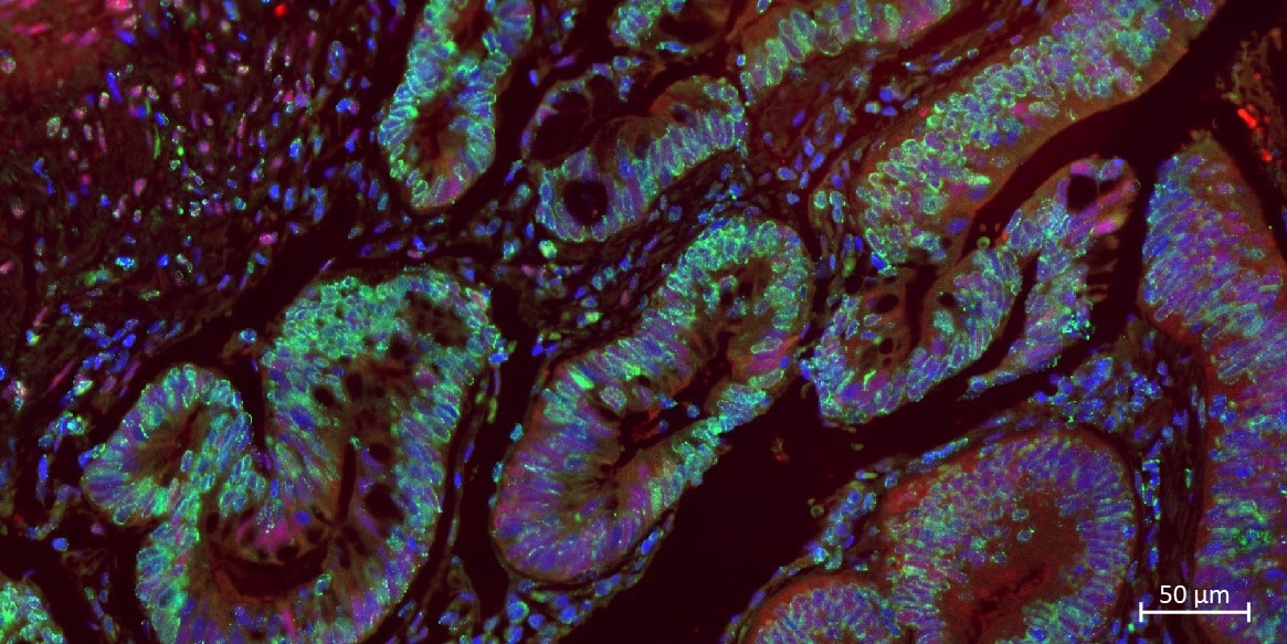
Detection of IL-8/CXCL8 by Immunohistochemistry
Effect of IL-8 in co-culture CM on proliferation, migration, and invasion of osteosarcoma cells.A Cell viability assay for OS cells treated with or without recombinant IL-8 (rIL-8, 10 ng/mL) using Cell Counting Kit-8. B, C Scratch and invasion assays for OS cells treated with or without co-culture CM. The scratch assay was performed for 12 h, and the invasion assay was conducted for 24 h. Scale bars represent 500 μm. D Cell viability assay for OS cells treated with or without co-culture CM and with or without anti-IL-8 antibodies (IL-8 Abs, 1 µg/mL) using Cell Counting Kit-8. E, F Scratch and invasion assays for OS cells treated with or without co-culture CM and with or without IL-8 Abs (100 ng/mL). The scratch assay was performed for 12 h, and the invasion assay was conducted for 24 h. Scale bars represent 500 μm. CM: conditioned medium, OS: osteosarcoma, ns: not significant. Data are presented as mean ± standard deviation. *p < 0.05 and **p < 0.01. All data were obtained from at least three independent experiments. Image collected and cropped by CiteAb from the following open publication (https://www.nature.com/articles/s41419-024-06487-y), licensed under a CC-BY license. Not internally tested by R&D Systems.
Detection of IL-8/CXCL8 by Immunohistochemistry
Effect of IL-8 in co-culture CM on lung metastasis. A Schematic showing the experimental design for OS tail vein injection. 143B-Luc cells were treated with DMEM or co-culture CM for 12 h prior to tail vein injection; 143B-Luc cells (1 × 10⁶/mouse) were injected into the tail vein of 6–7 week-old BALB/c nu/nu mice. B IVIS imaging and quantification of lung metastasis 14 days after the injection of tumors pretreated with or without co-culture CM. C Hematoxylin and eosin staining of lung sections with or without co-culture CM and quantification of lung colonies. Scale bars represent 1 mm and 500 µm. D Immunohistochemistry of Ki-67 positive cells in lung colonies with or without co-culture CM and quantification of the Ki-67 labeling index. Two colonies per sample were randomly selected, and quantification was performed for 10 colonies. Ki-67-positive cells in the colonies were counted and shown as the Ki-67 labeling index. Scale bars represent 100 µm. E Schematic showing the experimental design for OS tail vein injection using anti-IL-8 antibody. F IVIS imaging and quantification of lung metastasis 14 days after the injection of tumors pretreated with co-culture CM with or without IL-8 antibodies. G Hematoxylin and eosin staining of lung sections with co-culture CM with or without IL-8 Abs and quantification of lung colonies. Scale bars represent 1 mm and 500 µm. H Immunohistochemistry of Ki-67-positive cells in lung colonies with co-culture CM with or without IL-8 Abs and quantification of Ki-67 labeling index. Scale bars represent 100 µm. CM conditioned medium, DMEM Dulbecco’s modified Eagle’s medium, IVIS, in vivo imaging system, OS osteosarcoma. Data are presented as mean ± standard deviation; *p < 0.05, **p < 0.01. Each group contained five animals. Image collected and cropped by CiteAb from the following open publication (https://www.nature.com/articles/s41419-024-06487-y), licensed under a CC-BY license. Not internally tested by R&D Systems.
Detection of IL-8/CXCL8 by Immunohistochemistry
Effect of IL-8 in co-culture CM on primary growth. A Schematic showing the experimental design for subcutaneous transplantation of OS cells. 143B-Luc cells (2 × 10⁶/mouse) were subcutaneously transplanted in 6–7-week-old BALB/c nu/nu mice, and DMEM or co-culture CM was injected into the para-tumor thrice per week. B Tumor volume with or without co-culture CM measured thrice per week after tumor transplantation. C, D Tumor weight and image of the excised tumor with or without co-cultured CM 14 days after tumor transplantation. E, F Immunohistochemistry and quantification of Ki-67 and phospho-FAK in tumor sections with or without co-culture CM 14 days after tumor transplantation. Two fields of view per sample were randomly selected, and quantification was performed in 10 fields. E Ki-67-positive cells in the field were counted and indicated as Ki-67 labeling index. Scale bars represent 50 μm. G Schematic showing the experimental design for subcutaneous OS transplantation using anti-IL-8 antibodies (IL-8 Abs). H Tumor volume after pretreatment with co-culture CM with or without IL-8 Abs (10 µg/mouse) measured thrice per week after tumor transplantation. I and J Weight and image of the excised tumor pretreated with co-culture CM with or without IL-8 Abs 14 days after tumor transplantation. K, L Immunohistochemistry and quantification of Ki-67 and phospho-FAK in tumor sections with co-culture CM with or without IL-8 Abs 14 days after tumor transplantation. Scale bars represent 50 µm. CM conditioned medium, DMEM Dulbecco’s modified Eagle’s medium, FAK focal adhesion kinase, OS osteosarcoma. Data are presented as mean ± standard deviation; *p < 0.05, **p < 0.01. Each group contained five animals. Image collected and cropped by CiteAb from the following open publication (https://www.nature.com/articles/s41419-024-06487-y), licensed under a CC-BY license. Not internally tested by R&D Systems.
Detection of IL-8/CXCL8 by Flow Cytometry
Interaction between osteosarcoma and macrophages increased IL-8 production. A Cytokine arrays of 143B-Luc cells, human macrophages, and co-culture CM. B IL-8 production during the co-culture of OS cells (143B-Luc, SJSA-1) and macrophages (THP-1 Mφ, HMDMs). C IL-8 expression in macrophages stimulated with OS-CM. D IL-8 expression in OS cells stimulated with macrophage CM. E UMAP plot of OS lung metastases. F UMAP plot of Iba1, CD163, and IL-8 expression in myeloid cell clusters. G Violin plot depicting IL-8 expression in Iba1+/− or CD163+/− myeloid cell clusters. CM conditioned medium, HMDMs human monocyte-derived macrophages, OS osteosarcoma, THP-1 Mφ THP-1-derived macrophage. Data are presented as mean ± standard deviation. **p < 0.01. All data were obtained from at least three independent experiments. Image collected and cropped by CiteAb from the following open publication (https://www.nature.com/articles/s41419-024-06487-y), licensed under a CC-BY license. Not internally tested by R&D Systems.
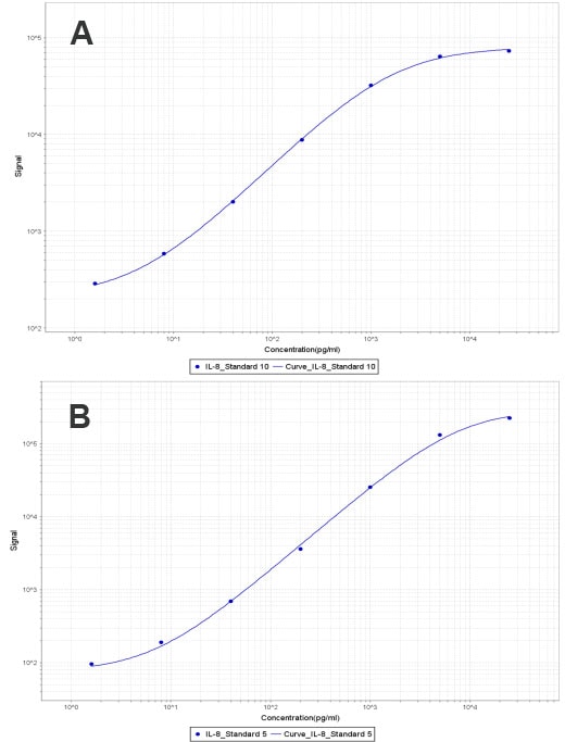
Human IL-8 / CXCL8 ELISA Standard Curve
Recombinant Human IL-8/CXCL8 (Catalog # 208-IL) was serially diluted and captured by Mouse Anti-Human IL-8/CXCL8 Monoclonal Antibody (Catalog # MAB208) coated on a Clear Polystyrene Microplate (Catalog # DY990). Goat Anti-Human IL-8/CXCL8 Antigen Affinity-purified Polyclonal Antibody (Catalog # AF-208-NA) was biotinylated and incubated with the protein captured on the plate. Detection of the standard curve was achieved by incubating Streptavidin-HRP (Catalog # DY998)Applications for Human IL-8/CXCL8 Antibody (6217)
Application
Recommended Usage
CyTOF-ready
Ready to be labeled using established conjugation methods. No BSA or other carrier proteins that could interfere with conjugation.
Immunocytochemistry
8-25 µg/mL
Sample: Immersion fixed human peripheral blood mononuclear cells (PBMCs) stimulated with PMA and ionomycin
Sample: Immersion fixed human peripheral blood mononuclear cells (PBMCs) stimulated with PMA and ionomycin
Intracellular Staining by Flow Cytometry
0.25 µg/106 cells
Sample: Human peripheral blood mononuclear cells treated with LPS, fixed with paraformaldehyde, and permeabilized with saponin
Sample: Human peripheral blood mononuclear cells treated with LPS, fixed with paraformaldehyde, and permeabilized with saponin
Simple Western
50 µg/mL
Sample: Human PBMC conditioned media treated with LPS
Sample: Human PBMC conditioned media treated with LPS
Western Blot
3 µg/mL
Sample: THP‑1 human acute monocytic leukemia cell line treated (+) with PMA and LPS
Sample: THP‑1 human acute monocytic leukemia cell line treated (+) with PMA and LPS
Neutralization
Measured by its ability to neutralize IL-8/CXCL8-induced chemotaxis in the BaF3 mouse pro‑B cell line transfected with human CXCR2. The Neutralization Dose (ND50) is typically 0.08-0.4 µg/mL in the presence of 20 ng/mL Recombinant Human IL-8/CXCL8.
Human IL-8/CXCL8 Sandwich Immunoassay
Please Note: Optimal dilutions of this antibody should be experimentally determined.
Reviewed Applications
Read 11 reviews rated 4.8 using MAB208 in the following applications:
Flow Cytometry Panel Builder
Bio-Techne Knows Flow Cytometry
Save time and reduce costly mistakes by quickly finding compatible reagents using the Panel Builder Tool.
Advanced Features
- Spectra Viewer - Custom analysis of spectra from multiple fluorochromes
- Spillover Popups - Visualize the spectra of individual fluorochromes
- Antigen Density Selector - Match fluorochrome brightness with antigen density

Formulation, Preparation, and Storage
Purification
Protein A or G purified from hybridoma culture supernatant
Reconstitution
Reconstitute at 0.5 mg/mL in sterile PBS. For liquid material, refer to CoA for concentration.
Loading...
Formulation
Lyophilized from a 0.2 μm filtered solution in PBS with Trehalose. *Small pack size (SP) is supplied either lyophilized or as a 0.2 µm filtered solution in PBS.
Shipping
Lyophilized product is shipped at ambient temperature. Liquid small pack size (-SP) is shipped with polar packs. Upon receipt, store immediately at the temperature recommended below.
Stability & Storage
Use a manual defrost freezer and avoid repeated freeze-thaw cycles.
- 12 months from date of receipt, -20 to -70 °C as supplied.
- 1 month, 2 to 8 °C under sterile conditions after reconstitution.
- 6 months, -20 to -70 °C under sterile conditions after reconstitution.
Calculators
Background: IL-8/CXCL8
Long Name
Interleukin 8
Alternate Names
CXCL8, GCP1, IL8, LAI, LECT, LUCT, LYNAP, MDNCF, MONAP, NAF, NAP1, NCF, TCF, TSG1
Gene Symbol
CXCL8
UniProt
Additional IL-8/CXCL8 Products
Product Documents for Human IL-8/CXCL8 Antibody (6217)
Certificate of Analysis
To download a Certificate of Analysis, please enter a lot or batch number in the search box below.
Note: Certificate of Analysis not available for kit components.
Product Specific Notices for Human IL-8/CXCL8 Antibody (6217)
For research use only
Citations for Human IL-8/CXCL8 Antibody (6217)
 Powered by Bioz
Customer Reviews for Human IL-8/CXCL8 Antibody (6217) (11)
4.8 out of 5
11 Customer Ratings
Have you used Human IL-8/CXCL8 Antibody (6217)?
Submit a review and receive an Amazon gift card!
$25/€18/£15/$25CAN/¥2500 Yen for a review with an image
$10/€7/£6/$10CAN/¥1110 Yen for a review without an image
Submit a review
Customer Images

Showing
1
-
5 of
11 reviews
Showing All
Filter By:
-
Application: Immunocytochemistry/ImmunofluorescenceSample Tested: Colon cancer tissueSpecies: HumanVerified Customer | Posted 08/13/2023IF of CXCL8 was done using dilution of 1:2000 in 2.5% horse serum/PBS and incubated at 4 degree overnight, and AF488 conjugated secondary antibody was used for 1h at RT.

-
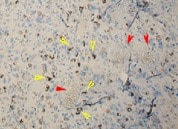
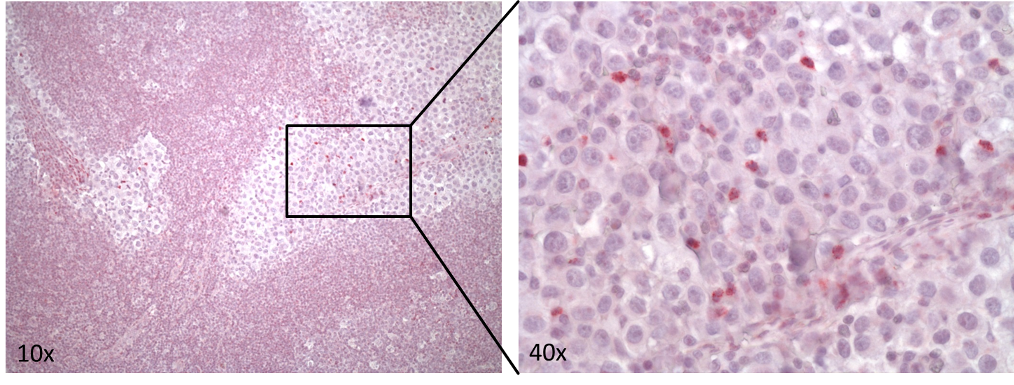
Application: ImmunohistochemistrySample Tested: Brain tumorSpecies: HumanVerified Customer | Posted 08/06/2021Yellow arrows on the picture indicate staining in tumor cells.
-
Application: Western BlotSample Tested: macrophageSpecies: HumanVerified Customer | Posted 07/30/2021
-
Application: Functional AssaySample Tested: Whole CellsSpecies: HumanVerified Customer | Posted 03/16/2020
-
Application: Block/NeutralizeSample Tested: Serum-free Cell Culture Media and Breast cancer cellsSpecies: HumanVerified Customer | Posted 05/27/2019
-
Application: ELISASample Tested: PlasmaSpecies: HumanVerified Customer | Posted 11/10/2018
-
Application: MSD assaySample Tested: Vitreous humorSpecies: Cynomolgus Monkey and PorcineVerified Customer | Posted 11/01/2018After biotinylation, used as a capture reagent according to the manufacturer’s protocol (Meso Scale Diagnostics LLC). A: Paired with SulfoTag-modified AF-208-NA as a detection antibody. Calibration curves with Recombinant Human IL-8 (208-IL-010/CF) is shown (dynamic range 1.6-25,000 pg/ml). B: Paired with SulfoTag-modified MAB-535 to detect porcine IL-8. Calibration curves with Recombinant Porcine IL-8 (535-IN/CF) is shown (dynamic range 1.6-25,000 pg/ml).
-
Application: Immunohistochemistry-ParaffinSample Tested: Human lymph node (melanoma)Species: HumanVerified Customer | Posted 08/30/2018Human FFPE metastatic melanoma lymph node sections stained with anti-human IL-8 MAB208 (5 µg/ml) over night at 4 °C. Detection with an Alkaline Phosphate/Red Chromogen System.Human FFPE metastatic melanoma lymph node sections were stained with anti-human IL-8 monoclonal antibody (MAB208) at 5 µg/ml followed by secondary goat anti-mouse biotinylated antibody and Streptavidin AP conjugate. Detection via an Alkaline Phosphatase/Red staining System. Blocking: 5 % goat Serum in TBS-T Antigen Retrieval: Citrat buffer Primary antibody incubation: over night at 4 °C
-
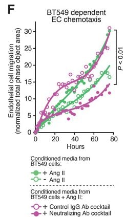
Application: Block/NeutralizeSample Tested: Breast cancer cellsSpecies: HumanVerified Customer | Posted 05/11/2018We used this IL8 neutralizing antibody (600 ng/mL) as part of an antibody cocktail containing several other antibodies to neutralize IL8 present in the Conditioned Medium (CM) from BT549 Cells treated with Angiotensin II. CM (+/- IL8 Neutralizing ab Cocktail) was used to further look at Endothelial (HUVEC) Cell migration using Incucyte Chemotaxis Assay. Cancer Res; 78(5) March 1, 2018 (Fig 6F)
-
Application: ELISASample Tested: SerumSpecies: HumanVerified Customer | Posted 04/03/2018
-
Application: Block/NeutralizeSample Tested: NeutrophilsSpecies: HumanVerified Customer | Posted 04/26/2017
There are no reviews that match your criteria.
Protocols
Find general support by application which include: protocols, troubleshooting, illustrated assays, videos and webinars.
- 7-Amino Actinomycin D (7-AAD) Cell Viability Flow Cytometry Protocol
- Appropriate Fixation of IHC/ICC Samples
- Cellular Response to Hypoxia Protocols
- ClariTSA™ Fluorophore Kits
- Detection & Visualization of Antibody Binding
- Extracellular Membrane Flow Cytometry Protocol
- Flow Cytometry Protocol for Cell Surface Markers
- Flow Cytometry Protocol for Staining Membrane Associated Proteins
- Flow Cytometry Staining Protocols
- Flow Cytometry Troubleshooting Guide
- ICC Cell Smear Protocol for Suspension Cells
- ICC Immunocytochemistry Protocol Videos
- ICC for Adherent Cells
- Immunocytochemistry (ICC) Protocol
- Immunocytochemistry Troubleshooting
- Immunofluorescence of Organoids Embedded in Cultrex Basement Membrane Extract
- Immunohistochemistry (IHC) and Immunocytochemistry (ICC) Protocols
- Intracellular Flow Cytometry Protocol Using Alcohol (Methanol)
- Intracellular Flow Cytometry Protocol Using Detergents
- Intracellular Nuclear Staining Flow Cytometry Protocol Using Detergents
- Intracellular Staining Flow Cytometry Protocol Using Alcohol Permeabilization
- Intracellular Staining Flow Cytometry Protocol Using Detergents to Permeabilize Cells
- Preparing Samples for IHC/ICC Experiments
- Preventing Non-Specific Staining (Non-Specific Binding)
- Primary Antibody Selection & Optimization
- Propidium Iodide Cell Viability Flow Cytometry Protocol
- Protocol for Liperfluo
- Protocol for VisUCyte™ HRP Polymer Detection Reagent
- Protocol for the Characterization of Human Th22 Cells
- Protocol for the Characterization of Human Th9 Cells
- Protocol for the Fluorescent ICC Staining of Cell Smears - Graphic
- Protocol for the Fluorescent ICC Staining of Cultured Cells on Coverslips - Graphic
- Protocol for the Preparation and Fluorescent ICC Staining of Cells on Coverslips
- Protocol for the Preparation and Fluorescent ICC Staining of Non-adherent Cells
- Protocol for the Preparation and Fluorescent ICC Staining of Stem Cells on Coverslips
- Protocol for the Preparation of a Cell Smear for Non-adherent Cell ICC - Graphic
- Protocol: Annexin V and PI Staining by Flow Cytometry
- Protocol: Annexin V and PI Staining for Apoptosis by Flow Cytometry
- R&D Systems Quality Control Western Blot Protocol
- TUNEL and Active Caspase-3 Detection by IHC/ICC Protocol
- The Importance of IHC/ICC Controls
- Troubleshooting Guide: Fluorokine Flow Cytometry Kits
- Troubleshooting Guide: Western Blot Figures
- Western Blot Conditions
- Western Blot Protocol
- Western Blot Protocol for Cell Lysates
- Western Blot Troubleshooting
- Western Blot Troubleshooting Guide
- View all Protocols, Troubleshooting, Illustrated assays and Webinars
FAQs for Human IL-8/CXCL8 Antibody (6217)
Showing
1
-
1 of
1 FAQ
Showing All
-
Q: Are mouse or rat CXCL8/IL-8 products offered?
A: Mouse and rat do not have a gene which encodes CXCL8/IL-8. In mouse, the functional homologs to human CXCL8/IL-8 are CXCL1/KC and CXCL2/MIP-2. In rat, the functional homolog to CXCL8/human IL-8 is CXCL3/CINC-2.


