
Key Product Details
Validated by
Species Reactivity
Validated:
Cited:
Applications
Validated:
Cited:
Label
Antibody Source
Product Specifications
Immunogen
Ala2-Phe470
Accession # AAH29923
Specificity
Clonality
Host
Isotype
Scientific Data Images for Human KLF4 Antibody
Detection of Human KLF4 by Western Blot.
Western blot shows lysates of HT-29 human colon adenocarcinoma cell line, SW480 human colorectal adenocarcinoma cell line, and HCT-116 human colorectal carcinoma cell line. PVDF membrane was probed with 0.5 µg/mL of Goat Anti-Human KLF4 Antigen Affinity-purified Polyclonal Antibody (Catalog # AF3640) followed by HRP-conjugated Anti-Goat IgG Secondary Antibody (HAF109). A specific band was detected for KLF4 at approximately 60 kDa (as indicated). This experiment was conducted under reducing conditions and using Immunoblot Buffer Group 5.
KLF4 in BG01V Human Stem Cells.
KLF4 was detected in immersion fixed BG01V human embryonic stem cells using 10 µg/mL Goat Anti-Human KLF4 Antigen Affinity-purified Polyclonal Antibody (Catalog # AF3640) for 3 hours at room temperature. Cells were stained with the NorthernLights™ 557-conjugated Anti-Goat IgG Secondary Antibody (red; NL001) and counterstained with DAPI (blue). View our protocol for Fluorescent ICC Staining of Cells on Coverslips.
Detection of KLF4-regulated Genes by Chromatin Immunoprecipitation.
BG01V human embryonic stem cells were fixed using formaldehyde, resuspended in lysis buffer, and sonicated to shear chromatin. KLF4/DNA complexes were immunoprecipitated using 5 µg Goat Anti-Human KLF4 Antigen Affinity-purified Polyclonal Antibody (Catalog # AF3640) or control antibody (AB-108-C) for 15 minutes in an ultrasonic bath, followed by Biotinylated Anti-Goat IgG Secondary Antibody (BAF109). Immunocomplexes were captured using 50 µL of MagCellect Streptavidin Ferrofluid (MAG999) and DNA was purified using chelating resin solution. TheB2Rpromoter was detected by standard PCR.
KLF4 in Human Colon.
KLF4 was detected in immersion fixed paraffin-embedded sections of human colon using 15 µg/mL Goat Anti-Human KLF4 Antigen Affinity-purified Polyclonal Antibody (Catalog # AF3640) overnight at 4 °C. Tissue was stained with the Anti-Goat HRP-DAB Cell & Tissue Staining Kit (brown; Catalog # CTS008) and counterstained with hematoxylin (blue). Specific labeling was localized to the nuclei of epithelial cells. View our protocol for Chromogenic IHC Staining of Paraffin-embedded Tissue Sections.
Detection of Human KLF4 by Simple WesternTM.
Simple Western lane view shows lysates of HT-29 human colon adenocarcinoma cell line, loaded at 0.2 mg/mL. A specific band was detected for KLF4 at approximately 63 kDa (as indicated) using 25 µg/mL of Goat Anti-Human KLF4 Antigen Affinity-purified Polyclonal Antibody (Catalog # AF3640) followed by 1:50 dilution of HRP-conjugated Anti-Goat IgG Secondary Antibody (HAF109). This experiment was conducted under reducing conditions and using the 12-230 kDa separation system. Non-specific interaction with the 230 kDa Simple Western standard may be seen with this antibody.
Detection of Mouse KLF4 by Knockdown Validated
KLF4 regulates the EndMT switch in CCM1 KO ECsA–DCultured lung‐derived WT and CCM1 KO ECs were lentivirally transduced with shRNA directed to either Klf4 (shKLF4) or control sequence (shCTRL). (A) qRT–PCR of mesenchymal (Fsp1, Id1), stem cell‐like (Sca1), and endothelial markers (VE‐cadherin and Claudin5) in WT shCTRL, WT shKLF4, CCM1 KO shCTRL, and CCM1 KO shKLF4 ECs. Data are mean ± SD (n = 3). Fold difference in gene expression is relative to WT shCTRL ECs. A two‐tailed unpaired t‐test was performed. Klf4: ****P < 0.00001, ***P = 0.0008, **P = 0.007; Fsp1: ***P = 0.0001, ****P = 3.9E‐05, ###P = 0.0002; Sca1: ***P = 0.0001, ****P < 0.00001; Id1: ***P = 0.0001; Ve‐cadherin: ***P = 0.0001; Claudin5: ****P = 1.37E‐05. (B) WB of EndMT markers in WT shCTRL, WT shKLF4, CCM1 KO shCTRL, and CCM1 KO shKLF4 ECs. Vinculin is the loading control. These data are representative of three independent observations. (C) Proliferation rate of WT shCTRL, WTshKLF4, CCM1 KO shCTRL, and CCM1 KO shKLF4 ECs cultured for 5 days. Columns represent mean ± SD (n = 8). A two‐tailed unpaired t‐test was performed. 3 days of culture: ***P = 0.0001, **P = 0.0045, ##P = 0.0017; 5 days of culture: ***P = 0.0001, **P = 0.0029, ##P = 0.0040. (D) Migration rate measured in a wound assay of WT shCTRL, CCM1 KO shCTRL, and CCM1 KO shKLF4 ECs. Mean ± SD is shown (n = 6). A two‐tailed unpaired t‐test was performed. ***P = 0.0004, ###P = 0.0009.ECultured lung‐derived WT ECs were lentivirally transduced with a full‐length murine Klf4 (LentiKLF4) or empty vector (Mock). qRT–PCR (left panel) and WB (right panel) of EndMT markers in Mock and LentiKLF4 ECs. qRT–PCR data are mean ± SD (n = 3) and the fold changes are relative to Mock ECs. A two‐tailed unpaired t‐test was performed. **P = 0.002, ***P = 0.0007, ###P = 0.0001. WB results are representative of three independent observations. Tubulin is the loading control.Source data are available online for this figure. Image collected and cropped by CiteAb from the and HDLM‑2 Human Hodgkin’s Lymphoma Cell Line (negative) Cells.")
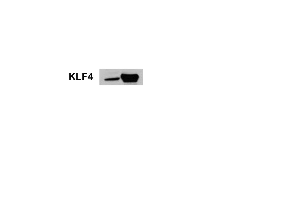
Detection of KLF4 in HT‑29 Human Colon Adenocarcinoma Cell Line (positive) and HDLM‑2 Human Hodgkin’s Lymphoma Cell Line (negative) Cells.
KLF4 was detected in immersion fixed HT‑29 Human Colon Adenocarcinoma Cell Line (positive) and HDLM‑2 Human Hodgkin’s Lymphoma Cell Line (negative) Cells using Goat Anti-Human KLF4 Antigen Affinity-purified Polyclonal Antibody (Catalog # AF3640) at 5 µg/mL for 3 hours at room temperature. Cells were stained using the NorthernLights™ 557-conjugated Anti-Goat IgG Secondary Antibody (red; Catalog # NL001) and counterstained with DAPI (blue). Specific staining was localized to cytoplasm. View our protocol for Fluorescent ICC Staining of Cells on Coverslips.
Detection of Human KLF4 by Knockdown Validated
Klf4 significantly rescues the effects of HDAC1 on leukemia cell proliferation. (a) Effects of ectopic expression of Klf4 on HDAC1-induced cell proliferation. * and ** indicate P<0.05 and P<0.01, respectively. (b) FACS analysis showing the effects of ectopic expression of Klf4 on HDAC1-induced cell cycle. * and ** indicate P<0.05 and P<0.01, respectively. (c) CCK8 analysis showing the effects of Klf4 knockdown on HDAC1 deficiency-mediated cell proliferation. * and ** indicate P<0.05 and P<0.01, respectively. (d) FACS analysis showing effects of Klf4 knockdown on HDAC1 deficiency-mediated cell cycle. * and ** indicate P<0.05 and P<0.01, respectively. (e) Western blotting analyses showing the effects of ectopic expression of Klf4 and HDAC1 on the expression levels of p21 and p27. GAPDH was used as the loading control. (f) Western blotting analyses showing the effects Klf4 and HDAC1 knockdown on the expression levels of p21 and p27. GAPDH was used as the loading control. (g) ChIP-PCR assays showing Klf4 binding at the promoter region of p21 and p27 after HDAC1 knockdown. Control or HDAC1-deficient cells were fixed and subjected to ChIP assay with anti-Klf4 antibody. ChIP-PCR primers were used for p21 and p27 promoters where shown in Supplementary Table S2. *, **, and *** indicate P<0.05, P<0.01, and P<0.001, respectively Image collected and cropped by CiteAb from the following publication (https://www.nature.com/articles/cddis2014433), licensed under a CC-BY license. Not internally tested by R&D Systems.Applications for Human KLF4 Antibody
Chromatin Immunoprecipitation (ChIP)
Sample: BG01V human embryonic stem cell chromatin, B2R promoter detected by standard PCR.
Immunocytochemistry
Sample: Immersion fixed BG01V human embryonic stem cells, HT 29 Human Colon Adenocarcinoma Cell Line (positive) and HDLM‑2 Human Hodgkin's Lymphoma Cell Line (negative) Cells
Immunohistochemistry
Sample:
Immersion fixed paraffin-embedded sections of human colon
Simple Western
Sample: HT‑29 human colon adenocarcinoma cell line
Western Blot
Sample: HT‑29 human colon adenocarcinoma cell line, SW480 human colorectal adenocarcinoma cell line, and HCT‑116 human colorectal carcinoma cell line
Reviewed Applications
Read 2 reviews rated 4 using AF3640 in the following applications:
Formulation, Preparation, and Storage
Purification
Reconstitution
Reconstitute at 0.2 mg/mL in sterile PBS. For liquid material, refer to CoA for concentration.
Formulation
Shipping
Stability & Storage
- 12 months from date of receipt, -20 to -70 °C as supplied.
- 1 month, 2 to 8 °C under sterile conditions after reconstitution.
- 6 months, -20 to -70 °C under sterile conditions after reconstitution.
Calculators
Background: KLF4
Long Name
Alternate Names
Entrez Gene IDs
Gene Symbol
UniProt
Additional KLF4 Products
Product Documents for Human KLF4 Antibody
Certificate of Analysis
To download a Certificate of Analysis, please enter a lot or batch number in the search box below.
Note: Certificate of Analysis not available for kit components.
Product Specific Notices for Human KLF4 Antibody
For research use only
Citations for Human KLF4 Antibody
 Powered by Bioz
Customer Reviews for Human KLF4 Antibody (2)
Have you used Human KLF4 Antibody?
Submit a review and receive an Amazon gift card!
$25/€18/£15/$25CAN/¥2500 Yen for a review with an image
$10/€7/£6/$10CAN/¥1110 Yen for a review without an image
Submit a review
Customer Images
-
Application: Western BlotSample Tested: A172 human glioblastoma cell lineSpecies: HumanVerified Customer | Posted 01/25/2018

-
Application: Chromatin ImmunoprecipitationSample Tested: See PMID 23159369Species: HumanVerified Customer | Posted 01/08/2015
There are no reviews that match your criteria.
Protocols
Find general support by application which include: protocols, troubleshooting, illustrated assays, videos and webinars.
- Antigen Retrieval Protocol (PIER)
- Antigen Retrieval for Frozen Sections Protocol
- Appropriate Fixation of IHC/ICC Samples
- Cellular Response to Hypoxia Protocols
- ChIP Protocol Video
- Chromatin Immunoprecipitation (ChIP) Protocol
- Chromatin Immunoprecipitation Protocol
- Chromogenic IHC Staining of Formalin-Fixed Paraffin-Embedded (FFPE) Tissue Protocol
- Chromogenic Immunohistochemistry Staining of Frozen Tissue
- ClariTSA™ Fluorophore Kits
- Detection & Visualization of Antibody Binding
- Fluorescent IHC Staining of Frozen Tissue Protocol
- Graphic Protocol for Heat-induced Epitope Retrieval
- Graphic Protocol for the Preparation and Fluorescent IHC Staining of Frozen Tissue Sections
- Graphic Protocol for the Preparation and Fluorescent IHC Staining of Paraffin-embedded Tissue Sections
- Graphic Protocol for the Preparation of Gelatin-coated Slides for Histological Tissue Sections
- ICC Cell Smear Protocol for Suspension Cells
- ICC Immunocytochemistry Protocol Videos
- ICC for Adherent Cells
- IHC Sample Preparation (Frozen sections vs Paraffin)
- Immunocytochemistry (ICC) Protocol
- Immunocytochemistry Troubleshooting
- Immunofluorescence of Organoids Embedded in Cultrex Basement Membrane Extract
- Immunofluorescent IHC Staining of Formalin-Fixed Paraffin-Embedded (FFPE) Tissue Protocol
- Immunohistochemistry (IHC) and Immunocytochemistry (ICC) Protocols
- Immunohistochemistry Frozen Troubleshooting
- Immunohistochemistry Paraffin Troubleshooting
- Preparing Samples for IHC/ICC Experiments
- Preventing Non-Specific Staining (Non-Specific Binding)
- Primary Antibody Selection & Optimization
- Protocol for Heat-Induced Epitope Retrieval (HIER)
- Protocol for Making a 4% Formaldehyde Solution in PBS
- Protocol for VisUCyte™ HRP Polymer Detection Reagent
- Protocol for the Fluorescent ICC Staining of Cell Smears - Graphic
- Protocol for the Fluorescent ICC Staining of Cultured Cells on Coverslips - Graphic
- Protocol for the Preparation & Fixation of Cells on Coverslips
- Protocol for the Preparation and Chromogenic IHC Staining of Frozen Tissue Sections
- Protocol for the Preparation and Chromogenic IHC Staining of Frozen Tissue Sections - Graphic
- Protocol for the Preparation and Chromogenic IHC Staining of Paraffin-embedded Tissue Sections
- Protocol for the Preparation and Chromogenic IHC Staining of Paraffin-embedded Tissue Sections - Graphic
- Protocol for the Preparation and Fluorescent ICC Staining of Cells on Coverslips
- Protocol for the Preparation and Fluorescent ICC Staining of Non-adherent Cells
- Protocol for the Preparation and Fluorescent ICC Staining of Stem Cells on Coverslips
- Protocol for the Preparation and Fluorescent IHC Staining of Frozen Tissue Sections
- Protocol for the Preparation and Fluorescent IHC Staining of Paraffin-embedded Tissue Sections
- Protocol for the Preparation of Gelatin-coated Slides for Histological Tissue Sections
- Protocol for the Preparation of a Cell Smear for Non-adherent Cell ICC - Graphic
- R&D Systems Quality Control Western Blot Protocol
- TUNEL and Active Caspase-3 Detection by IHC/ICC Protocol
- The Importance of IHC/ICC Controls
- Troubleshooting Guide: Immunohistochemistry
- Troubleshooting Guide: Western Blot Figures
- Western Blot Conditions
- Western Blot Protocol
- Western Blot Protocol for Cell Lysates
- Western Blot Troubleshooting
- Western Blot Troubleshooting Guide
- View all Protocols, Troubleshooting, Illustrated assays and Webinars
FAQs for Human KLF4 Antibody
-
Q: Can I use this antibody in rabbit samples?
A: AF3640 is against KLF4 and is derived from human. Unfortunately, it has not been tested with the rabbit samples. Therefore we don't know for sure whether this antibody will work in rabbit. Since it is a polyclonal antibody, the likelihood that this antibody will recognize the rabbit KLF4 is good.

