Axl (Ufo, Ark), Dtk (Sky, Tyro3, Rse, Brt) and Mer (human and mouse homologues of chicken c-Eyk) constitute a receptor tyrosine kinase subfamily. The extracellular domains of these proteins contain two Ig-like motifs and two fibronectin type III motifs. This characteristic topology is also found in neural cell adhesion molecules and in receptor tyrosine phosphatases. These receptors bind the vitamin K-dependent protein growth-arrest-specific gene 6 (Gas6) which is structurally related to the anticoagulation factor protein S. Binding of Gas6 induces receptor autophosphorylation and downstream signaling pathways that can lead to cell proliferation, migration or the prevention of apoptosis. Recent studies suggest that this family of tyrosine kinase receptors may be involved in hematopoiesis, embryonic development, tumorigenesis and regulation of testicular functions.

Key Product Details
Validated by
Knockout/Knockdown
Species Reactivity
Validated:
Human
Cited:
Human
Applications
Validated:
Knockout Validated, ELISA Capture (Matched Antibody Pair), Flow Cytometry, CyTOF-ready
Cited:
Immunohistochemistry-Paraffin, Western Blot, Flow Cytometry, Immunoprecipitation, Activation
Label
Unconjugated
Antibody Source
Monoclonal Mouse IgG2B Clone # 125518
Loading...
Product Specifications
Immunogen
S. frugiperda insect ovarian cell line Sf 21-derived recombinant human Mer
Met1-Ala499
Accession # AAB60430
Met1-Ala499
Accession # AAB60430
Specificity
Detects human Mer in direct ELISAs. In direct ELISAs, no cross-reactivity with recombinant human (rh) Axl, rhDtk, or recombinant mouse Mer is observed.
Clonality
Monoclonal
Host
Mouse
Isotype
IgG2B
Scientific Data Images for Human Mer Antibody (125518)
Detection of Mer in HepG2 Human Cell Line by Flow Cytometry.
HepG2 human hepatocellular carcinoma cell line was stained with Mouse Anti-Human Mer Monoclonal Antibody (Catalog # MAB8912, filled histogram) or isotype control antibody (Catalog # MAB0041, open histogram), followed by Phycoerythrin-conjugated Anti-Mouse IgG Secondary Antibody (Catalog # F0102B).
Detection of Mer in U937 Human Cell Line by Flow Cytometry.
U937 human histiocytic lymphoma cell line was stained with Mouse Anti-Human Mer Monoclonal Antibody (Catalog # MAB8912, filled histogram) or isotype control antibody (Catalog # MAB0041, open histogram), followed by Phycoerythrin-conjugated Anti-Mouse IgG Secondary Antibody (Catalog # F0102B).
Mer Specificity is Shown by Flow Cytometry in Knockout Cell Line.
Mer knockout HepG2 human hepatocellular carcinoma cell line was stained with Mouse Anti-Human Mer Monoclonal Antibody (Catalog # MAB8912, filled histogram) or isotype control antibody (Catalog # MAB0041, open histogram) followed by anti-Mouse IgG PE-conjugated secondary antibody (Catalog # F0102B). No staining in the Mer knockout HepG2 cell line was observed. View our protocol for Staining Membrane-associated Proteins.
Detection of Human Human Mer Antibody by Flow Cytometry
MR766 infection of unstimulated and decidualized T-HESC cell line.(a) T-HESC (left) and decidualized (d) T-HESC (right) were stained with ZIKV anti-dsRNA or E protein mAbs whereas the nuclei were stained with Hoechst. (b) Surface expression of AXL (red) and MER (blue) in T-HESC (left) and dT-HESC (right) was determined in uninfected cells by flow cytometry. The histograms of one experiment representative of 3 independently performed are shown. Double immunostaining for ZIKV E protein and calreticulin (c) or dsRNA and vimentin (d) in dT-HESC either uninfected or infected with MR766 at 72 h post-infection; Hoechst was used to stain nuclei. Arrows indicate localization of E protein in areas occupied by calreticulin. Image collected and cropped by CiteAb from the following publication (https://pubmed.ncbi.nlm.nih.gov/28281680), licensed under a CC-BY license. Not internally tested by R&D Systems.
Detection of Mer by Flow Cytometry
Expression of Mer on dendritic cell populations from the peripheral blood of normal individuals. (A) Gating strategy for the identification of dendritic cells subsets. (B) Expression of Mer on the surface of CD1c+ myeloid dendritic cells subpopulations from the peripheral blood of a representative normal individual. (C) Expression of Mer on the surface of pDCs on myeloid dendritic cells subpopulations from the peripheral blood of a representative normal individual. (D) Expression of Mer on dendritic subsets of normal individuals (n = 15). Horizontal bars represent mean values. Image collected and cropped by CiteAb from the following open publication (https://pubmed.ncbi.nlm.nih.gov/24650765), licensed under a CC-BY license. Not internally tested by R&D Systems.
Human Mer ELISA Standard Curve
Recombinant Human Mer Fc Chimera (Catalog # 891-MR) was serially diluted and captured by Mouse Anti-Human Mer Monoclonal Antibody (Catalog # MAB8912) coated on a Clear Polystyrene Microplate (Catalog # DY990). Goat Anti-Human Mer Antigen Affinity-purified Polyclonal Antibody (Catalog # AF891) was biotinylated and incubated with the protein captured on the plate. Detection of the standard curve was achieved by incubating Streptavidin-HRP (Catalog # DY998)Applications for Human Mer Antibody (125518)
Application
Recommended Usage
CyTOF-ready
Ready to be labeled using established conjugation methods. No BSA or other carrier proteins that could interfere with conjugation.
Flow Cytometry
0.25 µg/106 cells
Sample: HepG2 human hepatocellular carcinoma cell line and U937 human histiocytic lymphoma cell line
Sample: HepG2 human hepatocellular carcinoma cell line and U937 human histiocytic lymphoma cell line
Knockout Validated
0.25 µg/106 cells
Sample: Mer is specifically detected in HepG2 human hepatocellular carcinoma parental cell line but is not detectable in Mer knockout HepG2 cell line.
Sample: Mer is specifically detected in HepG2 human hepatocellular carcinoma parental cell line but is not detectable in Mer knockout HepG2 cell line.
Human Mer Sandwich Immunoassay
Please Note: Optimal dilutions of this antibody should be experimentally determined.
Reviewed Applications
Read 3 reviews rated 4.7 using MAB8912 in the following applications:
Flow Cytometry Panel Builder
Bio-Techne Knows Flow Cytometry
Save time and reduce costly mistakes by quickly finding compatible reagents using the Panel Builder Tool.
Advanced Features
- Spectra Viewer - Custom analysis of spectra from multiple fluorochromes
- Spillover Popups - Visualize the spectra of individual fluorochromes
- Antigen Density Selector - Match fluorochrome brightness with antigen density

Formulation, Preparation, and Storage
Purification
Protein A or G purified from hybridoma culture supernatant
Reconstitution
Reconstitute at 0.5 mg/mL in sterile PBS. For liquid material, refer to CoA for concentration.
Loading...
Formulation
Lyophilized from a 0.2 μm filtered solution in PBS with Trehalose. *Small pack size (SP) is supplied either lyophilized or as a 0.2 µm filtered solution in PBS.
Shipping
Lyophilized product is shipped at ambient temperature. Liquid small pack size (-SP) is shipped with polar packs. Upon receipt, store immediately at the temperature recommended below.
Stability & Storage
Use a manual defrost freezer and avoid repeated freeze-thaw cycles.
- 12 months from date of receipt, -20 to -70 °C as supplied.
- 1 month, 2 to 8 °C under sterile conditions after reconstitution.
- 6 months, -20 to -70 °C under sterile conditions after reconstitution.
Calculators
Background: Mer
References
- Nagata, K. et al. (1996) J. Biol. Chem. 22:30022.
- Crosier, K.E. and P.S Crosier (1997) Pathology 29:131.
Long Name
Receptor Tyrosine Protein Kinase Mer
Alternate Names
c-Eyk, C-mer, MerTK
Gene Symbol
MERTK
UniProt
Additional Mer Products
Product Documents for Human Mer Antibody (125518)
Certificate of Analysis
To download a Certificate of Analysis, please enter a lot or batch number in the search box below.
Note: Certificate of Analysis not available for kit components.
Product Specific Notices for Human Mer Antibody (125518)
For research use only
Citations for Human Mer Antibody (125518)
 Powered by Bioz
Customer Reviews for Human Mer Antibody (125518) (3)
4.7 out of 5
3 Customer Ratings
Have you used Human Mer Antibody (125518)?
Submit a review and receive an Amazon gift card!
$25/€18/£15/$25CAN/¥2500 Yen for a review with an image
$10/€7/£6/$10CAN/¥1110 Yen for a review without an image
Submit a review
Customer Images
Showing
1
-
3 of
3 reviews
Showing All
Filter By:
-
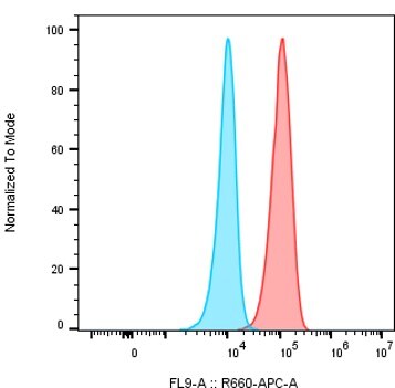
Application: Flow CytometrySample Tested: M2c macrophagesSpecies: HumanVerified Customer | Posted 06/20/2026Detection of MerTK in M2c macrophages. The cells were incubated with 2 μl of the human MerTK antibody (Red) or isotype control (blue) for 30 minutes on ice, followed by a secondary a-mouse IgG APCThe antibody was used to detect MerTK in M2c macrophages using 2 μl of the human MerTK antibody or isotype control for 30 minutes on ice, followed by a secondary a-mouse IgG APC at 1:300

-
Verified Customer | Posted 04/26/2017
-
Application: Functional AssaySample Tested: NCI-H1299Species: HumanVerified Customer | Posted 06/07/2016
There are no reviews that match your criteria.
Protocols
Find general support by application which include: protocols, troubleshooting, illustrated assays, videos and webinars.
- 7-Amino Actinomycin D (7-AAD) Cell Viability Flow Cytometry Protocol
- Extracellular Membrane Flow Cytometry Protocol
- Flow Cytometry Protocol for Cell Surface Markers
- Flow Cytometry Protocol for Staining Membrane Associated Proteins
- Flow Cytometry Staining Protocols
- Flow Cytometry Troubleshooting Guide
- Intracellular Flow Cytometry Protocol Using Alcohol (Methanol)
- Intracellular Flow Cytometry Protocol Using Detergents
- Intracellular Nuclear Staining Flow Cytometry Protocol Using Detergents
- Intracellular Staining Flow Cytometry Protocol Using Alcohol Permeabilization
- Intracellular Staining Flow Cytometry Protocol Using Detergents to Permeabilize Cells
- Propidium Iodide Cell Viability Flow Cytometry Protocol
- Protocol for Liperfluo
- Protocol for the Characterization of Human Th22 Cells
- Protocol for the Characterization of Human Th9 Cells
- Protocol: Annexin V and PI Staining by Flow Cytometry
- Protocol: Annexin V and PI Staining for Apoptosis by Flow Cytometry
- Troubleshooting Guide: Fluorokine Flow Cytometry Kits
- View all Protocols, Troubleshooting, Illustrated assays and Webinars
Loading...