MICB (MHC class I chain-related gene B) is a transmembrane glycoprotein that functions as a ligand for NKG2D. A closely related protein, MICA, shares 85% amino acid identity with MICB. These 2 proteins are distantly related to the MHC class I proteins. MICA and MICB (MICA/B) possess three extracellular immunoglobulin-like domains, but have no capacity to bind peptide or interact with beta 2-microglobulin. The genes encoding MICA/B are found within the major histocompatibility complex on human chromosome 6. The MICB locus is polymorphic with more than 15 recognized human alleles. MICA/B are minimally expressed on normal cells, but are frequently expressed on epithelial tumors and can be induced by bacterial and viral infections. MICA/B are ligands for NKG2D, an activating receptor expressed on NK cells, NKT cells, gamma delta T cells, and CD8+ alpha beta T cells. Recognition of MICA/B by NKG2D results in the activation of cytolytic activity and/or cytokine production by these effector cells. MICA/B recognition is involved in tumor surveillance, viral infections, and autoimmune diseases. The release of soluble forms of MICA/B from tumors down-regulates NKG2D surface expression on effector cells resulting in the impairment of anti-tumor immune response (1-7).

Key Product Details
Validated by
Knockout/Knockdown, Biological Validation
Species Reactivity
Validated:
Human
Cited:
Human, Mouse
Applications
Validated:
Knockout Validated, Western Blot, ELISA Capture (Matched Antibody Pair), Flow Cytometry, CyTOF-ready
Cited:
Immunohistochemistry, Western Blot, Neutralization, Flow Cytometry, Immunocytochemistry, ELISA Development, FACS
Label
Unconjugated
Antibody Source
Monoclonal Mouse IgG2B Clone # 236511
Loading...
Product Specifications
Immunogen
Mouse myeloma cell line NS0-derived recombinant human MICB
Ala23-Gly298
Accession # CAI18747
Ala23-Gly298
Accession # CAI18747
Specificity
Detects human MICB in direct ELISAs and Western blots. Does not cross-react with recombinant human MICA.
Clonality
Monoclonal
Host
Mouse
Isotype
IgG2B
Scientific Data Images for Human MICB Antibody (236511)
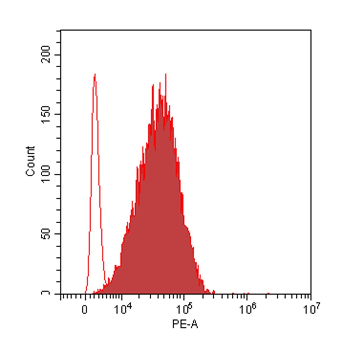
Detection of MICB in K562 Human Cell Line by Flow Cytometry.
K562 human chronic myelogenous leukemia cell line was stained with Mouse Anti-Human MICB Monoclonal Antibody (Catalog # MAB1599, filled histogram) or isotype control antibody (Catalog # MAB0041, open histogram), followed by Phycoerythrin-conjugated Anti-Mouse IgG Secondary Antibody (Catalog # F0102B).
MICB Specificity is Shown by Flow Cytometry in Knockout Cell Line.
MICB knockout K562 human myelogenous leukemia cell line was stained with Mouse Anti-Human MICB Monoclonal Antibody (Catalog # MAB1599, filled histogram) or isotype control antibody (Catalog # MAB0041, open histogram) followed by anti-Mouse IgG PE-conjugated secondary antibody (Catalog # F0102B). No staining in the MICB knockout K562 cell line was observed. View our protocol for Staining Membrane-associated Proteins.
Detection of Human MICB by Flow Cytometry
Multiple receptors and ligands are involved in NK cell-mediated lysis of activated CD4+ T cells.Role of (A) activating and (B) inhibitory NK receptors in NK cell degranulation. Left column: representative histograms (of n≥3) for surface expression of ligands on activated (thick black line) and resting CD4+ T cells (thin black line). Isotype-matched control Ig are represented by dashed line (activated CD4+ T) and filled histogram (resting CD4+ T). Middle- and right column: NK and CD4+ T cells were activated for 4 days in vitro as described, and co-cultured for 4 hours with 10 ug/mL mAb (or relevant isotype-matched control Ig). Degranulation is shown for CD56dim (middle column) and CD56bright (right column) NK cells. Representative histograms of surface expression of receptors on activated (thick black line) and resting NK cells (thin black line). Isotype-matched control Ig are represented by dashed line (activated NK) and filled histogram (resting NK). * P<0.05, ** P<0.005, *** P<0.001. (C) Sorted IL-2-activated CD56dim and CD56bright NK cells were co-cultured with 51Cr-labeled activated CD4+ T cells in a 51Cr-release assay with human IgG4 isotype control (•) or anti-NKG2A mAb (○). Data represents n = 3 experiments. Image collected and cropped by CiteAb from the following publication (https://pubmed.ncbi.nlm.nih.gov/22384114), licensed under a CC-BY license. Not internally tested by R&D Systems.
Detection of Mouse MICB by Flow Cytometry
Hydroxycitrate reduces MICA expression in activated T cells and multiple cancer cells. (A) MICA mRNA analyzed by quantitative RT-PCR in total RNA purified from HEK293 cells after more than 30 passages in glucose (Glc) and galactose (Gal). MICA expression is normalized to housekeeping gene RPLP0 and displayed as mean ± SEM from six independent experiments. (B) MICA surface expression analyzed by flow cytometry of Glc and Gal cells at basal levels. Dot plots are representative of at least three independent experiments. Grid is set to ∼5% of corresponding isotype control stainings. (C) Mitochondrial stress test on HEK293 cultivated in Glc or Gal under same conditions as in Figure 4A. The graph is baselined to measuring point three and displays mean ± SEM from two independent experiments. (D) MICA/B surface expression of peripheral blood lymphocytes (PBLs) activated for 3 days in Glc or Gal growth medium prior to 18 h treatment with FR901228 (20 ng/mL). Grids in dot plots are set to ∼5% of corresponding isotype control staining and dot plots are representative of seven different donors. The bar graph displays mean ± SEM of isotype-corrected MICA/B MFI ( delta MFI) from seven donors. Left panel is zoomed in on the difference between untreated Glc and Gal PBLs. (E,F) HEK293 MGAT5 knockout (KO) cells were treated with (E) 2DG (20 mM) or (F) hydroxycitrate (HC) (15 mM) in addition to PBS (UT), citrate (10 mM), or GlcNAc (25 mM) for 22–24 h. Bar graphs display MICA surface expression as mean ± SEM of delta MFI values from three independent experiments. Data of UT samples share values with UT samples in Figure 3H. (G) MICA surface expression in several cancer cell lines after 18 or 42 h treatment with HC (10 mM). delta MFI values are normalized to UT control and shown as mean ± SEM from at least three independent experiments. (H,I) MICA surface expression (H) and NKG2D-fc binding (I) in cancer cell lines after 2.5 h treatment with HC (10 mM) prior to 18 h stimulation with FR901228 (FR, 20 ng
Human MICB ELISA Standard Curve
Recombinant Human MICB Fc Chimera, aa 23-298 (Catalog # 1599-MB) was serially diluted and captured by Mouse Anti-Human MICB Monoclonal Antibody (Catalog # MAB1599) coated on a Clear Polystyrene Microplate (Catalog # DY990). Goat Anti-Human MICB Antigen Affinity-purified Polyclonal Antibody (Catalog # AF1599) was biotinylated and incubated with the protein captured on the plate. Detection of the standard curve was achieved by incubating Streptavidin-HRP (Catalog # DY998)Applications for Human MICB Antibody (236511)
Application
Recommended Usage
CyTOF-ready
Ready to be labeled using established conjugation methods. No BSA or other carrier proteins that could interfere with conjugation.
Flow Cytometry
0.25 µg/106 cells
Sample: K562 human chronic myelogenous leukemia cell line
Sample: K562 human chronic myelogenous leukemia cell line
Knockout Validated
MICB is specifically detected in K562 myelogenous leukemia parental cell line but is not detectable in MICB knockout K562 cell line.
Western Blot
1 µg/mL
Sample: Recombinant Human MICB Fc Chimera, aa 23-298 (Catalog # 1599-MB)
Sample: Recombinant Human MICB Fc Chimera, aa 23-298 (Catalog # 1599-MB)
Human MICB Sandwich Immunoassay
Please Note: Optimal dilutions of this antibody should be experimentally determined.
Reviewed Applications
Read 1 review rated 5 using MAB1599 in the following applications:
Flow Cytometry Panel Builder
Bio-Techne Knows Flow Cytometry
Save time and reduce costly mistakes by quickly finding compatible reagents using the Panel Builder Tool.
Advanced Features
- Spectra Viewer - Custom analysis of spectra from multiple fluorochromes
- Spillover Popups - Visualize the spectra of individual fluorochromes
- Antigen Density Selector - Match fluorochrome brightness with antigen density

Formulation, Preparation, and Storage
Purification
Protein A or G purified from hybridoma culture supernatant
Reconstitution
Reconstitute at 0.5 mg/mL in sterile PBS. For liquid material, refer to CoA for concentration.
Loading...
Formulation
Lyophilized from a 0.2 μm filtered solution in PBS with Trehalose. *Small pack size (SP) is supplied either lyophilized or as a 0.2 µm filtered solution in PBS.
Shipping
Lyophilized product is shipped at ambient temperature. Liquid small pack size (-SP) is shipped with polar packs. Upon receipt, store immediately at the temperature recommended below.
Stability & Storage
Use a manual defrost freezer and avoid repeated freeze-thaw cycles.
- 12 months from date of receipt, -20 to -70 °C as supplied.
- 1 month, 2 to 8 °C under sterile conditions after reconstitution.
- 6 months, -20 to -70 °C under sterile conditions after reconstitution.
Calculators
Background: MICB
References
- Groh, V. et al. (2001) Nature Immunol. 2:255.
- Stephens, H. (2001) Trends Immunol. 22:378.
- Bauer, S. et al. (1999) Science 285:727.
- Groh, V. et al. (2002) Nature 419:734.
- Steinle, A. et al. (2001) Immunogenetics 53:279.
- Pende, D. et al. (2002) Cancer Res. 62:6178.
- Salih, H. et al. (2003) Blood 102:1389.
Long Name
MHC Class I-related Protein B
Alternate Names
MHC class I chain-related protein B, MHC class I mic-B antigen, MHC class I polypeptide-related sequence B, MIC-B, PERB11.2MHC class I-like molecule PERB11.2-IMX, stress inducible class I homolog
Entrez Gene IDs
4277 (Human)
Gene Symbol
MICB
UniProt
Additional MICB Products
Product Documents for Human MICB Antibody (236511)
Certificate of Analysis
To download a Certificate of Analysis, please enter a lot or batch number in the search box below.
Note: Certificate of Analysis not available for kit components.
Product Specific Notices for Human MICB Antibody (236511)
For research use only
Related Research Areas
Citations for Human MICB Antibody (236511)
 Powered by Bioz
Customer Reviews for Human MICB Antibody (236511) (1)
5 out of 5
1 Customer Rating
Have you used Human MICB Antibody (236511)?
Submit a review and receive an Amazon gift card!
$25/€18/£15/$25CAN/¥2500 Yen for a review with an image
$10/€7/£6/$10CAN/¥1110 Yen for a review without an image
Submit a review
Customer Images
Showing
1
-
1 of
1 review
Showing All
Filter By:
-
Application: Flow CytometrySample Tested: T98G human glioblastoma cell lineSpecies: HumanVerified Customer | Posted 10/24/2022This antibody, followed by anti-mouse secondary antibodies, achieves a clean staining of surface MICB in glioma cells

There are no reviews that match your criteria.
Protocols
Find general support by application which include: protocols, troubleshooting, illustrated assays, videos and webinars.
- 7-Amino Actinomycin D (7-AAD) Cell Viability Flow Cytometry Protocol
- Cellular Response to Hypoxia Protocols
- Extracellular Membrane Flow Cytometry Protocol
- Flow Cytometry Protocol for Cell Surface Markers
- Flow Cytometry Protocol for Staining Membrane Associated Proteins
- Flow Cytometry Staining Protocols
- Flow Cytometry Troubleshooting Guide
- Intracellular Flow Cytometry Protocol Using Alcohol (Methanol)
- Intracellular Flow Cytometry Protocol Using Detergents
- Intracellular Nuclear Staining Flow Cytometry Protocol Using Detergents
- Intracellular Staining Flow Cytometry Protocol Using Alcohol Permeabilization
- Intracellular Staining Flow Cytometry Protocol Using Detergents to Permeabilize Cells
- Propidium Iodide Cell Viability Flow Cytometry Protocol
- Protocol for Liperfluo
- Protocol for the Characterization of Human Th22 Cells
- Protocol for the Characterization of Human Th9 Cells
- Protocol: Annexin V and PI Staining by Flow Cytometry
- Protocol: Annexin V and PI Staining for Apoptosis by Flow Cytometry
- R&D Systems Quality Control Western Blot Protocol
- Troubleshooting Guide: Fluorokine Flow Cytometry Kits
- Troubleshooting Guide: Western Blot Figures
- Western Blot Conditions
- Western Blot Protocol
- Western Blot Protocol for Cell Lysates
- Western Blot Troubleshooting
- Western Blot Troubleshooting Guide
- View all Protocols, Troubleshooting, Illustrated assays and Webinars
Loading...