UCH-L1/PGP9.5 Antibody (972119)
R&D Systems | Catalog # MAB60072

Key Product Details
Species Reactivity
Human, Mouse, Rat
Applications
Immunohistochemistry, Western Blot, Immunocytochemistry, Simple Western
Label
Unconjugated
Antibody Source
Monoclonal Mouse IgG2B Clone # 972119
Loading...
Product Specifications
Immunogen
E. coli-derived recombinant human UCH-L1/PGP9.5
Gln2-Ala223
Accession # P09936
Gln2-Ala223
Accession # P09936
Specificity
Detects human UCH-L1/PGP9.5 in direct ELISAs. Detects human, mouse, and rat in Western blot.
Clonality
Monoclonal
Host
Mouse
Isotype
IgG2B
Scientific Data Images for UCH-L1/PGP9.5 Antibody (972119)
Detection of Human, Mouse, and Rat UCH-L1/PGP9.5 by Western Blot.
Western blot shows lysates of A172 human glioblastoma cell line, Neuro-2A mouse neuroblastoma cell line, and PC-12 rat adrenal pheochromocytoma cell line. PVDF membrane was probed with 0.1 µg/mL of Mouse Anti-Human UCH-L1/PGP9.5 Monoclonal Antibody (Catalog # MAB60072) followed by HRP-conjugated Anti-Mouse IgG Secondary Antibody (Catalog # HAF018). A specific band was detected for UCH-L1/PGP9.5 at approximately 26 kDa (as indicated). This experiment was conducted under reducing conditions and using Immunoblot Buffer Group 1.
UCH-L1/PGP9.5 in A172 Human Cell Line.
UCH-L1/PGP9.5 was detected in immersion fixed A172 human glioblastoma cell line using Mouse Anti-Human UCH-L1/PGP9.5 Monoclonal Antibody (Catalog # MAB60072) at 0.3 µg/mL for 3 hours at room temperature. Cells were stained using the NorthernLights™ 557-conjugated Anti-Mouse IgG Secondary Antibody (red; Catalog # NL007) and counterstained with DAPI (blue). Specific staining was localized to cytoplasm. View our protocol for Fluorescent ICC Staining of Cells on Coverslips.
UCH-L1/PGP9.5 in Human Brain.
UCH-L1/PGP9.5 was detected in immersion fixed paraffin-embedded sections of human brain (caudate nucleus) using Mouse Anti-Human UCH-L1/PGP9.5 Monoclonal Antibody (Catalog # MAB60072) at 5 µg/mL for 1 hour at room temperature followed by incubation with the Anti-Mouse IgG VisUCyte™ HRP Polymer Antibody (Catalog # VC001). Tissue was stained using DAB (brown) and counterstained with hematoxylin (blue). Specific staining was localized to cytoplasm. View our protocol for IHC Staining with VisUCyte HRP Polymer Detection Reagents.
Detection of Human and Mouse UCH-L1/PGP9.5 by Simple WesternTM.
Simple Western lane view shows lysates of A172 human glioblastoma cell line and Neuro‑2A mouse neuroblastoma cell line, loaded at 0.2 mg/mL. A specific band was detected for UCH-L1/PGP9.5 at approximately 31 kDa (as indicated) using 1 µg/mL of Mouse Anti-Human UCH-L1/PGP9.5 Monoclonal Antibody (Catalog # MAB60072). This experiment was conducted under reducing conditions and using the 12-230 kDa separation system.
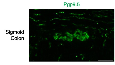
UCH-L1/PGP9.5 in Human Colon.
1:100 dilution in PFA-fixed, frozen human sigmoid colon. 0.2% Triton X-100 was included in primary antibody mix. Image from a verified customer review.Applications for UCH-L1/PGP9.5 Antibody (972119)
Application
Recommended Usage
Immunocytochemistry
0.3-25 µg/mL
Sample: Immersion fixed A172 human glioblastoma cell line
Sample: Immersion fixed A172 human glioblastoma cell line
Immunohistochemistry
5-25 µg/mL
Sample: Immersion fixed paraffin-embedded sections of human brain (caudate nucleus)
Sample: Immersion fixed paraffin-embedded sections of human brain (caudate nucleus)
Simple Western
1 µg/mL
Sample: A172 human glioblastoma cell line and Neuro‑2A mouse neuroblastoma cell line
Sample: A172 human glioblastoma cell line and Neuro‑2A mouse neuroblastoma cell line
Western Blot
0.1 µg/mL
Sample: A172 human glioblastoma cell line, Neuro‑2A mouse neuroblastoma cell line, and PC‑12 rat adrenal pheochromocytoma cell line
Sample: A172 human glioblastoma cell line, Neuro‑2A mouse neuroblastoma cell line, and PC‑12 rat adrenal pheochromocytoma cell line
Formulation, Preparation, and Storage
Purification
Protein A or G purified from hybridoma culture supernatant
Reconstitution
Reconstitute at 0.5 mg/mL in sterile PBS. For liquid material, refer to CoA for concentration.
Loading...
Formulation
Lyophilized from a 0.2 μm filtered solution in PBS with Trehalose. *Small pack size (SP) is supplied either lyophilized or as a 0.2 µm filtered solution in PBS.
Shipping
Lyophilized product is shipped at ambient temperature. Liquid small pack size (-SP) is shipped with polar packs. Upon receipt, store immediately at the temperature recommended below.
Stability & Storage
Use a manual defrost freezer and avoid repeated freeze-thaw cycles.
- 12 months from date of receipt, -20 to -70 °C as supplied.
- 1 month, 2 to 8 °C under sterile conditions after reconstitution.
- 6 months, -20 to -70 °C under sterile conditions after reconstitution.
Calculators
Background: UCH-L1/PGP9.5
Long Name
Ubiquitin C-terminal Hydrolase L1
Alternate Names
PARK5, PGP9.5, UCHL1
Gene Symbol
UCHL1
UniProt
Additional UCH-L1/PGP9.5 Products
Product Documents for UCH-L1/PGP9.5 Antibody (972119)
Certificate of Analysis
To download a Certificate of Analysis, please enter a lot or batch number in the search box below.
Note: Certificate of Analysis not available for kit components.
Product Specific Notices for UCH-L1/PGP9.5 Antibody (972119)
For research use only
Related Research Areas
Citations for UCH-L1/PGP9.5 Antibody (972119)
 Powered by Bioz
Customer Reviews for UCH-L1/PGP9.5 Antibody (972119) (1)
5 out of 5
1 Customer Rating
Have you used UCH-L1/PGP9.5 Antibody (972119)?
Submit a review and receive an Amazon gift card!
$25/€18/£15/$25CAN/¥2500 Yen for a review with an image
$10/€7/£6/$10CAN/¥1110 Yen for a review without an image
Submit a review
Customer Images
Showing
1
-
1 of
1 review
Showing All
Filter By:
-
Verified Customer | Posted 10/16/20251:100 dilution shows strong staining in human myenteric plexus1:100 dilution in PFA-fixed, frozen human sigmoid colon. 0.2% Triton X-100 was included in primary antibody mix.

There are no reviews that match your criteria.
Protocols
Find general support by application which include: protocols, troubleshooting, illustrated assays, videos and webinars.
- Antigen Retrieval Protocol (PIER)
- Antigen Retrieval for Frozen Sections Protocol
- Appropriate Fixation of IHC/ICC Samples
- Cellular Response to Hypoxia Protocols
- Chromogenic IHC Staining of Formalin-Fixed Paraffin-Embedded (FFPE) Tissue Protocol
- Chromogenic Immunohistochemistry Staining of Frozen Tissue
- ClariTSA™ Fluorophore Kits
- Detection & Visualization of Antibody Binding
- Fluorescent IHC Staining of Frozen Tissue Protocol
- Graphic Protocol for Heat-induced Epitope Retrieval
- Graphic Protocol for the Preparation and Fluorescent IHC Staining of Frozen Tissue Sections
- Graphic Protocol for the Preparation and Fluorescent IHC Staining of Paraffin-embedded Tissue Sections
- Graphic Protocol for the Preparation of Gelatin-coated Slides for Histological Tissue Sections
- ICC Cell Smear Protocol for Suspension Cells
- ICC Immunocytochemistry Protocol Videos
- ICC for Adherent Cells
- IHC Sample Preparation (Frozen sections vs Paraffin)
- Immunocytochemistry (ICC) Protocol
- Immunocytochemistry Troubleshooting
- Immunofluorescence of Organoids Embedded in Cultrex Basement Membrane Extract
- Immunofluorescent IHC Staining of Formalin-Fixed Paraffin-Embedded (FFPE) Tissue Protocol
- Immunohistochemistry (IHC) and Immunocytochemistry (ICC) Protocols
- Immunohistochemistry Frozen Troubleshooting
- Immunohistochemistry Paraffin Troubleshooting
- Preparing Samples for IHC/ICC Experiments
- Preventing Non-Specific Staining (Non-Specific Binding)
- Primary Antibody Selection & Optimization
- Protocol for Heat-Induced Epitope Retrieval (HIER)
- Protocol for Making a 4% Formaldehyde Solution in PBS
- Protocol for VisUCyte™ HRP Polymer Detection Reagent
- Protocol for the Fluorescent ICC Staining of Cell Smears - Graphic
- Protocol for the Fluorescent ICC Staining of Cultured Cells on Coverslips - Graphic
- Protocol for the Preparation & Fixation of Cells on Coverslips
- Protocol for the Preparation and Chromogenic IHC Staining of Frozen Tissue Sections
- Protocol for the Preparation and Chromogenic IHC Staining of Frozen Tissue Sections - Graphic
- Protocol for the Preparation and Chromogenic IHC Staining of Paraffin-embedded Tissue Sections
- Protocol for the Preparation and Chromogenic IHC Staining of Paraffin-embedded Tissue Sections - Graphic
- Protocol for the Preparation and Fluorescent ICC Staining of Cells on Coverslips
- Protocol for the Preparation and Fluorescent ICC Staining of Non-adherent Cells
- Protocol for the Preparation and Fluorescent ICC Staining of Stem Cells on Coverslips
- Protocol for the Preparation and Fluorescent IHC Staining of Frozen Tissue Sections
- Protocol for the Preparation and Fluorescent IHC Staining of Paraffin-embedded Tissue Sections
- Protocol for the Preparation of Gelatin-coated Slides for Histological Tissue Sections
- Protocol for the Preparation of a Cell Smear for Non-adherent Cell ICC - Graphic
- R&D Systems Quality Control Western Blot Protocol
- TUNEL and Active Caspase-3 Detection by IHC/ICC Protocol
- The Importance of IHC/ICC Controls
- Troubleshooting Guide: Immunohistochemistry
- Troubleshooting Guide: Western Blot Figures
- Western Blot Conditions
- Western Blot Protocol
- Western Blot Protocol for Cell Lysates
- Western Blot Troubleshooting
- Western Blot Troubleshooting Guide
- View all Protocols, Troubleshooting, Illustrated assays and Webinars
Loading...