
Key Product Details
Validated by
Biological Validation
Species Reactivity
Validated:
Human, Mouse
Cited:
Human, Mouse
Applications
Validated:
Western Blot, Intracellular Staining by Flow Cytometry, Immunocytochemistry, Simple Western, CyTOF-ready
Cited:
Western Blot, Flow Cytometry, Immunocytochemistry
Label
Unconjugated
Antibody Source
Monoclonal Mouse IgG2A Clone # 527327
Loading...
Product Specifications
Immunogen
E. coli-derived recombinant human RUNX3/CBFA3
Lys186-Tyr415
Accession # Q13761
Lys186-Tyr415
Accession # Q13761
Specificity
Detects human and mouse RUNX3/CBFA3 in Western blots.
Clonality
Monoclonal
Host
Mouse
Isotype
IgG2A
Scientific Data Images for RUNX3/CBFA3 Antibody (527327)
Detection of RUNX3/CBFA3 in Human PBMC by Flow Cytometry.
Human PBMC unstimulated (light orange filled histogram) or treated with 50ng/mL PMA (dark orange filled histogram) were stained with Mouse Anti-Human/Mouse RUNX3/CBFA3 Monoclonal Antibody (Catalog # MAB3765) or isotype control antibody (Catalog # MAB003, open histogram), followed by Phycoerythrin-conjugated Anti-Mouse IgG F(ab')2Secondary Antibody (Catalog # F0102B). To facilitate intracellular staining, cells were fixed with paraformaldehyde and permeabilized with saponin.
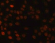
RUNX3/CBFA3 in Human PBMCs.
RUNX3/CBFA3 was detected in immersion fixed human peripheral blood mononuclear cells (PBMCs) using Mouse Anti-Human/Mouse RUNX3/CBFA3 Monoclonal Antibody (Catalog # MAB3765) at 10 µg/mL for 3 hours at room temperature. Cells were stained using the NorthernLights™ 557-conjugated Anti-Mouse IgG Secondary Antibody (red; Catalog # NL007) and counterstained with DAPI (blue). Specific staining was localized to nuclei. View our protocol for Fluorescent ICC Staining of Non-adherent Cells.
Detection of Human and Mouse RUNX3/CBFA3 by Western Blot.
Western blot shows lysates of Jurkat human acute T cell leukemia cell line, Daudi human Burkitt's lymphoma cell line, and DA3 mouse myeloma cell line. Gels were loaded with 30 µg of whole cell lysate (WCL), 20 µg of cytoplasmic (Cyto), and 10 µg of nuclear extracts (Nuc). PVDF Membrane was probed with 1 µg/mL of Mouse Anti-Human/Mouse RUNX3/CBFA3 Monoclonal Antibody (Catalog # MAB3765) followed by HRP-conjugated Anti-Mouse IgG Secondary Antibody (Catalog # HAF007). Specific bands were detected for RUNX3/CBFA3 at approximately 48 and 52 kDa (as indicated). This experiment was conducted under reducing conditions and using Immunoblot Buffer Group 1.
Detection of Human RUNX3/CBFA3 by Simple WesternTM.
Simple Western lane view shows lysates of Jurkat human acute T cell leukemia cell line and Daudi human Burkitt's lymphoma cell line, loaded at 0.5 mg/mL. A specific band was detected for RUNX3/CBFA3 at approximately 54 kDa (as indicated) using 10 µg/mL of Mouse Anti-Human/Mouse RUNX3/CBFA3 Monoclonal Antibody (Catalog # MAB3765). This experiment was conducted under reducing conditions and using the 12-230 kDa separation system.
Detection of RUNX3/CBFA3 by Flow Cytometry
RA and RAR alpha reciprocally regulate the expression of the positive and negative LC-regulating transcription factors, Runx3 and C/EBP beta. a Impact of RA and RAR alpha deficiency on Runx3 expression at mRNA level. b Impact of RA and RAR alpha deficiency on Runx3 expression at protein level. c Effect of enforced Runx3 expression on BM-derived LC differentiation in the presence and absence of RA. The data shown are gated for transduced Thy1.1+CD11c+ cells. d Impact of RA and RAR alpha deficiency on expression of Cebpb mRNA. e Impact of RA and RAR alpha deficiency on expression of C/EBP beta protein. f Effect of dnC/EBP beta on BM-derived LC differentiation in the presence and absence of RA. BM cells from WT or ∆RaraCD11c mice were cultured with GM-CSF and TGF beta 1 for 5 days (3 days following retroviral transduction) in the presence of At-RA (10 nM except in panels c and f where 0.1 nM was used) in a medium containing charcoal-treated FBS. Representative and combined data (n = 3–7) from at least 3 experiments are shown. Significant differences from controls by one-way ANOVA with Bonferroni adjustments (p < 0.05)* or between indicated groups by two-way ANOVA with Tukey adjustments (p < 0.05)**. Error bars are defined as s.e.m Image collected and cropped by CiteAb from the following open publication (https://pubmed.ncbi.nlm.nih.gov/30254197), licensed under a CC-BY license. Not internally tested by R&D Systems.
Detection of RUNX3/CBFA3 by Flow Cytometry
RA regulates CEBPb and RUNX3 expression and human langerin+ cell differentiation from blood monocytes. a Human LC differentiation from blood monocytes in regular vs. charcoal FBS. b At-RA promotes C/EBP beta + non-LC differentiation. c RA and RAR alpha antagonists reciprocally regulate human LC differentiation. d Effects of At-RA and BMS493 on the expression of human CEBPb and RUNX3. Human blood CD14+ monocytes were cultured in GM-CSF and TGF-beta 1 for 5–7 days in media containing charcoal FBS except in panel A where regular FBS was also used. At-RA was used at 1 nM, and BMS493 and Ro4153 were used at 500 nM. Representative and combined data (n = 5–7) from at least 5 experiments are shown. *Significant differences from control by Mann–Whitney U test (p < 0.05, unpaired, 2-sided). Error bars are defined as s.e.m. Langerhans cells (LC) and langerin-expressing conventional dendritic cells are made from distinct progenitors and enriched in the distinct microenvironments of the skin. Here the authors show that these immune cells are regulated by RAR alpha via simultaneous induction of LC-promoting Runx3 and repression of LC-inhibiting C/EBP beta Image collected and cropped by CiteAb from the following open publication (https://pubmed.ncbi.nlm.nih.gov/30254197), licensed under a CC-BY license. Not internally tested by R&D Systems.Applications for RUNX3/CBFA3 Antibody (527327)
Application
Recommended Usage
CyTOF-ready
Ready to be labeled using established conjugation methods. No BSA or other carrier proteins that could interfere with conjugation.
Immunocytochemistry
8-25 µg/mL
Sample: Immersion fixed human peripheral blood mononuclear cells (PBMCs) and DA3 mouse myeloma cell line
Sample: Immersion fixed human peripheral blood mononuclear cells (PBMCs) and DA3 mouse myeloma cell line
Intracellular Staining by Flow Cytometry
2.5 µg/106 cells
Sample: Human PBMC treated with PMA and mouse splenocytes fixed with paraformaldehyde and permeabilized with saponin
Sample: Human PBMC treated with PMA and mouse splenocytes fixed with paraformaldehyde and permeabilized with saponin
Simple Western
10 µg/mL
Sample: Jurkat human acute T cell leukemia cell line and Daudi human Burkitt's lymphoma cell line
Sample: Jurkat human acute T cell leukemia cell line and Daudi human Burkitt's lymphoma cell line
Western Blot
1 µg/mL
Sample: Jurkat human acute T cell leukemia cell line, Daudi human Burkitt's lymphoma cell line, and DA3 mouse myeloma cell line
Sample: Jurkat human acute T cell leukemia cell line, Daudi human Burkitt's lymphoma cell line, and DA3 mouse myeloma cell line
Reviewed Applications
Read 1 review rated 5 using MAB3765 in the following applications:
Flow Cytometry Panel Builder
Bio-Techne Knows Flow Cytometry
Save time and reduce costly mistakes by quickly finding compatible reagents using the Panel Builder Tool.
Advanced Features
- Spectra Viewer - Custom analysis of spectra from multiple fluorochromes
- Spillover Popups - Visualize the spectra of individual fluorochromes
- Antigen Density Selector - Match fluorochrome brightness with antigen density

Formulation, Preparation, and Storage
Purification
Protein A or G purified from hybridoma culture supernatant
Reconstitution
Reconstitute at 0.5 mg/mL in sterile PBS. For liquid material, refer to CoA for concentration.
Loading...
Formulation
Lyophilized from a 0.2 μm filtered solution in PBS with Trehalose. *Small pack size (SP) is supplied either lyophilized or as a 0.2 µm filtered solution in PBS.
Shipping
Lyophilized product is shipped at ambient temperature. Liquid small pack size (-SP) is shipped with polar packs. Upon receipt, store immediately at the temperature recommended below.
Stability & Storage
Use a manual defrost freezer and avoid repeated freeze-thaw cycles.
- 12 months from date of receipt, -20 to -70 °C as supplied.
- 1 month, 2 to 8 °C under sterile conditions after reconstitution.
- 6 months, -20 to -70 °C under sterile conditions after reconstitution.
Calculators
Background: RUNX3/CBFA3
Long Name
Runt-related Transcription Factor 3
Alternate Names
AML2, CBFA3, PEBP2A3, PEBP2AC
Gene Symbol
RUNX3
UniProt
Additional RUNX3/CBFA3 Products
Product Documents for RUNX3/CBFA3 Antibody (527327)
Certificate of Analysis
To download a Certificate of Analysis, please enter a lot or batch number in the search box below.
Note: Certificate of Analysis not available for kit components.
Product Specific Notices for RUNX3/CBFA3 Antibody (527327)
For research use only
Related Research Areas
Citations for RUNX3/CBFA3 Antibody (527327)
 Powered by Bioz
Customer Reviews for RUNX3/CBFA3 Antibody (527327) (1)
5 out of 5
1 Customer Rating
Have you used RUNX3/CBFA3 Antibody (527327)?
Submit a review and receive an Amazon gift card!
$25/€18/£15/$25CAN/¥2500 Yen for a review with an image
$10/€7/£6/$10CAN/¥1110 Yen for a review without an image
Submit a review
Customer Images
Showing
1
-
1 of
1 review
Showing All
Filter By:
-
Application: Immunocytochemistry/ImmunofluorescenceSample Tested: Peripheral blood mononuclear cells (PBMCs)Species: MouseVerified Customer | Posted 12/18/2021

There are no reviews that match your criteria.
Protocols
Find general support by application which include: protocols, troubleshooting, illustrated assays, videos and webinars.
- 7-Amino Actinomycin D (7-AAD) Cell Viability Flow Cytometry Protocol
- Appropriate Fixation of IHC/ICC Samples
- Cellular Response to Hypoxia Protocols
- ClariTSA™ Fluorophore Kits
- Detection & Visualization of Antibody Binding
- Extracellular Membrane Flow Cytometry Protocol
- Flow Cytometry Protocol for Cell Surface Markers
- Flow Cytometry Protocol for Staining Membrane Associated Proteins
- Flow Cytometry Staining Protocols
- Flow Cytometry Troubleshooting Guide
- ICC Cell Smear Protocol for Suspension Cells
- ICC Immunocytochemistry Protocol Videos
- ICC for Adherent Cells
- Immunocytochemistry (ICC) Protocol
- Immunocytochemistry Troubleshooting
- Immunofluorescence of Organoids Embedded in Cultrex Basement Membrane Extract
- Immunohistochemistry (IHC) and Immunocytochemistry (ICC) Protocols
- Intracellular Flow Cytometry Protocol Using Alcohol (Methanol)
- Intracellular Flow Cytometry Protocol Using Detergents
- Intracellular Nuclear Staining Flow Cytometry Protocol Using Detergents
- Intracellular Staining Flow Cytometry Protocol Using Alcohol Permeabilization
- Intracellular Staining Flow Cytometry Protocol Using Detergents to Permeabilize Cells
- Preparing Samples for IHC/ICC Experiments
- Preventing Non-Specific Staining (Non-Specific Binding)
- Primary Antibody Selection & Optimization
- Propidium Iodide Cell Viability Flow Cytometry Protocol
- Protocol for Liperfluo
- Protocol for VisUCyte™ HRP Polymer Detection Reagent
- Protocol for the Characterization of Human Th22 Cells
- Protocol for the Characterization of Human Th9 Cells
- Protocol for the Fluorescent ICC Staining of Cell Smears - Graphic
- Protocol for the Fluorescent ICC Staining of Cultured Cells on Coverslips - Graphic
- Protocol for the Preparation and Fluorescent ICC Staining of Cells on Coverslips
- Protocol for the Preparation and Fluorescent ICC Staining of Non-adherent Cells
- Protocol for the Preparation and Fluorescent ICC Staining of Stem Cells on Coverslips
- Protocol for the Preparation of a Cell Smear for Non-adherent Cell ICC - Graphic
- Protocol: Annexin V and PI Staining by Flow Cytometry
- Protocol: Annexin V and PI Staining for Apoptosis by Flow Cytometry
- R&D Systems Quality Control Western Blot Protocol
- TUNEL and Active Caspase-3 Detection by IHC/ICC Protocol
- The Importance of IHC/ICC Controls
- Troubleshooting Guide: Fluorokine Flow Cytometry Kits
- Troubleshooting Guide: Western Blot Figures
- Western Blot Conditions
- Western Blot Protocol
- Western Blot Protocol for Cell Lysates
- Western Blot Troubleshooting
- Western Blot Troubleshooting Guide
- View all Protocols, Troubleshooting, Illustrated assays and Webinars
Loading...
Associated Pathways


