Epidermal growth factor receptor (EGFR, also known as ErbB1 and HER1) is the founding member of the ErbB family of receptor tyrosine kinases. Ligand binding induces receptor dimerization and autophosphorylation on multiple tyrosine residues. Phosphorylation at Tyr 1068 allows binding of the SH2 domain of the cytosolic adaptor Grb2. This binding results in Ras activation.
Human phospho-EGFR (Y1068) Antibody (338324)
R&D Systems | Catalog # MAB3570
 by Western Blot.")
Key Product Details
Validated by
Biological Validation
Species Reactivity
Validated:
Human
Cited:
Human
Applications
Validated:
Western Blot, Intracellular Staining by Flow Cytometry, CyTOF-ready
Cited:
Immunohistochemistry, Western Blot, Immunocytochemistry
Label
Unconjugated
Antibody Source
Monoclonal Mouse IgG2A Clone # 338324
Loading...
Product Specifications
Immunogen
Phosphopeptide containing human EGFR Y1068 site
Specificity
Detects human EGFR phosphorylated at Y1068.
Clonality
Monoclonal
Host
Mouse
Isotype
IgG2A
Scientific Data Images for Human phospho-EGFR (Y1068) Antibody (338324)
Detection of Human Phospho-EGFR (Y1068) by Western Blot.
Western blot shows lysates of A431 human epithelial carcinoma cell line untreated (-) or treated (+) with 100 ng/mL Recombinant Human EGF (Catalog # 236-EG) for 5 minutes. PVDF membrane was probed with 1 µg/mL of Human Phospho-EGFR (Y1068) Monoclonal Antibody (Catalog # MAB3570), followed by HRP-conjugated Anti-Mouse IgG Secondary Antibody (Catalog # HAF007). A specific band was detected for Phospho-EGFR (Y1068) at approximately 190 kDa (as indicated). This experiment was conducted under reducing conditions and using Immunoblot Buffer Group 1.
Detection of Phospho-EGFR in EGF-treated A431 Human Cell Line by Flow Cytometry.
A431 human epithelial carcinoma cell line was untreated (open histogram) or treated for 5 minutes with 100 ng/mL Recombinant Human EGF (Catalog # 236-EG, filled histogram) then stained with Human Phospho--EGFR (Y1068) Monoclonal Antibody (Catalog # MAB3570), followed by Phycoerythrin-conjugated Anti-Mouse IgG F(ab')2Secondary Antibody (Catalog # F0102B). Mouse IgG2A(Catalog # MAB003, data not shown) was used as an isotype control. To facilitate intracellular staining, cells were fixed with paraformaldehyde and permeabilized with saponin.Applications for Human phospho-EGFR (Y1068) Antibody (338324)
Application
Recommended Usage
CyTOF-ready
Ready to be labeled using established conjugation methods. No BSA or other carrier proteins that could interfere with conjugation.
Intracellular Staining by Flow Cytometry
2.5 µg/106 cells
Sample: A431 human epithelial carcinoma cell line treated with Recombinant Human EGF (Catalog # 236-EG), fixed with paraformaldehyde, and permeabilized with saponin
Sample: A431 human epithelial carcinoma cell line treated with Recombinant Human EGF (Catalog # 236-EG), fixed with paraformaldehyde, and permeabilized with saponin
Western Blot
1 µg/mL
Sample: A431 human epithelial carcinoma cell line treated with Recombinant Human EGF (Catalog # 236-EG)
Sample: A431 human epithelial carcinoma cell line treated with Recombinant Human EGF (Catalog # 236-EG)
Reviewed Applications
Read 2 reviews rated 4.5 using MAB3570 in the following applications:
Flow Cytometry Panel Builder
Bio-Techne Knows Flow Cytometry
Save time and reduce costly mistakes by quickly finding compatible reagents using the Panel Builder Tool.
Advanced Features
- Spectra Viewer - Custom analysis of spectra from multiple fluorochromes
- Spillover Popups - Visualize the spectra of individual fluorochromes
- Antigen Density Selector - Match fluorochrome brightness with antigen density

Formulation, Preparation, and Storage
Purification
Protein A or G purified from hybridoma culture supernatant
Reconstitution
Reconstitute at 0.5 mg/mL in sterile PBS. For liquid material, refer to CoA for concentration.
Loading...
Formulation
Lyophilized from a 0.2 μm filtered solution in PBS with Trehalose. *Small pack size (SP) is supplied either lyophilized or as a 0.2 µm filtered solution in PBS.
Shipping
Lyophilized product is shipped at ambient temperature. Liquid small pack size (-SP) is shipped with polar packs. Upon receipt, store immediately at the temperature recommended below.
Stability & Storage
Use a manual defrost freezer and avoid repeated freeze-thaw cycles.
- 12 months from date of receipt, -20 to -70 °C as supplied.
- 1 month, 2 to 8 °C under sterile conditions after reconstitution.
- 6 months, -20 to -70 °C under sterile conditions after reconstitution.
Calculators
Background: EGFR
Long Name
Epidermal Growth Factor Receptor
Alternate Names
EGF R, ErbB, ErbB1, HER-1
Gene Symbol
EGFR
Additional EGFR Products
Product Documents for Human phospho-EGFR (Y1068) Antibody (338324)
Certificate of Analysis
To download a Certificate of Analysis, please enter a lot or batch number in the search box below.
Note: Certificate of Analysis not available for kit components.
Product Specific Notices for Human phospho-EGFR (Y1068) Antibody (338324)
For research use only
Related Research Areas
Citations for Human phospho-EGFR (Y1068) Antibody (338324)
 Powered by Bioz
Customer Reviews for Human phospho-EGFR (Y1068) Antibody (338324) (2)
4.5 out of 5
2 Customer Ratings
Have you used Human phospho-EGFR (Y1068) Antibody (338324)?
Submit a review and receive an Amazon gift card!
$25/€18/£15/$25CAN/¥2500 Yen for a review with an image
$10/€7/£6/$10CAN/¥1110 Yen for a review without an image
Submit a review
Customer Images

Showing
1
-
2 of
2 reviews
Showing All
Filter By:
-
Application: MicroarraySample Tested: EDTA PlasmaSpecies: HumanVerified Customer | Posted 12/07/2020Antibody was printed on custom arrays and incubated with fluorescently labeled human EDTA plasma

-
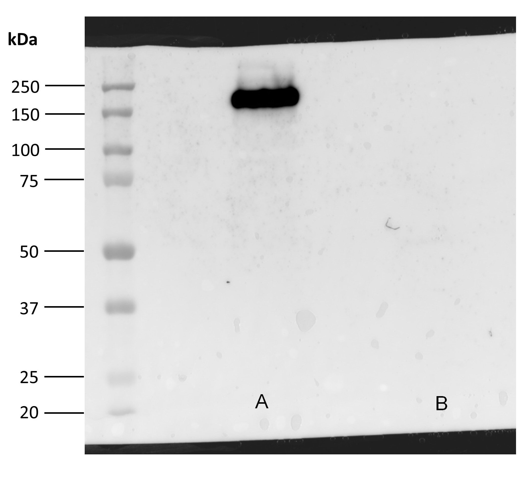
Application: Western BlotSample Tested: EGF-Stimulated A431 Cell lysate; EGF-Stimulated rat neuronsSpecies: HumanVerified Customer | Posted 10/18/2016A: EGF-Stimulated A431 Cell lysate, 20 μg; B: EGF-Stimulated rat neurons, 20 μg. MAB3570 has a high specificity for human phosphorylated EGFR, being unable to identify the rat isoform.
There are no reviews that match your criteria.
Protocols
Find general support by application which include: protocols, troubleshooting, illustrated assays, videos and webinars.
- 7-Amino Actinomycin D (7-AAD) Cell Viability Flow Cytometry Protocol
- Cellular Response to Hypoxia Protocols
- Extracellular Membrane Flow Cytometry Protocol
- Flow Cytometry Protocol for Cell Surface Markers
- Flow Cytometry Protocol for Staining Membrane Associated Proteins
- Flow Cytometry Staining Protocols
- Flow Cytometry Troubleshooting Guide
- Intracellular Flow Cytometry Protocol Using Alcohol (Methanol)
- Intracellular Flow Cytometry Protocol Using Detergents
- Intracellular Nuclear Staining Flow Cytometry Protocol Using Detergents
- Intracellular Staining Flow Cytometry Protocol Using Alcohol Permeabilization
- Intracellular Staining Flow Cytometry Protocol Using Detergents to Permeabilize Cells
- Propidium Iodide Cell Viability Flow Cytometry Protocol
- Protocol for Liperfluo
- Protocol for the Characterization of Human Th22 Cells
- Protocol for the Characterization of Human Th9 Cells
- Protocol: Annexin V and PI Staining by Flow Cytometry
- Protocol: Annexin V and PI Staining for Apoptosis by Flow Cytometry
- R&D Systems Quality Control Western Blot Protocol
- Troubleshooting Guide: Fluorokine Flow Cytometry Kits
- Troubleshooting Guide: Western Blot Figures
- Western Blot Conditions
- Western Blot Protocol
- Western Blot Protocol for Cell Lysates
- Western Blot Troubleshooting
- Western Blot Troubleshooting Guide
- View all Protocols, Troubleshooting, Illustrated assays and Webinars
Loading...
Associated Pathways


