Angiogensin I Converting Enzyme-2 (ACE-2), also called ACEH (ACE homolog), is a type I transmembrane zinc protease that cleaves angiotensins I and II to produce vasodilatory and anti-proliferative peptides. The balance between ACE-1 and ACE-2 activity is critical for maintaining cardiovascular, renal, and pulmonary function (1). ACE-2 also functions as the cellular uptake receptor for the SARS coronoavirus. Within the extracellular domain, human ACE-2 shares 83% aa sequence identity with mouse and rat ACE-2. Human ACE-2 has about 40% amino acid identity to the N- and C-terminal domains of human somatic ACE. The predicted human ACE-2 protein sequence consists of 805 amino acids, including a N-terminal signal peptide, a single catalytic domain, a C-terminal membrane anchor, and a short cytoplasmic tail. ACE-2 mRNA is found at high levels in testis, kidney and heart and at moderate levels in colon, small intestine and ovary. Classical ACE inhibitors such as captopril and lisinopril do not inhibit ACE-2 activity. Novel peptide inhibitors of ACE-2 do not inhibit ACE activity (2). Genetic data from Drosophila, mice and rats show that ACE-2 is an essential regulator of heart function in vivo (3). ACE-2 isoforms of 75 kDa and 120 kDa are differentially expressed between lung and kidney, respectively, and a shed soluble form is generated by TACE/ADAM17 mediated cleavage.

Key Product Details
Species Reactivity
Validated:
Human, Rat, Hamster
Cited:
Human, Primate - Chlorocebus aethiops (African Green Monkey)
Applications
Validated:
Immunohistochemistry, Western Blot, Flow Cytometry, CyTOF-ready
Cited:
Immunohistochemistry, Western Blot, Flow Cytometry, Immunocytochemistry
Label
Unconjugated
Antibody Source
Monoclonal Mouse IgG2A Clone # 535919
Loading...
Product Specifications
Immunogen
Mouse myeloma cell line NS0-derived recombinant human ACE-2
Gln18-Ser740
Accession # Q9BYF1
Gln18-Ser740
Accession # Q9BYF1
Specificity
Detects human ACE-2 in direct ELISAs.
Clonality
Monoclonal
Host
Mouse
Isotype
IgG2A
Scientific Data Images for ACE-2 Antibody (535919)
Detection of Human ACE‑2 by Western Blot.
Western blot shows lysates of human kidney and human testis. PVDF membrane was probed with 2 µg/mL of Mouse Anti-Human/Rat/Hamster ACE‑2 Monoclonal Antibody (Catalog # MAB9332) followed by HRP-conjugated Anti-Mouse IgG Secondary Antibody (HAF018). A specific band was detected for ACE‑2 at approximately 120 kDa (as indicated). This experiment was conducted under reducing conditions and using Western Blot Buffer Group 1.
Detection of ACE-2 in HEK293 Human Cell Line Transfected with Human ACE-2 and eGFP by Flow Cytometry.
HEK293 human embryonic kidney cell line transfected with (A) human ACE-2 or (B) irrelevant protein, and eGFP was stained with Mouse Anti-Human ACE-2 Monoclonal Antibody (Catalog # MAB9332) followed by Allophycocyanin-conjugated Anti-Mouse IgG Secondary Antibody (F0101B). Quadrant markers were set based on Mouse IgG2A Isotype Control (MAB003). Staining was performed using our Staining Membrane-associated Proteins protocol.
ACE‑2 in Rat Kidney.
ACE‑2 was detected in immersion fixed frozen sections of rat kidney using Mouse Anti-Human ACE‑2 Monoclonal Antibody (Catalog # MAB9332) at 1 µg/mL overnight at 4 °C. Before incubation with the primary antibody, tissue was subjected to heat-induced epitope retrieval using Antigen Retrieval Reagent-Basic (CTS013). Tissue was stained using the NorthernLights™ 557-conjugated Anti-Mouse IgG Secondary Antibody (red; NL007). Specific staining was localized to convoluted tubules. Staining was performed using our protocol for Fluorescent IHC Staining of Frozen Tissue Sections.
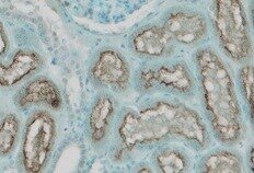
ACE‑2 in Hamster Lung.
ACE‑2 was detected in immersion fixed paraffin-embedded sections of hamster lung using Mouse Anti-Human ACE‑2 Monoclonal Antibody (Catalog # MAB9332) at 10 µg/mL for 1 hour at room temperature followed by incubation with the Anti-Mouse IgG VisUCyte™ HRP Polymer Antibody (VC001). Before incubation with the primary antibody, tissue was subjected to heat-induced epitope retrieval using Antigen Retrieval Reagent-Basic (CTS013). Tissue was stained using DAB (brown) and counterstained with hematoxylin (blue). Specific staining was localized to respiratory bronchioles. Staining was performed using our protocol for IHC Staining with VisUCyte HRP Polymer Detection Reagents.
Detection of Human Human/Rat/Hamster ACE-2 Antibody by Western Blot
Sialic acid has modest effect on Spike binding and viral entry. (B) Western blot for purified Fc-proteins from HEK293T probed with anti-Fc, anti-RBD or anti-ACE2 Ab. CD44-Fc is positive control. Image collected and cropped by CiteAb from the following publication (https://pubmed.ncbi.nlm.nih.gov/33103998), licensed under a CC-BY license. Not internally tested by R&D Systems.Applications for ACE-2 Antibody (535919)
Application
Recommended Usage
CyTOF-ready
Ready to be labeled using established conjugation methods. No BSA or other carrier proteins that could interfere with conjugation.
Flow Cytometry
0.25 µg/106 cells
Sample: HEK293 Human Cell Line Transfected with Human ACE-2 and eGFP
Sample: HEK293 Human Cell Line Transfected with Human ACE-2 and eGFP
Immunohistochemistry
1-25 µg/mL
Sample: Frozen rat kidney and paraffin-embedded hamster lung sections.
Sample: Frozen rat kidney and paraffin-embedded hamster lung sections.
Western Blot
2 µg/mL
Sample: Human kidney and human testis
Sample: Human kidney and human testis
Reviewed Applications
Read 1 review rated 5 using MAB9332 in the following applications:
Flow Cytometry Panel Builder
Bio-Techne Knows Flow Cytometry
Save time and reduce costly mistakes by quickly finding compatible reagents using the Panel Builder Tool.
Advanced Features
- Spectra Viewer - Custom analysis of spectra from multiple fluorochromes
- Spillover Popups - Visualize the spectra of individual fluorochromes
- Antigen Density Selector - Match fluorochrome brightness with antigen density

Formulation, Preparation, and Storage
Purification
Protein A or G purified from hybridoma culture supernatant
Reconstitution
Reconstitute at 0.5 mg/mL in sterile PBS. For liquid material, refer to CoA for concentration.
Loading...
Formulation
Lyophilized from a 0.2 μm filtered solution in PBS with Trehalose. *Small pack size (SP) is supplied either lyophilized or as a 0.2 µm filtered solution in PBS.
Shipping
Lyophilized product is shipped at ambient temperature. Liquid small pack size (-SP) is shipped with polar packs. Upon receipt, store immediately at the temperature recommended below.
Stability & Storage
Use a manual defrost freezer and avoid repeated freeze-thaw cycles.
- 12 months from date of receipt, -20 to -70 °C as supplied.
- 1 month, 2 to 8 °C under sterile conditions after reconstitution.
- 6 months, -20 to -70 °C under sterile conditions after reconstitution.
Calculators
Background: ACE-2
References
- Tipnis, S.R. et al. (2000) J. Biol. Chem. 275:33238.
- Crackower, M.A. et al. (2002) Nature 417:822.
- Huang, L. et al. (2003) J. Biol. Chem. 278:15532.
Long Name
Angiotensin I Converting Enzyme 2
Alternate Names
ACE2, ACEH
Entrez Gene IDs
Gene Symbol
ACE2
UniProt
Additional ACE-2 Products
Product Documents for ACE-2 Antibody (535919)
Certificate of Analysis
To download a Certificate of Analysis, please enter a lot or batch number in the search box below.
Note: Certificate of Analysis not available for kit components.
Product Specific Notices for ACE-2 Antibody (535919)
For research use only
Related Research Areas
Citations for ACE-2 Antibody (535919)
 Powered by Bioz
Customer Reviews for ACE-2 Antibody (535919) (1)
5 out of 5
1 Customer Rating
Have you used ACE-2 Antibody (535919)?
Submit a review and receive an Amazon gift card!
$25/€18/£15/$25CAN/¥2500 Yen for a review with an image
$10/€7/£6/$10CAN/¥1110 Yen for a review without an image
Submit a review
Customer Images
Showing
1
-
1 of
1 review
Showing All
Filter By:
-
Application: ImmunohistochemistrySample Tested: Kidney tissueSpecies: RatVerified Customer | Posted 09/10/2021

There are no reviews that match your criteria.
Protocols
Find general support by application which include: protocols, troubleshooting, illustrated assays, videos and webinars.
- 7-Amino Actinomycin D (7-AAD) Cell Viability Flow Cytometry Protocol
- Antigen Retrieval Protocol (PIER)
- Antigen Retrieval for Frozen Sections Protocol
- Appropriate Fixation of IHC/ICC Samples
- Cellular Response to Hypoxia Protocols
- Chromogenic IHC Staining of Formalin-Fixed Paraffin-Embedded (FFPE) Tissue Protocol
- Chromogenic Immunohistochemistry Staining of Frozen Tissue
- ClariTSA™ Fluorophore Kits
- Detection & Visualization of Antibody Binding
- Extracellular Membrane Flow Cytometry Protocol
- Flow Cytometry Protocol for Cell Surface Markers
- Flow Cytometry Protocol for Staining Membrane Associated Proteins
- Flow Cytometry Staining Protocols
- Flow Cytometry Troubleshooting Guide
- Fluorescent IHC Staining of Frozen Tissue Protocol
- Graphic Protocol for Heat-induced Epitope Retrieval
- Graphic Protocol for the Preparation and Fluorescent IHC Staining of Frozen Tissue Sections
- Graphic Protocol for the Preparation and Fluorescent IHC Staining of Paraffin-embedded Tissue Sections
- Graphic Protocol for the Preparation of Gelatin-coated Slides for Histological Tissue Sections
- IHC Sample Preparation (Frozen sections vs Paraffin)
- Immunofluorescent IHC Staining of Formalin-Fixed Paraffin-Embedded (FFPE) Tissue Protocol
- Immunohistochemistry (IHC) and Immunocytochemistry (ICC) Protocols
- Immunohistochemistry Frozen Troubleshooting
- Immunohistochemistry Paraffin Troubleshooting
- Intracellular Flow Cytometry Protocol Using Alcohol (Methanol)
- Intracellular Flow Cytometry Protocol Using Detergents
- Intracellular Nuclear Staining Flow Cytometry Protocol Using Detergents
- Intracellular Staining Flow Cytometry Protocol Using Alcohol Permeabilization
- Intracellular Staining Flow Cytometry Protocol Using Detergents to Permeabilize Cells
- Preparing Samples for IHC/ICC Experiments
- Preventing Non-Specific Staining (Non-Specific Binding)
- Primary Antibody Selection & Optimization
- Propidium Iodide Cell Viability Flow Cytometry Protocol
- Protocol for Heat-Induced Epitope Retrieval (HIER)
- Protocol for Liperfluo
- Protocol for Making a 4% Formaldehyde Solution in PBS
- Protocol for VisUCyte™ HRP Polymer Detection Reagent
- Protocol for the Characterization of Human Th22 Cells
- Protocol for the Characterization of Human Th9 Cells
- Protocol for the Preparation & Fixation of Cells on Coverslips
- Protocol for the Preparation and Chromogenic IHC Staining of Frozen Tissue Sections
- Protocol for the Preparation and Chromogenic IHC Staining of Frozen Tissue Sections - Graphic
- Protocol for the Preparation and Chromogenic IHC Staining of Paraffin-embedded Tissue Sections
- Protocol for the Preparation and Chromogenic IHC Staining of Paraffin-embedded Tissue Sections - Graphic
- Protocol for the Preparation and Fluorescent IHC Staining of Frozen Tissue Sections
- Protocol for the Preparation and Fluorescent IHC Staining of Paraffin-embedded Tissue Sections
- Protocol for the Preparation of Gelatin-coated Slides for Histological Tissue Sections
- Protocol: Annexin V and PI Staining by Flow Cytometry
- Protocol: Annexin V and PI Staining for Apoptosis by Flow Cytometry
- R&D Systems Quality Control Western Blot Protocol
- TUNEL and Active Caspase-3 Detection by IHC/ICC Protocol
- The Importance of IHC/ICC Controls
- Troubleshooting Guide: Fluorokine Flow Cytometry Kits
- Troubleshooting Guide: Immunohistochemistry
- Troubleshooting Guide: Western Blot Figures
- Western Blot Conditions
- Western Blot Protocol
- Western Blot Protocol for Cell Lysates
- Western Blot Troubleshooting
- Western Blot Troubleshooting Guide
- View all Protocols, Troubleshooting, Illustrated assays and Webinars
Loading...
Associated Pathways

