
Key Product Details
Species Reactivity
Validated:
Human
Cited:
Human, Rat, Xenograft
Applications
Validated:
Immunohistochemistry, Western Blot, Neutralization, Simple Western, Immunoprecipitation
Cited:
Immunohistochemistry, Immunohistochemistry-Frozen, Western Blot, Neutralization, Dot Blot
Label
Unconjugated
Antibody Source
Polyclonal Goat IgG
Loading...
Product Specifications
Immunogen
S. frugiperda insect ovarian cell line Sf 21-derived recombinant human Serpin E1/PAI‑1
Gly21-Pro402
Accession # P05121
Gly21-Pro402
Accession # P05121
Specificity
Detects human Serpin E1/PAI‑1 in direct ELISAs and Western blots. In direct ELISAs, approximately 15% cross-reactivity with recombinant mouse Serpin E1 is observed.
Clonality
Polyclonal
Host
Goat
Isotype
IgG
Scientific Data Images for Human Serpin E1/PAI-1 Antibody
Detection of Human Serpin E1/PAI‑1 by Western Blot.
Western blot shows lysates of HUVEC human umbilical vein endothelial cells. PVDF membrane was probed with 1 µg/mL of Goat Anti-Human Serpin E1/PAI-1 Antigen Affinity-purified Polyclonal Antibody (Catalog # AF1786) followed by HRP-conjugated Anti-Goat IgG Secondary Antibody (Catalog # HAF017). A specific band was detected for Serpin E1/PAI-1 at approximately 45 kDa (as indicated). This experiment was conducted under reducing conditions and using Immunoblot Buffer Group 1.
Serpin E1/PAI‑1 in Human Liver Cancer Tissue.
Serpin E1/PAI-1 was detected in immersion fixed paraffin-embedded sections of human liver cancer tissue using Goat Anti-Human Serpin E1/PAI-1 Antigen Affinity-purified Polyclonal Antibody (Catalog # AF1786) at 15 µg/mL overnight at 4 °C. Tissue was stained using the Anti-Goat HRP-DAB Cell & Tissue Staining Kit (brown; Catalog # CTS008) and counterstained with hematoxylin (blue). View our protocol for Chromogenic IHC Staining of Paraffin-embedded Tissue Sections.
Detection of Human Serpin E1/PAI‑1 by Simple WesternTM.
Simple Western lane view shows lysates of HUVEC human umbilical vein endothelial cells, loaded at 0.2 mg/mL. A specific band was detected for Serpin E1/PAI‑1 at approximately 54 kDa (as indicated) using 2.5 µg/mL of Goat Anti-Human Serpin E1/PAI‑1 Antigen Affinity-purified Polyclonal Antibody (Catalog # AF1786) followed by 1:50 dilution of HRP-conjugated Anti-Goat IgG Secondary Antibody (Catalog # HAF109). This experiment was conducted under reducing conditions and using the 12-230 kDa separation system.
Neutralization of Serpin E1/ PAI‑1 Activity by Human Serpin E1/PAI‑1 Antibody.
Recombinant Human u-Plasminogen Activator (uPA)/Urokinase (0.1 µg/mL, Catalog # 1310-SE) activity is measured in the presence of Recombinant Human Serpin E1 (1.35 µg/mL, Catalog # 1786-PI) that has been preincubated with increasing concentrations of Goat Anti-Human Serpin E1/PAI-1 Antigen Affinity-purified Polyclonal Antibody (Catalog # AF1786). The ND50 is typically 10 µg/mL.
Detection of Serpin E1/PAI-1 by Immunohistochemistry
The expression of PAI-1 and LRP1 in human ESCC tissues. A Immunohistochemical staining for PAI-1 in human ESCC tissues. Representative images are shown: PAI-1 low-intensity in the stroma (left) and PAI-1 high-intensity in the stroma (right). Scale bars = 100 μm. b High-power fields of immunohistochemical staining for PAI-1 in same cases as Fig. 6a. Scale bars = 20 μm. Arrow heads: vascular endothelial cell. c Immunohistochemical staining for LRP1 in cancer nest of human ESCC tissues. Representative images are shown: LRP1 low-intensity in cancer nest (left) and LRP1 high-intensity in cancer nest (right). Scale bars = 100 μm. d Immunohistochemical staining for LRP1 in the stroma of human ESCC tissues. Representative images are shown: LRP1 low-intensity in the stroma (left) and LRP1 high-intensity in the stroma (right). Scale bars = 20 μm. e Stromal cells, including CD204+ TAMs, expressed LRP1 in human ESCC tissue. Double immunofluorescence was performed using anti-LRP1 (green) and anti-CD204 (red) antibodies in formalin-fixed paraffin-embedded section of human ESCC tissue. LRP1 was expressed in both cancer cells and stromal cells, and some of the LRP1-positive stromal cells were CD204+ macrophages (arrow heads). Nuclei were stained with DAPI (blue). Scale bar = 20 μm. CA cancer nest. ST stroma. Image collected and cropped by CiteAb from the following open publication (https://pubmed.ncbi.nlm.nih.gov/33311557), licensed under a CC-BY license. Not internally tested by R&D Systems.
Detection of Serpin E1/PAI-1 by Immunohistochemistry
The expression of PAI-1 and LRP1 in human ESCC tissues. A Immunohistochemical staining for PAI-1 in human ESCC tissues. Representative images are shown: PAI-1 low-intensity in the stroma (left) and PAI-1 high-intensity in the stroma (right). Scale bars = 100 μm. b High-power fields of immunohistochemical staining for PAI-1 in same cases as Fig. 6a. Scale bars = 20 μm. Arrow heads: vascular endothelial cell. c Immunohistochemical staining for LRP1 in cancer nest of human ESCC tissues. Representative images are shown: LRP1 low-intensity in cancer nest (left) and LRP1 high-intensity in cancer nest (right). Scale bars = 100 μm. d Immunohistochemical staining for LRP1 in the stroma of human ESCC tissues. Representative images are shown: LRP1 low-intensity in the stroma (left) and LRP1 high-intensity in the stroma (right). Scale bars = 20 μm. e Stromal cells, including CD204+ TAMs, expressed LRP1 in human ESCC tissue. Double immunofluorescence was performed using anti-LRP1 (green) and anti-CD204 (red) antibodies in formalin-fixed paraffin-embedded section of human ESCC tissue. LRP1 was expressed in both cancer cells and stromal cells, and some of the LRP1-positive stromal cells were CD204+ macrophages (arrow heads). Nuclei were stained with DAPI (blue). Scale bar = 20 μm. CA cancer nest. ST stroma. Image collected and cropped by CiteAb from the following open publication (https://pubmed.ncbi.nlm.nih.gov/33311557), licensed under a CC-BY license. Not internally tested by R&D Systems.Applications for Human Serpin E1/PAI-1 Antibody
Application
Recommended Usage
Immunohistochemistry
5-15 µg/mL
Sample: Immersion fixed paraffin-embedded sections of human breast cancer tissue and immersion fixed paraffin-embedded sections of human liver cancer tissue
Sample: Immersion fixed paraffin-embedded sections of human breast cancer tissue and immersion fixed paraffin-embedded sections of human liver cancer tissue
Immunoprecipitation
25 µg/mL
Sample: Conditioned cell culture medium spiked with Recombinant Human Serpin E1/PAI‑1 (Catalog # 1786-PI), see our available Western blot detection antibodies
Sample: Conditioned cell culture medium spiked with Recombinant Human Serpin E1/PAI‑1 (Catalog # 1786-PI), see our available Western blot detection antibodies
Simple Western
2.5 µg/mL
Sample: HUVEC human umbilical vein endothelial cells
Sample: HUVEC human umbilical vein endothelial cells
Western Blot
1 µg/mL
Sample: HUVEC human umbilical vein endothelial cells
Sample: HUVEC human umbilical vein endothelial cells
Neutralization
Measured by its ability to neutralize Recombinant Human Serpin E1 (1.35 µg/mL, Catalog # 1786-PI) inhibition of Recombinant Human u‑Plasminogen Activator (uPA)/Urokinase (0.1 µg/mL, Catalog # 1310-SE) cleavage of the fluorogenic peptide substrate Z-GGR-AMC (100 µM). The Neutralization Dose (ND50) is typically 10 µg/mL.
Reviewed Applications
Read 1 review rated 4 using AF1786 in the following applications:
Formulation, Preparation, and Storage
Purification
Antigen Affinity-purified
Reconstitution
Reconstitute at 0.2 mg/mL in sterile PBS. For liquid material, refer to CoA for concentration.
Loading...
Formulation
Lyophilized from a 0.2 μm filtered solution in PBS with Trehalose. *Small pack size (SP) is supplied either lyophilized or as a 0.2 µm filtered solution in PBS.
Shipping
Lyophilized product is shipped at ambient temperature. Liquid small pack size (-SP) is shipped with polar packs. Upon receipt, store immediately at the temperature recommended below.
Stability & Storage
Use a manual defrost freezer and avoid repeated freeze-thaw cycles.
- 12 months from date of receipt, -20 to -70 °C as supplied.
- 1 month, 2 to 8 °C under sterile conditions after reconstitution.
- 6 months, -20 to -70 °C under sterile conditions after reconstitution.
Calculators
Background: Serpin E1/PAI-1
References
- Silverman, G.A. et al. (2001) J. Biol. Chem. 276:33293.
- Stefansson, S. et al. (2003) Curr. Pharm. Des. 9:1545.
- Duffy, M.J. (2002) Clin. Chem. 48:1194.
- Juhan-Vague, I. et al. (2003) J. Thromb. Haemost. 1:1575.
- Harbeck, N. et al. (2002) Clin. Breast Cancer 3:196.
- Pannekoek, H. et al. (1986) EMBO J. 5:2539.
- Ginsburg, D. et al. (1986) J. Clin. Invest. 78:1673.
- Wang, Z. et al. (1996) Biochemistry 35:16443.
- Stromqvist, M. et al. (1994) Protein Expr. Purif. 5:309.
Long Name
Plasminogen Activator Inhibitor
Alternate Names
Nexin, PAI-1, PAI1, PLANH1
Gene Symbol
SERPINE1
UniProt
Additional Serpin E1/PAI-1 Products
Product Documents for Human Serpin E1/PAI-1 Antibody
Certificate of Analysis
To download a Certificate of Analysis, please enter a lot or batch number in the search box below.
Note: Certificate of Analysis not available for kit components.
Product Specific Notices for Human Serpin E1/PAI-1 Antibody
For research use only
Citations for Human Serpin E1/PAI-1 Antibody
 Powered by Bioz
Customer Reviews for Human Serpin E1/PAI-1 Antibody (1)
4 out of 5
1 Customer Rating
Have you used Human Serpin E1/PAI-1 Antibody?
Submit a review and receive an Amazon gift card!
$25/€18/£15/$25CAN/¥2500 Yen for a review with an image
$10/€7/£6/$10CAN/¥1110 Yen for a review without an image
Submit a review
Customer Images
Showing
1
-
1 of
1 review
Showing All
Filter By:
-
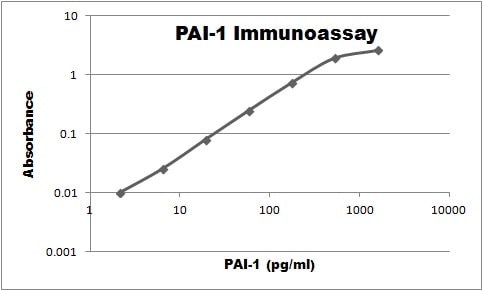
Application: ELISASample Tested: SerumSpecies: HumanVerified Customer | Posted 11/07/2018Combined with MAB1786 this antibody was used to make an immunoassay that was able to detect 3pg/ml of PAI-1.

There are no reviews that match your criteria.
Protocols
Find general support by application which include: protocols, troubleshooting, illustrated assays, videos and webinars.
- Antigen Retrieval Protocol (PIER)
- Antigen Retrieval for Frozen Sections Protocol
- Appropriate Fixation of IHC/ICC Samples
- Cellular Response to Hypoxia Protocols
- Chromogenic IHC Staining of Formalin-Fixed Paraffin-Embedded (FFPE) Tissue Protocol
- Chromogenic Immunohistochemistry Staining of Frozen Tissue
- ClariTSA™ Fluorophore Kits
- Detection & Visualization of Antibody Binding
- Fluorescent IHC Staining of Frozen Tissue Protocol
- Graphic Protocol for Heat-induced Epitope Retrieval
- Graphic Protocol for the Preparation and Fluorescent IHC Staining of Frozen Tissue Sections
- Graphic Protocol for the Preparation and Fluorescent IHC Staining of Paraffin-embedded Tissue Sections
- Graphic Protocol for the Preparation of Gelatin-coated Slides for Histological Tissue Sections
- IHC Sample Preparation (Frozen sections vs Paraffin)
- Immunofluorescent IHC Staining of Formalin-Fixed Paraffin-Embedded (FFPE) Tissue Protocol
- Immunohistochemistry (IHC) and Immunocytochemistry (ICC) Protocols
- Immunohistochemistry Frozen Troubleshooting
- Immunohistochemistry Paraffin Troubleshooting
- Immunoprecipitation Protocol
- Preparing Samples for IHC/ICC Experiments
- Preventing Non-Specific Staining (Non-Specific Binding)
- Primary Antibody Selection & Optimization
- Protocol for Heat-Induced Epitope Retrieval (HIER)
- Protocol for Making a 4% Formaldehyde Solution in PBS
- Protocol for VisUCyte™ HRP Polymer Detection Reagent
- Protocol for the Preparation & Fixation of Cells on Coverslips
- Protocol for the Preparation and Chromogenic IHC Staining of Frozen Tissue Sections
- Protocol for the Preparation and Chromogenic IHC Staining of Frozen Tissue Sections - Graphic
- Protocol for the Preparation and Chromogenic IHC Staining of Paraffin-embedded Tissue Sections
- Protocol for the Preparation and Chromogenic IHC Staining of Paraffin-embedded Tissue Sections - Graphic
- Protocol for the Preparation and Fluorescent IHC Staining of Frozen Tissue Sections
- Protocol for the Preparation and Fluorescent IHC Staining of Paraffin-embedded Tissue Sections
- Protocol for the Preparation of Gelatin-coated Slides for Histological Tissue Sections
- R&D Systems Quality Control Western Blot Protocol
- TUNEL and Active Caspase-3 Detection by IHC/ICC Protocol
- The Importance of IHC/ICC Controls
- Troubleshooting Guide: Immunohistochemistry
- Troubleshooting Guide: Western Blot Figures
- Western Blot Conditions
- Western Blot Protocol
- Western Blot Protocol for Cell Lysates
- Western Blot Troubleshooting
- Western Blot Troubleshooting Guide
- View all Protocols, Troubleshooting, Illustrated assays and Webinars
Loading...
Associated Pathways
